Introduction
Gastric cancer (GC) is a common malignant tumor with
high morbidity and mortality rates (1). The radical treatment for advanced GC
is surgical resection. However, 40–60% of patients with GC develop
recurrence and metastasis after surgery and the 5-year overall
survival rate of patients with advanced GC is only 10–21% (2,3).
Therefore, the molecular mechanisms underlying the occurrence and
metastasis of GC, as well as the search for potential diagnostic
and therapeutic targets, have become hotspots in the basic research
field of the molecular diagnosis and treatment of GC.
Circular RNAs (circRNAs/circs) are a class of
non-coding RNAs, which have a special closed ring structure and are
widely present in most organisms (4). circRNAs can regulate gene function
at several levels, such as epigenetic, transcriptional and
post-transcriptional levels (5).
MicroRNAs (miRNAs/miRs) comprise a group of non-coding RNA
molecules, 20-25 nucleotides in length, which can bind to the
3′-untranslated region (UTR) of target genes, causing their
degradation or inhibiting their translation (6). Some studies have revealed that
circRNAs and miRNAs are abnormally expressed in various tumor
tissues and cells, and they may participate in the whole process of
tumor development (7–9). For example, circ_BTG3 associated
nuclear protein-mediated miR-503/La ribonucleoprotein 1,
translational regulator signaling was shown to promote lung cancer
development and progression (10). Moreover, circ_5692 targets
miR-328-5p to inhibit the occurrence of liver cancer (11). Another study reported that
circ_homeodomain interacting protein kinase 3 promoted colorectal
cancer development and metastasis by sponging miR-7 (12). circ_0067934 is a newly identified
circRNA. A systematic meta-analysis based on an integrated dataset
pre-processed from three microarrays suggested that circ_0067934
may represent an authentic biomarker for GC screening (13). Previous studies have shown that
circ_0067934 may be involved in several types of cancer, including
breast cancer, non-small cell lung cancer (NSCLC) and bladder
cancer (14–16). However, the role of circ_0067934
in GC remains elusive.
Therefore, the aim of the present study was to
investigate the possible role of circ_0067934 in the proliferation,
invasion and migration of GC cells, as well as elucidate its
underlying mechanism.
Materials and methods
Bioinformatic analysis
The starBase database (version 3.0; http://starbase.sysu.edu.cn/index.php/)
was used to predict the binding site of circ_0067934 and miR-5047,
miR-1301-3p, miR-670-5p, miR-345-3p, miR-545-3p and miR-3605-5p. In
addition, the binding site of miR-1301-3p and KIF23 was predicted
using the starBase database.
Cell culture
The GES-1 normal gastric mucosal cell line, and the
GC cell lines, AGS and MKN-45 (China Infrastructure of Cell Line
Resources, Institute of Basic Medical Sciences, Chinese Academy of
Medical Sciences), were cultured in RPMI-1640 medium (Gibco; Thermo
Fisher Scientific, Inc.) containing 11 mM glucose, 10% (v/v) FBS
(cat. no. 10099141; Gibco; Thermo Fisher Scientific, Inc.), 50 µM
β-mercaptoethanol (cat. no. M3148; MilliporeSigma) and 10 mM HEPES
(cat. no. H1090; Beijing Solarbio Science & Technology Co.,
Ltd.) at 37°C with 5% CO2.
Cell transfection
Short hairpin RNA (shRNA/sh)-circ_0067934-1/2 and
corresponding negative control (NC) shRNA (sh-NC), miR-1301-3p
mimic and its NC (mimic-NC), a pcDNA3.1 expression vector
containing full-length human KIF23 (Ov-KIF23) and corresponding NCs
(Ov-NC) were obtained from Shanghai GenePharma Co., Ltd. The
sequence for the miR-1301-3p mimic was
5′-UUGCAGCUGCCUGGGAGUGACUUC-3′. The NC shRNA sequence was
5′-UCACAACCUCCUAGAAAGAGUAGA-3′. The AGS cell suspension was
inoculated into a 6-well plate and cultured in an incubator with 5%
CO2 at 37°C for 24 h. The cultured cells were
transfected with 100 nM of these recombinants using 2.5 µl/ml
Lipofectamine® 2000 transfection reagent (Thermo Fisher
Scientific, Inc.) at 37°C for 48 h in accordance with the
manufacturer's instructions. After transfection, the transfection
efficacy of the cells was detected via reverse
transcription-quantitative (RT-q)PCR.
Cell proliferation assay
AGS cells from each group were collected after
transfection for 48 h. After being counted, cells (1×10³
cells/well) were seeded into a 96-well cell culture plate and
cultured in an incubator with 5% CO2 at 37°C. After 24,
48 and 72 h, 10 µl Cell Counting Kit-8 (CCK-8) reagent (Beyotime
Institute of Biotechnology) was added to each well in accordance
with the manufacturer's instructions. Then, the cells were
incubated at 37°C in the dark for 2 h. The absorbance of each well
was measured at 450 nm with a microplate reader (Bio-Rad
Laboratories, Inc.). Each experiment was repeated ≥3 times. In
total, six duplicate wells were examined in each group.
EdU cell proliferation assay
AGS cells were seeded into 6-well plates (500
cells/well) and incubated at room temperature overnight. EdU
solution (Beyotime Institute of Biotechnology) was added and the
cells were incubated for 4 h at 37°C. After the working fluid was
removed, the cells were fixed with 4% paraformaldehyde for 15 min
at room temperature. The cells were then permeated with 0.5% Triton
X-100 for 15 min. After the addition of Click reaction solution,
the cells were incubated in the dark for 30 min at 37°C and imaged
under a fluorescence microscope (Leica Microsystems GmbH;
magnification, ×200).
Western blotting
AGS cells were harvested and total protein was
extracted using RIPA lysis buffer (Beyotime Institute of
Biotechnology). Total protein was quantified using a protein
concentration determination kit (cat. no. P0012; Beyotime Institute
of Biotechnology). Proteins (30 µg/lane) were separated via
SDS-PAGE (15%) and were subsequently transferred onto PVDF
membranes (MilliporeSigma). After blocking with 5% BSA (Beyotime
Institute of Biotechnology) at room temperature for 2 h, the
membranes were then incubated with the following primary antibodies
(Abcam) at 4°C overnight: Anti-Ki67 (cat. no. ab15580; 1:1,000),
anti-proliferating cell nuclear antigen (anti-PCNA; cat. no.
ab92552; 1:1,000), anti-MMP2 (cat. no. ab92536; 1:1,000), anti-MMP9
(cat. no. ab76003; 1:1,000) and anti-KIF23 (cat. no. ab174304;
1:1,000). After the incubation with the primary antibodies, the
membranes were washed with TBS containing Tween-20 (0.1%) and
incubated at room temperature for 1.5 h with HRP-conjugated
secondary antibodies [Goat Anti-Mouse IgG(H+L), 1:2,000, cat. no.
SA00001-1; Goat Anti-Rabbit IgG(H+L), 1:2,000, cat. no. SA00001-2;
ProteinTech Group, Inc.]. Protein bands were visualized using the
Odyssey Western Blot Analysis system (LI-COR Biosciences) and
semi-quantified using ImageJ software, version 7.6.5 (National
Institutes of Health).
Transwell assay
The invasive ability of the cells was examined using
a Transwell invasion assay. The Transwell chambers (Costar;
Corning, Inc.) were first coated with 0.1 ml Matrigel
(Becton-Dickinson and Company) at 37°C for 1 h. AGS cells
(5×10⁵ cells/well) were collected and suspended at
2×10⁵ cells/ml in serum-free DMEM. Cell suspensions were
placed into the upper compartment of a Transwell chamber and
cultured in 5% CO2 at 37°C. Medium supplemented with 10%
FBS was added into the lower compartment of the Transwell chamber.
After 24 h, the non-invaded cells on the upper face of the
Transwell membrane were wiped off with a cotton swab. The cells
invading to the lower surface of the filter were fixed with 4%
paraformaldehyde and stained with 0.1% crystal violet solution for
30 min at room temperature. Images were captured using a light
microscope at ×100 magnification.
Wound healing assay
AGS cells were cultured in 6-well plates
(6×10⁴ cells/well) and incubated to 80–90% confluence in
RPMI-1640 medium with 10% FBS at 37°C (17). A linear scratch was created in the
cell monolayer using a 200-µl pipette tip. The cells were
subsequently cultured under standard conditions in RPMI-1640 medium
containing 2% FBS (Procell Life Science & Technology Co., Ltd.)
for 48 h at 37°C in 5% CO2. The area occupied by cells
migrating into the scratch was evaluated using an inverted
microscope at ×100 magnification. The migration rate was calculated
based on the formula: (Wound width at 0 h - wound width at 24
h)/wound width at 0 h × 100%. In total, five fields were randomly
selected for analysis in each well.
Nuclear and cytoplasmic separation
experiment
AGS cells were lysed and centrifuged for 5 min at
500 × g at 4°C. Then, the cytoplasmic components retained in the
supernatant were collected. The precipitated nuclear components
were further cleaved. The collected cytoplasmic components and
nuclear lysates were mixed with 2X lysis/binding solution and then
pumped through a filter tube. The extracted cytoplasmic and nuclear
RNA were reverse-transcribed, and the expression level of
circ_0067934 was detected via RT-qPCR. U6 was the positive control
for the detection of nuclear RNA expression, and GAPDH was the
positive control for the detection of cytoplasmic RNA expression
(18).
Luciferase reporter assay
The circ_0067934 3′-UTR, containing wild-type (WT)
or mutant (MUT) target sites for miR-1301-3p, was amplified via PCR
and inserted into a pGL3 vector (Promega Corporation) to form the
reporter vector circ_0067934-WT or circ_0067934-MUT. Then, the DNA
sequence of KIF23 3′-UTR that included the putative binding sites
of miR-1301-3p was subcloned into a pGL3 vector (Promega
Corporation) to create a luciferase plasmid containing the WT
sequence of KIF23 3′-UTR. The miR-1301-3p binding sites were
mutated, and the MUT KIF23 3′-UTR sequence was subcloned into the
pGL3 vector so as to create a mutant reporter vector. AGS cells
were co-transfected with luciferase reporter vectors by using
Lipofectamine® 2000 (Invitrogen; Thermo Fisher
Scientific, Inc.) for 48 h. At 48 h after transfection, the
relative luciferase activities were measured by using a
Dual-Luciferase Reporter Assay (Promega Corporation) and luciferase
activities were normalized against Renilla luciferase.
RT-qPCR analysis
Total RNA from cell samples was extracted using
TRIzol® reagent (Thermo Fisher Scientific, Inc.) and
reverse-transcribed into cDNA using a commercial RevertAid™ cDNA
Synthesis kit (Takara Bio, Inc.) at 42°C for ~1 h and 90°C for 5
min. The SYBR Premix Ex Taq™ II kit (Thermo Fisher Scientific,
Inc.) was applied for qPCR. The PCR reaction mixture contained 3 mM
MgCl2, 0.5 µM forward and reverse primers, 2 µl SYBR
Green PCR master mix and 2 µl cDNA on a QuantStudio 3 Real-Time PCR
system (Applied Biosystems; Thermo Fisher Scientific, Inc.). The
primers used are shown in Table
I. The thermocycling conditions of the qPCR were as follows:
Initial denaturation for 5 min at 95°C, followed by 40 cycles for
30 sec at 95°C, annealing at 65°C for 45 sec and extension at 72°C
for 1 min. A final extension step at 72°C for 7 min was performed
in each PCR assay. The results were measured using the
2−ΔΔCq method (19).
U6 served as an endogenous control for miRNAs and GAPDH was used as
an endogenous control for circ_0067934 and KIF23.
 | Table I.Sequences of primers for reverse
transcription-quantitative PCR. |
Table I.
Sequences of primers for reverse
transcription-quantitative PCR.
| Gene | Sequence |
|---|
| circ_0067934 | F:
5′-TAGCAGTTCCCCAATCCTTG-3′ |
|
| R:
5′-CACAAATTCCCATCATTCCC-3′ |
| miR-5047 | F:
5′-GCCTAGACGAGACACAGTGC-3′ |
|
| R:
5′-GCCAAGACCTTACAACCGCA-3′ |
| miR-1301-3p | F:
5′-GCCCGCTTGCAGCTGCCTGGGAG-3′ |
|
| R:
5′-GTGCAGGGTCCGAGGT-3′ |
| miR-670-5p | F:
5′-AAACCCATGACACAGCACAA-3′ |
|
| R:
5′-ATTGCTTTGCCTCCAGAAGA-3′ |
| miR-345-3p | F:
5′-GCCGAGAGGGGTCTGGAGA-3′ |
|
| R:
5′-CTCAACTGGTGTCGTGGA-3′ |
| miR-545-3p | F:
5′-TGGCTCAGTTCAGCAGGAAC-3′ |
|
| R:
5′-CATTACTGGATCTATCAACAGG-3′ |
| KIF23 | F:
5′-AGACAGAAGGCGAGGGATG-3′ |
|
| R:
5′-GGAGACGAATTGGTGGTGC-3′ |
| U6 | F:
5′-CTCGCTTCGGCAGCACATATA-3′ |
|
| R:
5′-ACGCTTCACGAATTTGAGTGTC-3′ |
| GAPDH | F:
5′-GTCAAGGCTGAGAACGGGAA-3′ |
|
| R:
5′-AAATGAGCCCCAGCCTTCTC-3′ |
Statistical analysis
Statistical analysis was performed using GraphPad
Prism 5 (GraphPad Software, Inc.). All data are presented as the
mean ± SD of three independent experiments. Statistical differences
between two groups were determined using an unpaired two-tailed
Student's t-test, while one-way followed by Tukey's post hoc test
was used to analyze data in >2 groups. P<0.05 was considered
to indicate a statistically significant difference.
Results
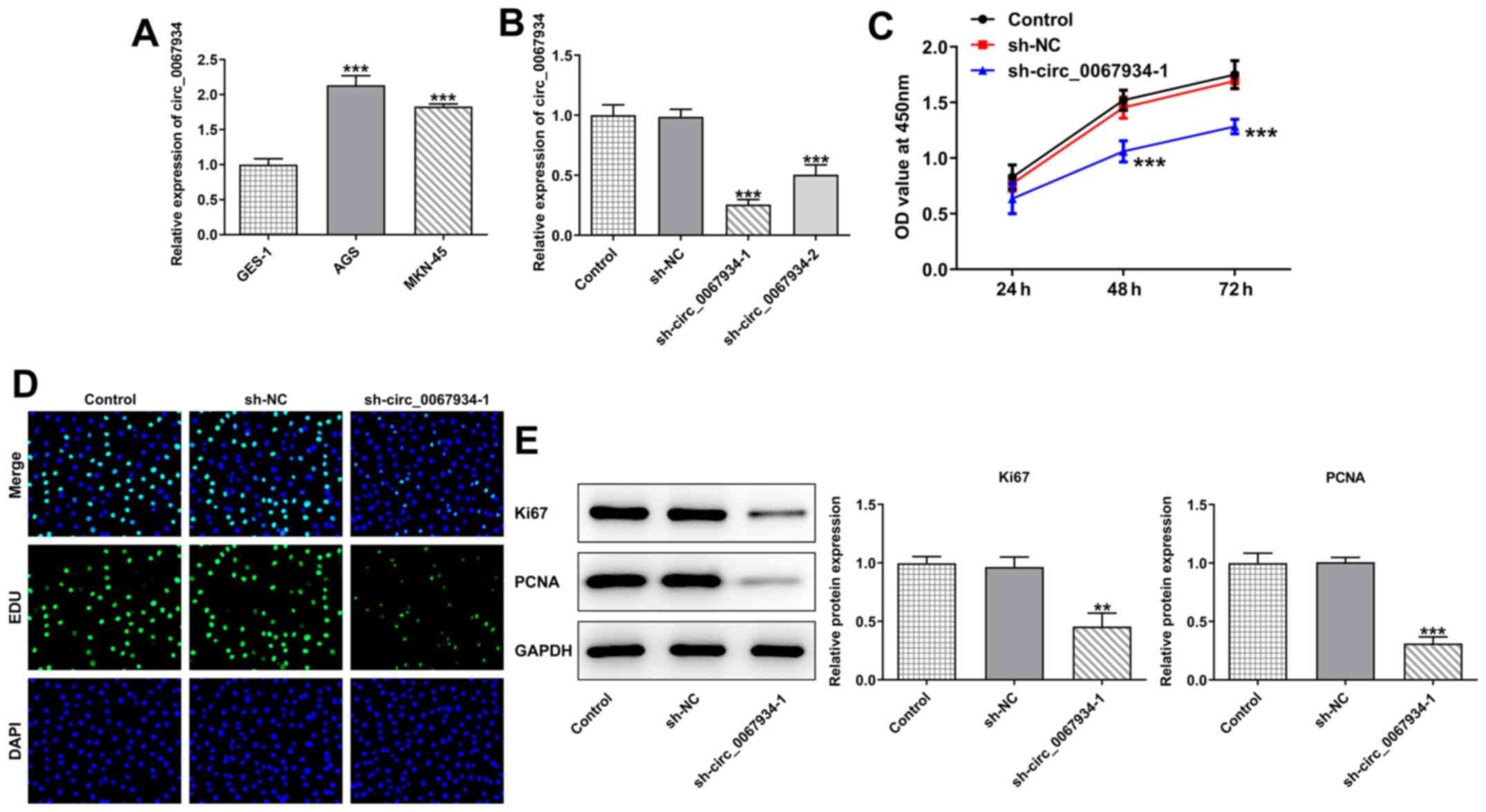
circ_0067934 silencing inhibits AGS
cell proliferation
To investigate the role of circ_0067934 in GC cells,
the expression level of circ_0067934 in GC cells was firstly
detected via RT-qPCR. The results demonstrated that the expression
level of circ_0067934 was upregulated in the GC cell lines, AGS and
MKN-45, compared with the normal gastric mucosa cells, GES-1
(Fig. 1A). AGS cells showed the
highest expression of circ_0067934, so these cells were selected
for subsequent experiments.
Cells were then transfected with sh-circ_0067934-1/2
to knock down the expression of circ_0067934 (Fig. 1B). Compared with the sh-NC group,
the cell proliferation ability of the sh-circ_0067934-1 group was
significantly decreased (Fig. 1C and
D). In addition, the results of western blotting revealed that
the expression levels of Ki67 and PCNA were downregulated following
transfection with sh-circ_0067934 (Fig. 1E).
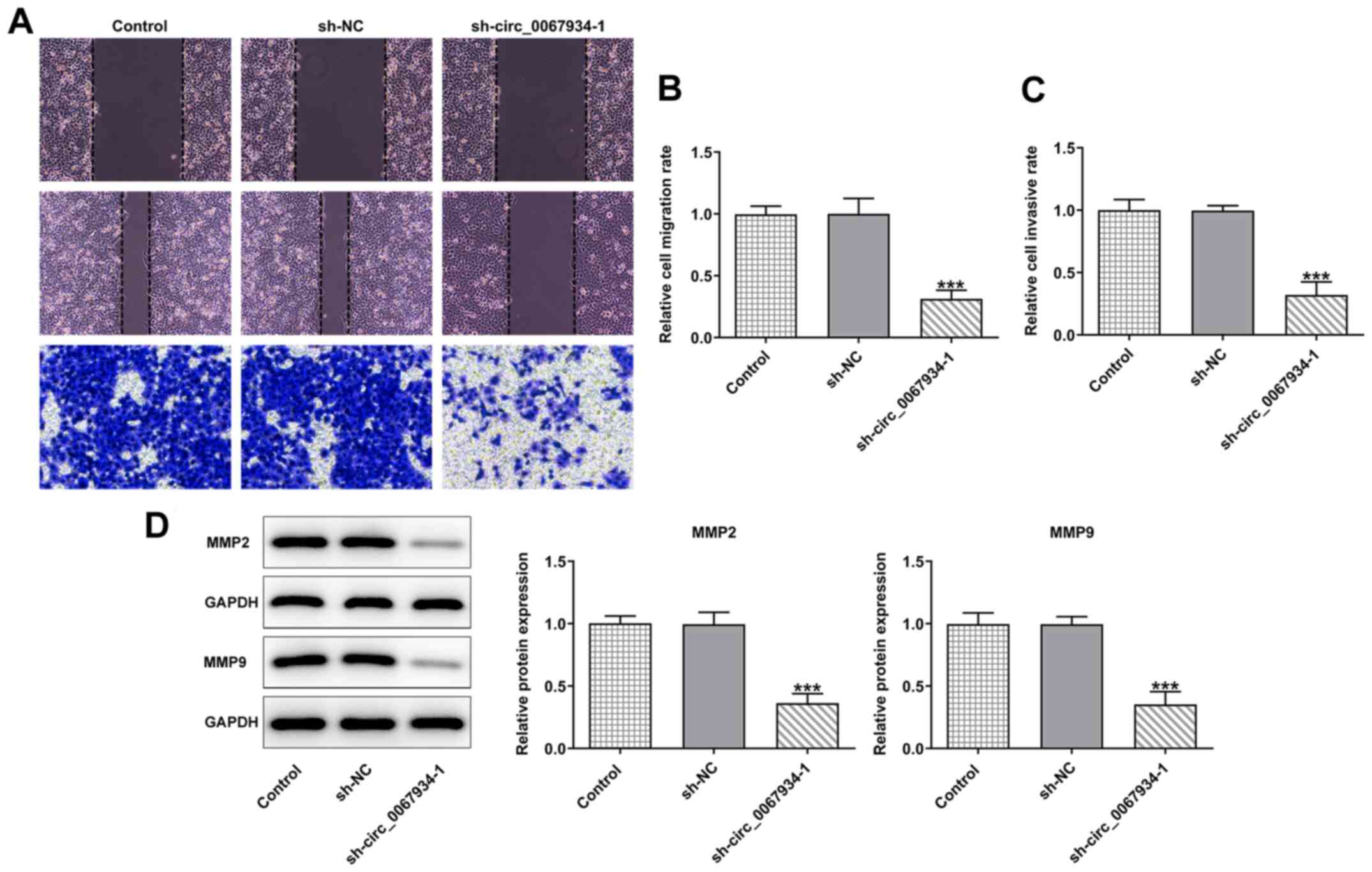
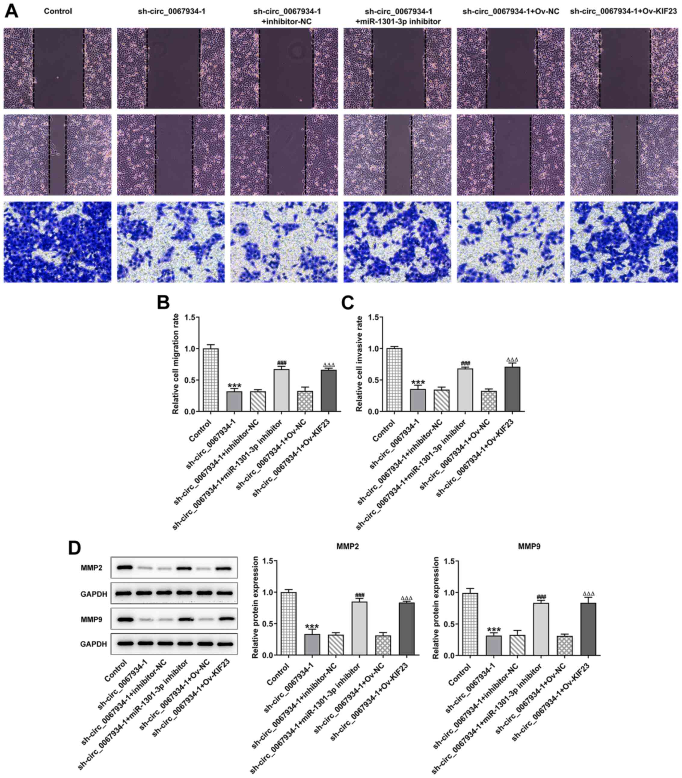
circ_0067934 silencing inhibits AGS
cell migration and invasion
As shown in Fig.
2A-C, following transfection with sh-circ_0067934, the
migratory and invasive abilities of AGS cells were decreased
compared with the control and sh-NC groups. Additionally, western
blotting revealed that the expression levels of the
migration-related proteins, MMP2 and MMP9, were significantly
downregulated compared with those in the control and sh-NC groups
(Fig. 2D), indicating that
circ_0067934 silencing inhibited AGS cell migration and
invasion.
circ_0067934 targets miR-1301-3p
Next, the expression level of circ_0067934 in the
cytoplasm and nucleus of GC cells was examined via RT-qPCR. The
results demonstrated that circ_0067934 was mainly present in the
cytoplasm of AGS cells (Fig. 3A).
The binding sequences of circ_0067934 with miR-5047, miR-1301-3p,
miR-670-5p, miR-345-3p, miR-545-3p and miR-3605-5p were predicted
using the starBase database (Fig.
3B). Moreover, the RT-qPCR results demonstrated that the
expression levels of miR-1301-3p and miR-345-3p were significantly
decreased in GC cells compared with control cells (Fig. 3C-H). As shown in Fig. 3I and J, transfection with
sh-circ_0067934 promoted the expression levels of miR-1301-3p more
significantly than miR-345-3p. Thus, miR-1301-3p was selected for
the subsequent experiments.
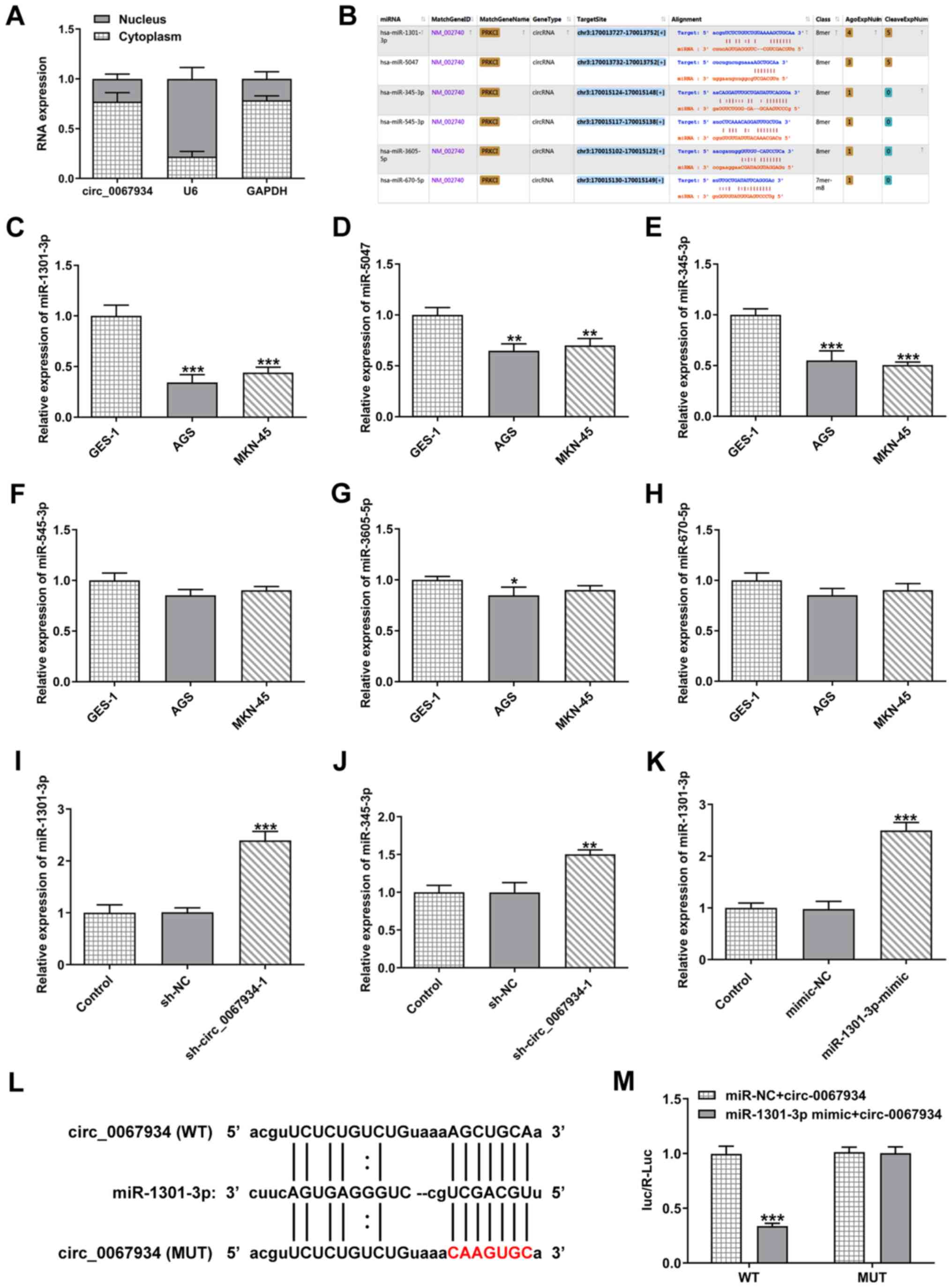 | Figure 3.circ_0067934 targets miR-1301-3p. (A)
Expression level of circ_0067934 in the cytoplasm and nucleus of
AGS cells was detected via RT-qPCR. (B) The starBase database was
used to predict the binding site of circ_0067934 with miR-5047,
miR-1301-3p, miR-670-5p, miR-345-3p, miR-545-3p and miR-3605-5p.
The expression levels of (C) miR-1301-3p, (D) miR-5047, (E)
miR-345-3p, (F) miR-545-3p, (G) miR-3605-5p and (H) miR-670-5p in
gastric cancer cell lines was identified via RT-qPCR. The
expression levels of (I) miR-1301-3p and (J) miR-345-3p were
detected after transfection with sh-circ_0067934. (K) miR-1301-3p
mimics increased miR-1301-3p expression in AGS cells. (L) The
predictive binding sequence between circ-0067934 and miR-1301-3p.
The red letters in the circ-0067934 showed the mutated sites. (M)
Luciferase activity was determined using a luciferase reporter
assay. Data are expressed as the mean ± SD. *P<0.05,
**P<0.01, ***P<0.001 vs. GES-1 cells, sh-NC or mimic-NC.
RT-qPCR, reverse transcription-quantitative PCR; circ, circular
RNA; sh, short hairpin RNA; NC, negative control; miR, microRNA;
WT, wild-type; MUT, mutant. |
To determine the effect of miR-1301-3p on AGS cells,
miR-1301-3p overexpression vector was transfected into AGS cells
(Fig. 3K). Furthermore, the
luciferase vectors containing the WT and MUT sequences (shown in
red letters) of circ-0067934 were constructed (Fig. 3L) and the results indicated that
miR-1301-3p was a direct target of circ_0067934 (Fig. 3M).
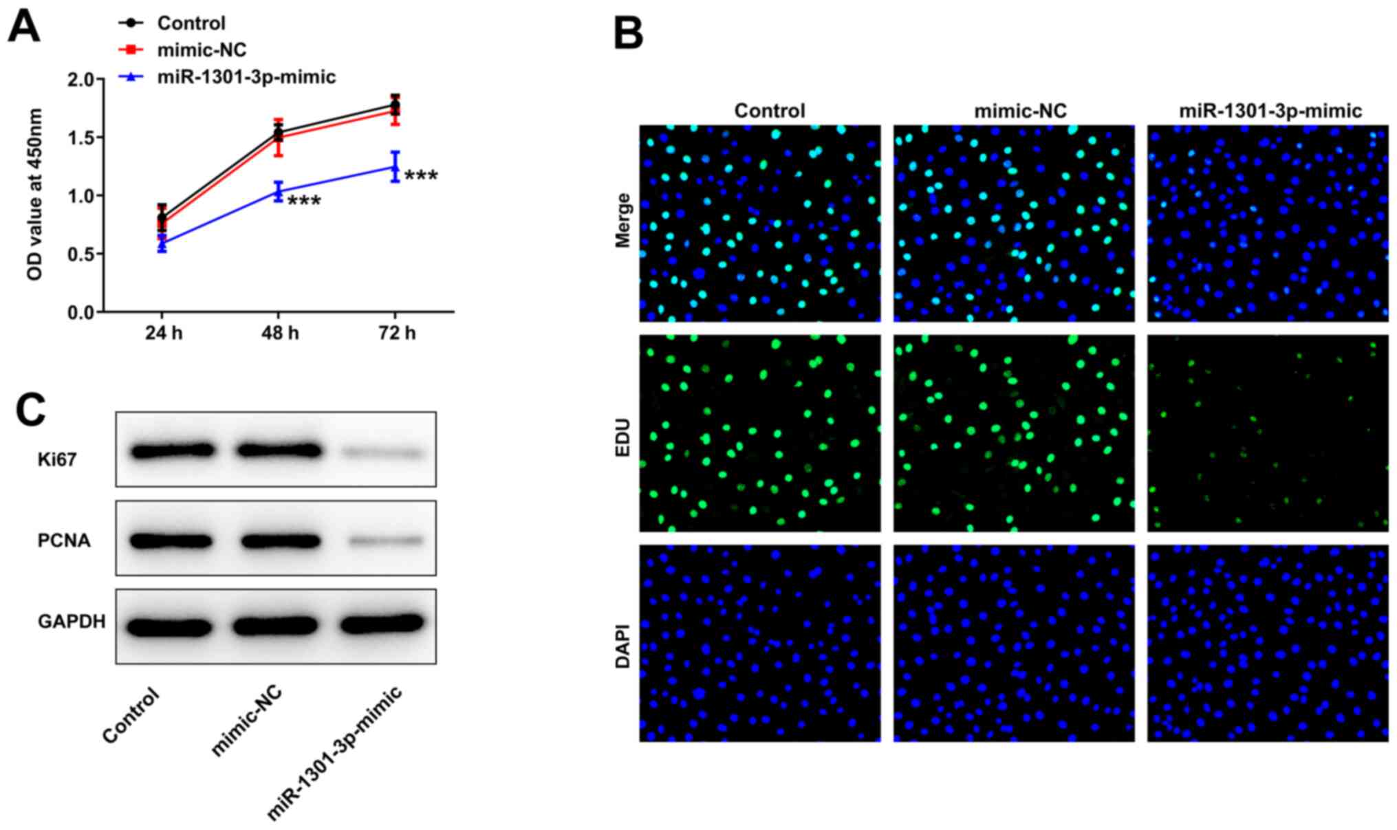
miR-1301-3p overexpression suppresses
AGS cell proliferation
As presented in Fig.
4A, the optical density (OD) value of miR-1301-3p
mimic-transfected cells was significantly lower compared with that
of the mimic NC and control groups. Moreover, EdU staining revealed
that transfection with miR-1301-3p mimic markedly inhibited cell
proliferation (Fig. 4B).
Furthermore, the protein expression levels of Ki67 and PCNA were
notably suppressed by miR-1301-3p mimic in comparison with those in
the NC and control groups (Fig.
4C).
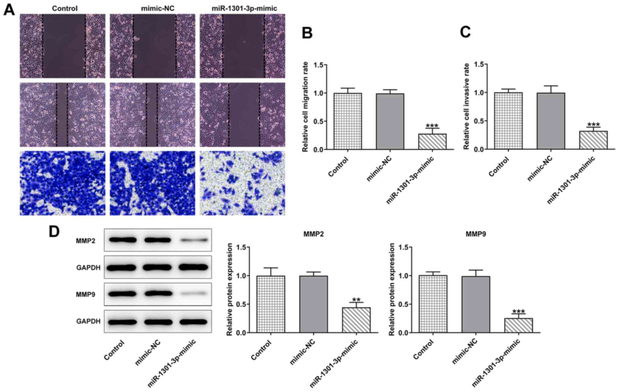
miR-1301-3p mimic represses the
migration and invasion of AGS cells
To further investigate the role of miR-1301-3p in
AGS cells, wound healing and Transwell assays were conducted to
measure cell migration and invasion. As shown in Fig. 5A-C, the cell migratory and
invasive abilities were weakened following transfection with
miR-1301-3p mimic compared with that in the mimic NC group. In
addition, according to the results of the western blotting assay,
the expression levels of the migration-related proteins, MMP2 and
MMP9, were significantly decreased following transfection with
miR-1301-3p mimic compared with the mimic NC or control groups
(Fig. 5D).
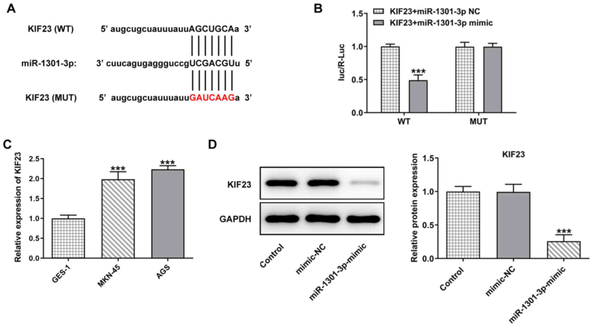
miR-1301-3p targets KIF23
The starBase database predicted the targeted binding
of miR-1301-3p to KIF23 (Fig.
6A), and luciferase reporter assay verified the association
between miR-1301-3p and KIF23 (Fig.
6B). Moreover, it was found that KIF23 expression was
significantly upregulated in GC cells lines compared with the
normal gastric mucosa cells (Fig.
6C). The results of western blotting identified that the
expression of KIF23 was decreased following transfection with
miR-1301-3p mimic compared with the mimic NC group (Fig. 6D). These results indicated that
miR-1301-3p targets KIF23 in AGS cells.
KIF23 overexpression promotes AGS cell
proliferation
To determine the role of KIF23 in GC cells, the
miR-1301-3p inhibitor and KIF23 overexpression vector were
transfected into AGS cells (Fig.
7A-C). The level of cell proliferation was detected using a
CCK-8 assay and the results revealed that the OD value was
significantly enhanced following transfection with miR-1301-3p
inhibitor or Ov-KIF23 in circ_0067934-silenced cells compared with
their NC groups (Fig. 7D). In
addition, compared with the sh-circ_0067934 + inhibitor-NC group
and sh-circ_0067934 + Ov-NC group, miR-1301-3p inhibitor and
Ov-KIF23 markedly increased the percentage of living cells
(Fig. 7E). Consistently, the
western blotting results demonstrated that miR-1301-3p silencing
and KIF23 overexpression increased the protein expression levels of
Ki67 and PCNA in cells transfected with sh-circ_0067934 (Fig. 7F).
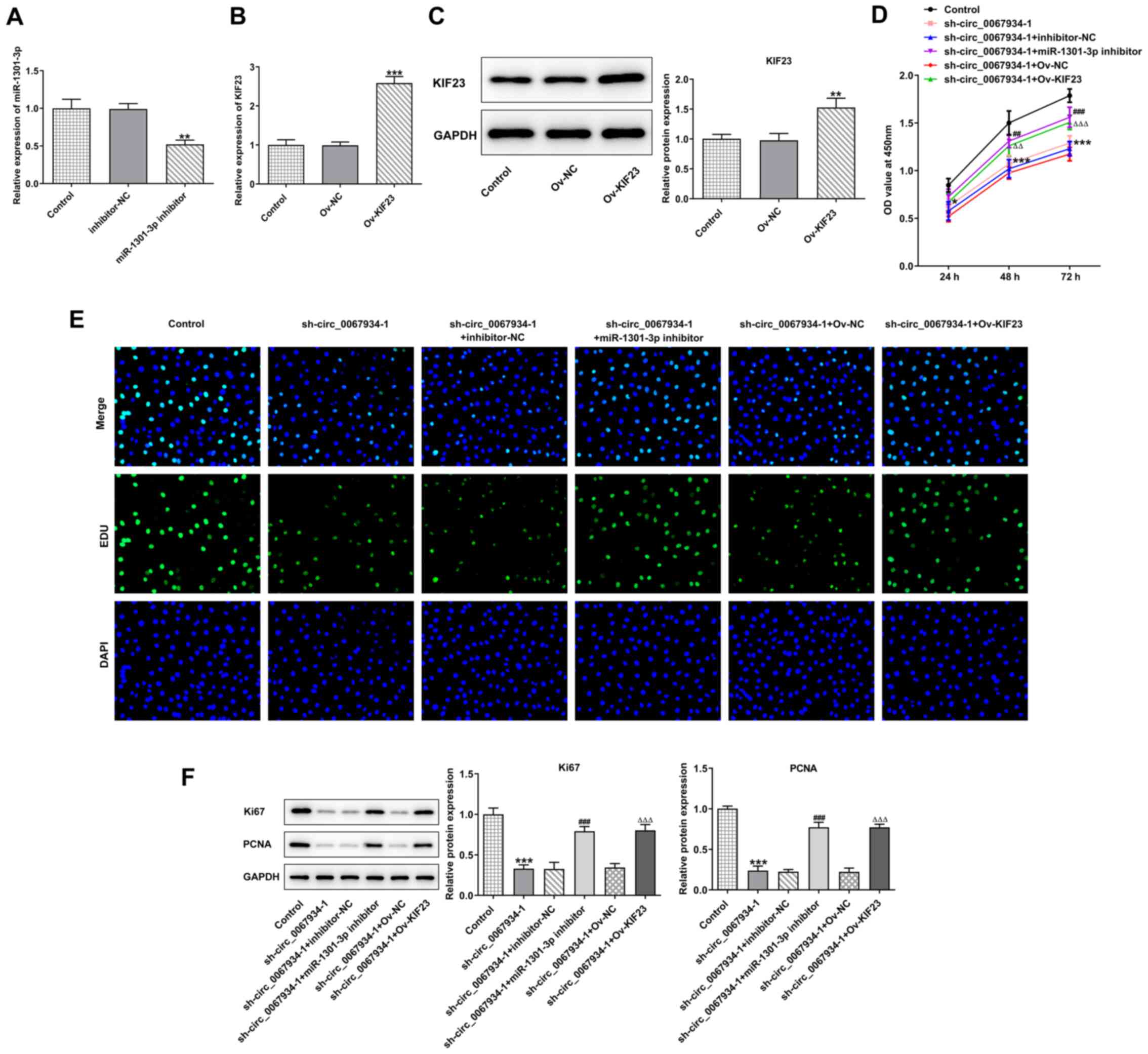 | Figure 7.Effects of KIF23 on proliferation of
AGS cells. (A) miR-1301-3p expression was detected via reverse
transcription-quantitative PCR in cells transfected with
miR-1301-3p inhibitor. KIF23 (B) mRNA and (C) protein expression
was measured after transfection with Ov-KIF23. (D) A Cell Counting
Kit-8 assay was applied for investigating cell proliferation. (E)
Cell proliferative capacity was evaluated using an EdU assay.
Original magnification, ×200. (F) Western blotting was conducted to
measure the protein expression levels of Ki67 and PCNA. Data are
expressed as the mean ± SD. **P<0.01, ***P<0.001 vs. Control
or corresponding NCs; ##P<0.01,
###P<0.001 vs. sh-circ_0067934-1 + inhibitor-NC
groups; △△P<0.01, △△△P<0.001 vs.
sh-circ_0067934-1 + Ov-NC. NC, negative control; miR, microRNA;
KIF23, kinesin family member 23; OD, optical density; Ov,
overexpression; sh, short hairpin RNA; circ, circular RNA; PCNA,
proliferating cell nuclear antigen. |
KIF23 overexpression facilitates AGS
cell migration and invasion
As shown in Fig.
8A-C, KIF23 overexpression or miR-1301-3p knockdown promoted
cell migration and invasion compared with the sh-circ_0067934-1 +
Ov-NC and sh-circ_0067934 + inhibitor-NC groups. In addition, the
western blotting results demonstrated that the protein expression
levels of MMP2 and MMP9 were significantly increased following
transfection with miR-1301-3p inhibitor or Ov-KIF23 in
circ_0067934-silenced cells compared with their corresponding NC
(Fig. 8D).
Discussion
GC is the fourth most common type of cancer, and its
fatality rate is the second highest worldwide (20). The prognosis of patients with GC
is poor and the 5-year relative survival rate for patients with
advanced-stage disease is only 20% (21). Therefore, it is crucial to study
the mechanisms underlying the pathogenesis and development of GC,
as well as to identify novel therapeutic targets for this type of
cancer.
circ_0067934 has been shown to act as an oncogene in
a variety of cancer types (22–24). Although it has been reported that
the expression of circ_0067934 is increased in GC tissues (13), its biological effect on GC cells
is yet to be elucidated. In the present study, the effect of
circ_0067934 on the proliferation, migration and invasion of GC
cells was investigated by silencing circ_0067934. It was recently
revealed that circRNAs may serve as key regulators in the
development of human diseases, including cancer (25). It was also reported that
downregulation of circ_0067934 decreased the proliferative ability
of NSCLC cells and, thus, it may serve as a predictive marker for
the prognosis of NSCLC and a target for the treatment of this
disease (26).
In the present study, following transfection with
sh-circ_0067934, CCK-8 assays and EdU staining were used to
evaluate cell proliferation, and western blotting was used to
detect the protein expression levels of Ki67 and PCNA, which are
known to be closely associated with cell proliferation (27). Ki67 is a common cell proliferation
status marker, which has high sensitivity in determining the
proliferative activity of tumor cells and is associated with the
occurrence, development, proliferation and metastasis of tumors
(28). PCNA is synthesized in and
localizes to the nucleus, and it is closely involved in the
proliferation and invasion of tumor cells. PCNA has also been found
to participate in the synthesis and metabolism of RNA and DNA in
tumor cells, and it is closely associated with the differentiation
and invasion of tumor cells, as well as with tumor metastasis,
recurrence and prognosis (29).
The current results demonstrated that circ_0067934 expression was
higher in GC cells, and the expression levels of Ki67 and PCNA were
downregulated after silencing of circ_0067934, which was consistent
with previous findings (16,22,30). The wound healing assay
demonstrated that the knockdown of circ_0067934 could reduce the
migratory ability of GC cells, and the expression levels of the
migration-related proteins, MMP2 and MMP9, were also downregulated.
MMP2 and MMP9 are closely associated with cell migration (31). Overexpression of MMP2 and MMP9 can
promote the degradation of the extracellular matrix and improve the
cell migratory ability (32,33).
Recently, circRNAs have been considered to be
members of the competing endogenous RNA family due to their
abundant conserved miRNA response elements (34). Some studies have shown that
miR-1301-3p was associated with a variety of cancer types,
including prostate (35), breast
(36) and colon cancer (37). According to data from the starBase
database, the current study predicted that circ_0067934 targeted
miR-1301-3p. The present study identified that the expression level
of miR-1301-3p was significantly increased after circ_0067934 was
silenced, while cell proliferation and migration were
decreased.
KIF23 is a kinesin-like motor protein that serves
crucial roles in cytokinesis (38). Previous studies have reported the
oncogenic roles of KIF23 in several types of cancer, including GC
(39–41). In the present study, the starBase
database also predicted that miR-1301-3p could bind to KIF23, and
when KIF23 was overexpressed, the cell proliferation, migratory and
invasive abilities were enhanced. However, the current study
primarily focused on the role of circ_0067934 and the mechanism
involved in circ_0067934/miR-1301-3p/KIF23 in GC cells, and thus,
did not investigate the regulation network of
circ_0067934/miR-1301-3p/KIF23 in normal gastric mucosal cells.
Future studies will further investigate the relationship of the
three genes in normal gastric mucosal cells. In addition, due to
the lack of samples from patients with GC, this study did not
include clinical sample testing. Moreover, only one GC cell line,
AGS, was used to examine the role of circ_0067934, miR-1301-3p and
KIF23, and so, this study cannot determine the correlation of these
circRNA, miRNA and KIF23 gene expressions in GC cells or patient
samples.
In conclusion, the present study provided evidence
that circ_0067934 can inhibit the proliferation, migration and
invasion of GC cells via the miR-1301-3p/KIF23 axis, which may
provide a theoretical and factual basis for the targeted therapy of
GC.
Acknowledgements
Not applicable.
Funding
Funding: No funding was received.
Availability of data and materials
The datasets used and/or analyzed during the current
study are available from the corresponding author on reasonable
request.
Authors' contributions
JX, NS and JZ designed the experiments and made
considerable contributions to the manuscript writing. JX, NS, WH,
NZ and XL performed the experiments and analyzed the data. JX and
NS revised the manuscript and guided the experiments. JX and NS
confirm the authenticity of all the raw data. All authors have read
and approved the final manuscript.
Ethics approval and consent to
participate
Not applicable.
Patient consent for publication
Not applicable.
Competing interests
The authors declare that they have no competing
interests.
References
|
1
|
Song Z, Wu Y, Yang J, Yang D and Fang X:
Progress in the treatment of advanced gastric cancer. Tumour Biol.
Jul 3–2017.(Epub ahead of print). doi: 10.1177/1010428317714626.
View Article : Google Scholar
|
|
2
|
Li JH, Zhang SW, Liu J, Shao MZ and Chen
L: Review of clinical investigation on recurrence of gastric cancer
following curative resection. Chin Med J (Engl). 125:1479–1495.
2012.PubMed/NCBI
|
|
3
|
Suzuki H and Mori H: Gastric cancer after
Helicobacter pylori eradication. Gan To Kagaku Ryoho.
45:1123–1127. 2018.(In Japanese). PubMed/NCBI
|
|
4
|
Hansen TB, Wiklund ED, Bramsen JB,
Villadsen SB, Statham AL, Clark SJ and Kjems J: miRNA-dependent
gene silencing involving Ago2-mediated cleavage of a circular
antisense RNA. EMBO J. 30:4414–4422. 2011. View Article : Google Scholar : PubMed/NCBI
|
|
5
|
Shi X, Zhang W, Nian X, Lu X, Li Y, Liu F,
Wang F, He B, Zhao L, Zhu Y, et al: The previously uncharacterized
lncRNA APP promotes prostate cancer progression by acting as a
competing endogenous RNA. Int J Cancer. 146:475–486. 2020.
View Article : Google Scholar : PubMed/NCBI
|
|
6
|
Khan S, Ayub H, Khan T and Wahid F:
MicroRNA biogenesis, gene silencing mechanisms and role in breast,
ovarian and prostate cancer. Biochimie. 167:12–24. 2019. View Article : Google Scholar : PubMed/NCBI
|
|
7
|
Wang J, Su Z, Lu S, Fu W, Liu Z, Jiang X
and Tai S: LncRNA HOXA-AS2 and its molecular mechanisms in human
cancer. Clin Chim Acta. 485:229–233. 2018. View Article : Google Scholar : PubMed/NCBI
|
|
8
|
Peng WX, Koirala P and Mo YY:
LncRNA-mediated regulation of cell signaling in cancer. Oncogene.
36:5661–5667. 2017. View Article : Google Scholar : PubMed/NCBI
|
|
9
|
Wu M, Wang G, Tian W, Deng Y and Xu Y:
MiRNA-based therapeutics for lung cancer. Curr Pharm Des.
23:5989–5996. 2018. View Article : Google Scholar : PubMed/NCBI
|
|
10
|
Han J, Zhao G, Ma X, Dong Q, Zhang H, Wang
Y and Cui J: CircRNA circ-BANP-mediated miR-503/LARP1 signaling
contributes to lung cancer progression. Biochem Biophys Res Commun.
503:2429–2435. 2018. View Article : Google Scholar : PubMed/NCBI
|
|
11
|
Liu Z, Yu Y, Huang Z, Kong Y, Hu X, Xiao
W, Quan J and Fan X: CircRNA-5692 inhibits the progression of
hepatocellular carcinoma by sponging miR-328-5p to enhance DAB2IP
expression. Cell Death Dis. 10:9002019. View Article : Google Scholar : PubMed/NCBI
|
|
12
|
Zeng K, Chen X, Xu M, Liu X, Hu X, Xu T,
Sun H, Pan Y, He B and Wang S: CircHIPK3 promotes colorectal cancer
growth and metastasis by sponging miR-7. Cell Death Dis. 9:4172018.
View Article : Google Scholar : PubMed/NCBI
|
|
13
|
Ding HX, Xu Q, Wang BG, Lv Z and Yuan Y:
MetaDE-cased analysis of circRNA expression profiles involved in
gastric cancer. Dig Dis Sci. 65:2884–2895. 2020. View Article : Google Scholar : PubMed/NCBI
|
|
14
|
Wang JM, Li XJ and Wang J: Circular RNA
circ_0067934 functions as an oncogene in breast cancer by targeting
Mcl-1. Eur Rev Med Pharmacol Sci. 23:9499–9505. 2019.PubMed/NCBI
|
|
15
|
Zou Q, Wang T, Li B, Li G, Zhang L, Wang B
and Sun S: Overexpression of circ-0067934 is associated with
increased cellular proliferation and the prognosis of non-small
cell lung cancer. Oncol Lett. 16:5551–5556. 2018.PubMed/NCBI
|
|
16
|
Liu Q, Zhou Q and Zhong P: circ_0067934
increases bladder cancer cell proliferation, migration and invasion
through suppressing miR-1304 expression and increasing Myc
expression levels. Exp Ther Med. 19:3751–3759. 2020.PubMed/NCBI
|
|
17
|
Wu Y, Zhang L, Zhang L, Wang Y, Li H, Ren
X, Wei F, Yu W, Liu T, Wang X, et al: Long non-coding RNA HOTAIR
promotes tumor cell invasion and metastasis by recruiting EZH2 and
repressing E-cadherin in oral squamous cell carcinoma. Int J Oncol.
46:2586–2594. 2015. View Article : Google Scholar : PubMed/NCBI
|
|
18
|
Yu F, Pillman KA, Neilsen CT, Toubia J,
Lawrence DM, Tsykin A, Gantier MP, Callen DF, Goodall GJ and
Bracken CP: Naturally existing isoforms of miR-222 have distinct
functions. Nucleic Acids Res. 45:11371–11385. 2017. View Article : Google Scholar : PubMed/NCBI
|
|
19
|
Wang GL, Xia XL, Li XL, He FH and Li JL:
Identification and expression analysis of the MSP130-related-2 gene
from Hyriopsis cumingii. Genet Mol Res. 14:4903–4913. 2015.
View Article : Google Scholar : PubMed/NCBI
|
|
20
|
Smyth EC, Nilsson M, Grabsch HI, van
Grieken NC and Lordick F: Gastric cancer. Lancet. 396:635–648.
2020. View Article : Google Scholar : PubMed/NCBI
|
|
21
|
Sitarz R, Skierucha M, Mielko J, Offerhaus
GJA, Maciejewski R and Polkowski WP: Gastric cancer: Epidemiology,
prevention, classification, and treatment. Cancer Manag Res.
10:239–248. 2018. View Article : Google Scholar : PubMed/NCBI
|
|
22
|
Hu C, Wang Y, Li A, Zhang J, Xue F and Zhu
L: Overexpressed circ_0067934 acts as an oncogene to facilitate
cervical cancer progression via the miR-545/EIF3C axis. J Cell
Physiol. 234:9225–9232. 2019. View Article : Google Scholar : PubMed/NCBI
|
|
23
|
Zhu Q, Lu G, Luo Z, Gui F, Wu J, Zhang D
and Ni Y: CircRNA circ_0067934 promotes tumor growth and metastasis
in hepatocellular carcinoma through regulation of
miR-1324/FZD5/Wnt/β-catenin axis. Biochem Biophys Res Commun.
497:626–632. 2018. View Article : Google Scholar : PubMed/NCBI
|
|
24
|
Cui XL, Wang XD, Lin SK, Miao CM, Wu M and
Wei JG: Circular RNA circ_0067934 functions as an oncogene in
glioma by targeting CSF1. Eur Rev Med Pharmacol Sci. 23:8449–8455.
2019.PubMed/NCBI
|
|
25
|
Patop IL and Kadener S: circRNAs in
Cancer. Curr Opin Genet Dev. 48:121–127. 2018. View Article : Google Scholar : PubMed/NCBI
|
|
26
|
Wang J and Li H: CircRNA circ_0067934
silencing inhibits the proliferation, migration and invasion of
NSCLC cells and correlates with unfavorable prognosis in NSCLC. Eur
Rev Med Pharmacol Sci. 22:3053–3060. 2018.PubMed/NCBI
|
|
27
|
Juríková M, Danihel L, Polák Š and Varga
I: Ki67, PCNA, and MCM proteins: Markers of proliferation in the
diagnosis of breast cancer. Acta Histochem. 118:544–552. 2016.
View Article : Google Scholar : PubMed/NCBI
|
|
28
|
Stevanovic L, Choschzick M, Moskovszky L
and Varga Z: Variability of predictive markers (hormone receptors,
Her2, Ki67) and intrinsic subtypes of breast cancer in four
consecutive years 2015-2018. J Cancer Res Clin Oncol.
145:2983–2994. 2019. View Article : Google Scholar : PubMed/NCBI
|
|
29
|
Boehm EM, Gildenberg MS and Washington MT:
The many roles of PCNA in eukaryotic DNA replication. Enzymes.
39:231–254. 2016. View Article : Google Scholar : PubMed/NCBI
|
|
30
|
Zhao M, Ma W and Ma C: Circ_0067934
promotes non-small cell lung cancer development by regulating
miR-1182/KLF8 axis and activating Wnt/β-catenin pathway. Biomed
Pharmacother. 129:1104612020. View Article : Google Scholar : PubMed/NCBI
|
|
31
|
Simbulan-Rosenthal CM, Dougherty R, Vakili
S, Ferraro AM, Kuo LW, Alobaidi R, Aljehane L, Gaur A, Sykora P,
Glasgow E, et al: CRISPR-Cas9 Knockdown and Induced Expression of
CD133 Reveal Essential Roles in Melanoma Invasion and Metastasis.
Cancers (Basel). 11:E14902019. View Article : Google Scholar
|
|
32
|
Cui F, Wang S, Lao I, Zhou C, Kong H,
Bayaxi N, Li J, Chen Q, Zhu T and Zhu H: miR-375 inhibits the
invasion and metastasis of colorectal cancer via targeting SP1 and
regulating EMT-associated genes. Oncol Rep. 36:487–493. 2016.
View Article : Google Scholar : PubMed/NCBI
|
|
33
|
Park GB, Chung YH and Kim D: Induction of
galectin-1 by TLR-dependent PI3K activation enhances
epithelial-mesenchymal transition of metastatic ovarian cancer
cells. Oncol Rep. 37:3137–3145. 2017. View Article : Google Scholar : PubMed/NCBI
|
|
34
|
Zhong Y, Du Y, Yang X, Mo Y, Fan C, Xiong
F, Ren D, Ye X, Li C, Wang Y, et al: Circular RNAs function as
ceRNAs to regulate and control human cancer progression. Mol
Cancer. 17:792018. View Article : Google Scholar : PubMed/NCBI
|
|
35
|
Song XL, Huang B, Zhou BW, Wang C, Liao
ZW, Yu Y and Zhao SC: miR-1301-3p promotes prostate cancer stem
cell expansion by targeting SFRP1 and GSK3β. Biomed Pharmacother.
99:369–374. 2018. View Article : Google Scholar : PubMed/NCBI
|
|
36
|
Peng X, Yan B and Shen Y: MiR-1301-3p
inhibits human breast cancer cell proliferation by regulating cell
cycle progression and apoptosis through directly targeting ICT1.
Breast Cancer. 25:742–752. 2018. View Article : Google Scholar : PubMed/NCBI
|
|
37
|
Wang L, Zhao Y, Xu M, Zhou F and Yan J:
Serum miR-1301-3p, miR-335-5p, miR-28-5p, and their target B7-H3
may serve as novel biomarkers for colorectal cancer. J BUON.
24:1120–1127. 2019.PubMed/NCBI
|
|
38
|
Sun X, Jin Z, Song X, Wang J, Li Y, Qian
X, zhang Y and Yin Y: Evaluation of KIF23 variant 1 expression and
relevance as a novel prognostic factor in patients with
hepatocellular carcinoma. BMC Cancer. 15:9612015. View Article : Google Scholar : PubMed/NCBI
|
|
39
|
Liu Y, Chen H, Dong P, Xie G, Zhou Y, Ma
Y, Yuan X, Yang J, Han L, Chen L, et al: KIF23 activated
Wnt/β-catenin signaling pathway through direct interaction with
Amer1 in gastric cancer. Aging (Albany NY). 12:8372–8396. 2020.
View Article : Google Scholar : PubMed/NCBI
|
|
40
|
Kato T, Wada H, Patel P, Hu HP, Lee D,
Ujiie H, Hirohashi K, Nakajima T, Sato M, Kaji M, et al:
Overexpression of KIF23 predicts clinical outcome in primary lung
cancer patients. Lung Cancer. 92:53–61. 2016. View Article : Google Scholar : PubMed/NCBI
|
|
41
|
Li XL, Ji YM, Song R, Li XN and Guo LS:
KIF23 promotes gastric cancer by stimulating cell proliferation.
Dis Markers. 2019:97519232019. View Article : Google Scholar : PubMed/NCBI
|