Spandidos Publications Logo
  • About
    • About Spandidos
    • Aims and Scopes
    • Abstracting and Indexing
    • Editorial Policies
    • Reprints and Permissions
    • Job Opportunities
    • Terms and Conditions
    • Contact
  • Journals
    • All Journals
    • Oncology Letters
      • Oncology Letters
      • Information for Authors
      • Editorial Policies
      • Editorial Board
      • Aims and Scope
      • Abstracting and Indexing
      • Bibliographic Information
      • Archive
    • International Journal of Oncology
      • International Journal of Oncology
      • Information for Authors
      • Editorial Policies
      • Editorial Board
      • Aims and Scope
      • Abstracting and Indexing
      • Bibliographic Information
      • Archive
    • Molecular and Clinical Oncology
      • Molecular and Clinical Oncology
      • Information for Authors
      • Editorial Policies
      • Editorial Board
      • Aims and Scope
      • Abstracting and Indexing
      • Bibliographic Information
      • Archive
    • Experimental and Therapeutic Medicine
      • Experimental and Therapeutic Medicine
      • Information for Authors
      • Editorial Policies
      • Editorial Board
      • Aims and Scope
      • Abstracting and Indexing
      • Bibliographic Information
      • Archive
    • International Journal of Molecular Medicine
      • International Journal of Molecular Medicine
      • Information for Authors
      • Editorial Policies
      • Editorial Board
      • Aims and Scope
      • Abstracting and Indexing
      • Bibliographic Information
      • Archive
    • Biomedical Reports
      • Biomedical Reports
      • Information for Authors
      • Editorial Policies
      • Editorial Board
      • Aims and Scope
      • Abstracting and Indexing
      • Bibliographic Information
      • Archive
    • Oncology Reports
      • Oncology Reports
      • Information for Authors
      • Editorial Policies
      • Editorial Board
      • Aims and Scope
      • Abstracting and Indexing
      • Bibliographic Information
      • Archive
    • Molecular Medicine Reports
      • Molecular Medicine Reports
      • Information for Authors
      • Editorial Policies
      • Editorial Board
      • Aims and Scope
      • Abstracting and Indexing
      • Bibliographic Information
      • Archive
    • World Academy of Sciences Journal
      • World Academy of Sciences Journal
      • Information for Authors
      • Editorial Policies
      • Editorial Board
      • Aims and Scope
      • Abstracting and Indexing
      • Bibliographic Information
      • Archive
    • International Journal of Functional Nutrition
      • International Journal of Functional Nutrition
      • Information for Authors
      • Editorial Policies
      • Editorial Board
      • Aims and Scope
      • Abstracting and Indexing
      • Bibliographic Information
      • Archive
    • International Journal of Epigenetics
      • International Journal of Epigenetics
      • Information for Authors
      • Editorial Policies
      • Editorial Board
      • Aims and Scope
      • Abstracting and Indexing
      • Bibliographic Information
      • Archive
    • Medicine International
      • Medicine International
      • Information for Authors
      • Editorial Policies
      • Editorial Board
      • Aims and Scope
      • Abstracting and Indexing
      • Bibliographic Information
      • Archive
  • Articles
  • Information
    • Information for Authors
    • Information for Reviewers
    • Information for Librarians
    • Information for Advertisers
    • Conferences
  • Language Editing
Spandidos Publications Logo
  • About
    • About Spandidos
    • Aims and Scopes
    • Abstracting and Indexing
    • Editorial Policies
    • Reprints and Permissions
    • Job Opportunities
    • Terms and Conditions
    • Contact
  • Journals
    • All Journals
    • Biomedical Reports
      • Information for Authors
      • Editorial Policies
      • Editorial Board
      • Aims and Scope
      • Abstracting and Indexing
      • Bibliographic Information
      • Archive
    • Experimental and Therapeutic Medicine
      • Information for Authors
      • Editorial Policies
      • Editorial Board
      • Aims and Scope
      • Abstracting and Indexing
      • Bibliographic Information
      • Archive
    • International Journal of Epigenetics
      • Information for Authors
      • Editorial Policies
      • Editorial Board
      • Aims and Scope
      • Abstracting and Indexing
      • Bibliographic Information
      • Archive
    • International Journal of Functional Nutrition
      • Information for Authors
      • Editorial Policies
      • Editorial Board
      • Aims and Scope
      • Abstracting and Indexing
      • Bibliographic Information
      • Archive
    • International Journal of Molecular Medicine
      • Information for Authors
      • Editorial Policies
      • Editorial Board
      • Aims and Scope
      • Abstracting and Indexing
      • Bibliographic Information
      • Archive
    • International Journal of Oncology
      • Information for Authors
      • Editorial Policies
      • Editorial Board
      • Aims and Scope
      • Abstracting and Indexing
      • Bibliographic Information
      • Archive
    • Medicine International
      • Information for Authors
      • Editorial Policies
      • Editorial Board
      • Aims and Scope
      • Abstracting and Indexing
      • Bibliographic Information
      • Archive
    • Molecular and Clinical Oncology
      • Information for Authors
      • Editorial Policies
      • Editorial Board
      • Aims and Scope
      • Abstracting and Indexing
      • Bibliographic Information
      • Archive
    • Molecular Medicine Reports
      • Information for Authors
      • Editorial Policies
      • Editorial Board
      • Aims and Scope
      • Abstracting and Indexing
      • Bibliographic Information
      • Archive
    • Oncology Letters
      • Information for Authors
      • Editorial Policies
      • Editorial Board
      • Aims and Scope
      • Abstracting and Indexing
      • Bibliographic Information
      • Archive
    • Oncology Reports
      • Information for Authors
      • Editorial Policies
      • Editorial Board
      • Aims and Scope
      • Abstracting and Indexing
      • Bibliographic Information
      • Archive
    • World Academy of Sciences Journal
      • Information for Authors
      • Editorial Policies
      • Editorial Board
      • Aims and Scope
      • Abstracting and Indexing
      • Bibliographic Information
      • Archive
  • Articles
  • Information
    • For Authors
    • For Reviewers
    • For Librarians
    • For Advertisers
    • Conferences
  • Language Editing
Login Register Submit
  • This site uses cookies
  • You can change your cookie settings at any time by following the instructions in our Cookie Policy. To find out more, you may read our Privacy Policy.

    I agree
Search articles by DOI, keyword, author or affiliation
Search
Advanced Search
presentation
Biomedical Reports
Join Editorial Board Propose a Special Issue
Print ISSN: 2049-9434 Online ISSN: 2049-9442
Journal Cover
January-February 2013 Volume 1 Issue 1

Full Size Image

Sign up for eToc alerts
Recommend to Library

Journals

International Journal of Molecular Medicine

International Journal of Molecular Medicine

International Journal of Molecular Medicine is an international journal devoted to molecular mechanisms of human disease.

International Journal of Oncology

International Journal of Oncology

International Journal of Oncology is an international journal devoted to oncology research and cancer treatment.

Molecular Medicine Reports

Molecular Medicine Reports

Covers molecular medicine topics such as pharmacology, pathology, genetics, neuroscience, infectious diseases, molecular cardiology, and molecular surgery.

Oncology Reports

Oncology Reports

Oncology Reports is an international journal devoted to fundamental and applied research in Oncology.

Experimental and Therapeutic Medicine

Experimental and Therapeutic Medicine

Experimental and Therapeutic Medicine is an international journal devoted to laboratory and clinical medicine.

Oncology Letters

Oncology Letters

Oncology Letters is an international journal devoted to Experimental and Clinical Oncology.

Biomedical Reports

Biomedical Reports

Explores a wide range of biological and medical fields, including pharmacology, genetics, microbiology, neuroscience, and molecular cardiology.

Molecular and Clinical Oncology

Molecular and Clinical Oncology

International journal addressing all aspects of oncology research, from tumorigenesis and oncogenes to chemotherapy and metastasis.

World Academy of Sciences Journal

World Academy of Sciences Journal

Multidisciplinary open-access journal spanning biochemistry, genetics, neuroscience, environmental health, and synthetic biology.

International Journal of Functional Nutrition

International Journal of Functional Nutrition

Open-access journal combining biochemistry, pharmacology, immunology, and genetics to advance health through functional nutrition.

International Journal of Epigenetics

International Journal of Epigenetics

Publishes open-access research on using epigenetics to advance understanding and treatment of human disease.

Medicine International

Medicine International

An International Open Access Journal Devoted to General Medicine.

Journal Cover
January-February 2013 Volume 1 Issue 1

Full Size Image

Sign up for eToc alerts
Recommend to Library

  • Article
  • Citations
    • Cite This Article
    • Download Citation
    • Create Citation Alert
    • Remove Citation Alert
    • Cited By
  • Similar Articles
    • Related Articles (in Spandidos Publications)
    • Similar Articles (Google Scholar)
    • Similar Articles (PubMed)
  • Download PDF
  • Download XML
  • View XML
Article

Excess iron undermined bone load-bearing capacity through tumor necrosis factor-α-dependent osteoclastic activation in mice

  • Authors:
    • Junping Li
    • Yanli Hou
    • Shuping Zhang
    • Hong Ji
    • Haiqin Rong
    • Guangbo Qu
    • Sijin Liu
  • View Affiliations / Copyright

    Affiliations: Shandong Institute of Endocrine and Metabolic Diseases, Jinan 250062, P.R. China, State Key Laboratory of Environmental Chemistry and Ecotoxicology, Research Center for Eco-Environmental Sciences, Chinese Academy of Sciences, Beijing 100085, P.R. China
  • Pages: 85-88
    |
    Published online on: August 28, 2012
       https://doi.org/10.3892/br.2012.6
  • Expand metrics +
Metrics: Total Views: 0 (Spandidos Publications: | PMC Statistics: )
Metrics: Total PDF Downloads: 0 (Spandidos Publications: | PMC Statistics: )
Cited By (CrossRef): 0 citations Loading Articles...

This article is mentioned in:


Abstract

Iron overload has been associated with bone mass loss. To elucidate the effects of excess iron on bone metabolism, an iron-overloading mouse model was established by administering iron-dextran at 250 mg/kg to female BALB/c mice. After 4 weeks, the mice were sacrificed and the biomechanical properties of the femurs were examined. The results suggested a notable decrease of the maximal bending stress and the modulus of bending elasticity in the femurs obtained from the excess iron‑treated mice compared to the control mice. The levels of the serum osteocalcin, C‑telopeptide of type I collagen (CTX‑1) and tumor necrosis factor‑α (TNF‑α) were measured in order to investigate the underlying mechanism responsible for the excess iron‑induced bone strength reduction. Overall, the results suggested that iron overload resulted in a marked reduction of bone load‑bearing capacity through a TNF‑triggered osteoclast differentiation and resorption mechanism.

Introduction

Bone mass is maintained by the balance of bone formation and resorption, involving a number of regulatory pathways. Bone formation is performed by osteoblasts, while bone resorption is carried out by osteoclasts. Disruptions of these processes are likely to induce hyperosteogeny or osteopenia (1,2). Numerous clinical studies reported that patients with osteoporosis showed iron accumulation in the bone, potentially leading to pathological damages, such as occurred femoral neck fractures in patients of African descent (3–5).

Ovariectomized rats with an increased iron level showed a higher tendency to have osteoporosis (6). Tsay et al(7) reported that the iron overload (IO)-induced bone loss in mice was correlated with the inflammatory bone resorption and oxidative stress. Using in vitro cell assays, iron treatment was demonstrated to inhibit osteoblast formation, proliferation and mineralization (8–10), while promoting osteoclast differentiation and increased osteoclastic function (11). Therefore, IO is believed to disrupt bone metabolism and induce bone loss.

Iron accumulation in tissue is likely to provoke the formation of reactive oxygen species (ROS) (12), while the iron excess is likely to induce an increased expression of tumor necrosis factor-α (TNF-α) (13). However, the contribution of oxidative stress to osteoporosis was also recognized (14,15). ROS induced increased TNF-α expression and activated several signaling systems involved in the osteoclastic differentiation, especially the nuclear factor-κB (NF-κB) pathway. TNF-α was found to synergize strongly with the receptor activator of NF-κB ligand in osteoclast formation and activation (16,17). Therefore, osteoclastic hyperresorption was highly involved in ROS- and TNF-α-induced bone loss. However, whether IO is linked to induced bone function impairment has yet to be investigated.

In this study, a mouse model with IO was developed and was used to determine whether the excess iron undermined bone strength and load-bearing capacity through TNF-α induction and osteoclastic function promotion.

Materials and methods

Animal experiments

Six-week-old female BALB/c mice were fed under sterile conditions and divided into two groups. The iron excess (i.e., IO), group was injected with iron-dextran (Sigma, St. Louis, MO, USA) at 250 mg/kg. The control mice were injected with equal amounts of phosphate-buffered saline (PBS) twice a week for four weeks. Subsequent to sacrificing the animals at 48 h after the final injection, blood and femur samples were collected for analysis.

Assessments of bone metabolic markers

Blood samples were collected from the heart and allowed to clot at room temperature. Serum samples were separated from the blood by centrifugation at 3,000 rpm for 10 min. Serum osteocalcin, C-telopeptide of type I collagen (CTX-1) and TNF-α were measured by ELISA (Rapidbio; RapidBio, West Hills, CA, USA; R&D Systems, Minneapolis, MN, USA), according to the manufacturer’s instructions.

Bone biomechanical analysis

Biomechanical properties of femurs were measured by the three-point bending test performed using a universal material test machine (AG-1S; Shimadzu Co., Kyoto, Japan) at room temperature, as described in previous studies (18–20). Briefly, the diaphysis was placed on two supports with a span of 10 mm, while the loading pin was centered above two supports at a displacement rate of 2 mm/min, until fracture occurred.

Statistical analysis

The two-tailed Student’s t-test was used to analyze experimental data. The data were presented as the means ± standard error (SE). P<0.05 was considered to indicate a statistically significant difference.

Results and Discussion

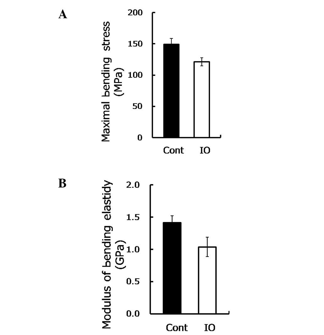
Mechanical loading sends signals which are essential for bone mass maintenance and strength depending on the stimulus (21). To determine whether IO undermines the bone strength and function, the biomechanical parameters of the bone were measured. Maximal bending stress is the maximum load per unit of bone area in the plastic deformation stage, while the modulus of bending elasticity involves the resistance to elastic deformation of the bone, reflecting the inner strength of the bone, without size effect. As shown in Fig. 1A, the maximal bending stress was reduced approximately by 20% (P=0.03) under IO conditions, while the modulus of bending elasticity was reduced by 30% (P=0.05, Fig. 1B). These findings suggested that IO led to a significant reduction of bone load-bearing capacity and thus increased the risk of fractures.

Figure 1.

Excess iron impaired biomechanical properties of femurs from mice with IO are shown. (A) Maximal bending stress was measured by the three-point bending test for femurs from mice with IO treatment and control mice (n=8–9). (B) Modulus of bending elasticity using the three-point bending test for femurs upon IO condition (n=8–9) is shown.

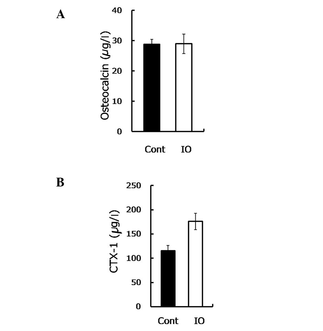
Bone load-bearing capacity is dependent on bone formation, as well as bone resorption. To gain insight into the mechanism underlying the IO-mediated impairment to bone load-bearing capacity, the osteocalcin and CTX-1 were measured. Osteocalcin is secreted by osteoblasts in the maturation stage to sustain mineralization and may be used as a marker of bone formation in the evaluation of bone mass. The findings of this study showed no statistically significant difference in the osteocalcin content between the mice with IO and the control group (28.82 vs. 28.99, P=0.963, Fig. 2A). In contrast to these findings, in their study, Yamasaki et al(8) demonstrated that excess iron reduced the mineralization of rat calvarial osteoblast-like cells by showing a decreased number of mineralized nodules (9,10). This discrepancy may be due to the in vitro sensitivity of osteoblasts being higher compared to the in vivo, when directly exposed to iron. Thus, consistent with another in vivo study (7), our results demonstrated that IO had little effect on bone formation.

Figure 2.

Excess iron provoked bone resorption. (A) Serum osteocalcin assayed by ELISA in the groups (n=9) is shown. (B) CTX-1 assessed by ELISA in the two groups (n=5–9) is shown.

CTX-1 is the degraded product of type I collagen with an enhanced osteoclastic activity, used to evaluate the bone resorptive process. In the present study, the CTX-1 content was observed to be notably increased (∼50%) in the IO group compared to the control group (P=0.008, Fig. 2B), suggesting an elevated bone resorption.

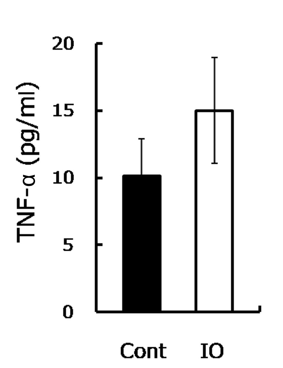
The increased bone resorption indicated that the osteoclastic activity was stimulated. Since IO was demonstrated to induce the TNF-α expression that further triggers osteoclastic function (13,22,23), the TNF-α level was measured. Consistent with a previous study (13), the TNF-α level in the IO mice was approximately 1.5-fold higher compared to the control group (Fig. 3), as shown. TNF-α was demonstrated to be involved in the enhancement of the development of osteopenia and osteoporosis (14,15). TNF-α promoted osteoclastogenesis through synergizing with a permissive level of the receptor activator of NF-κB ligand (RANKL), as well as through directly affecting osteoclasts (16,17). In the current study, a high serum TNF-α level was observed in mice with IO. The increased TNF-α is likely to promote the osteoclastic function and enhance inflammatory osteolysis, resulting in advanced bone resorption (24). Therefore, upon IO exposure, although increased TNF-α was not involved in mineralization inhibition, it accelerated bone loss and eventually reduced bone strength.

Figure 3.

Excess iron increased serum TNF-α. Serum TNF-α measured by ELISA from mice with IO treatment and the control mice (n=8) is shown.

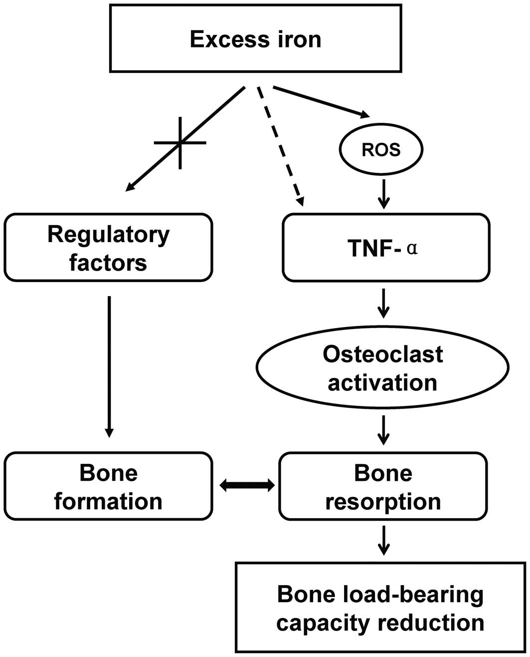
In conclusion, the findings of the present study have shown that IO undermined bone load-bearing capacity through the enhancement of TNF-α secretion, which facilitated osteoclastic differentiation and promoted the bone-resorbing activity (Fig. 4). Therefore, decreasing the iron burden is likely to be a promising approach for the treatment of bone metabolic diseases, such as osteopenia and osteoporosis.

Figure 4.

A schematic delineation of the mechanism responsible for IO-triggered impairment to bone strength is shown. IO largely increased serum TNF-α content by stimulating inflammatory cells through ROS induction. Consequently, increased TNF-α promoted osteoclastogenesis, then activated osteoclastic function stimulated bone resorption. However, excess iron had little effect on bone formation. As a result, bone mass was reduced and bone load-bearing capacity was undermined.

Acknowledgements

This study was financed by grants from the Chinese Academy of Sciences (KZCX2-EW-404) and the National Natural Science Foundation of China (nos. 21077128, 20921063 and 21177151). The authors would like to thank the laboratory staff members for their assistance with the experiments and reagents.

References

1. 

Mountzios G, Dimopoulos MA, Bamias A, Papadopoulos G, Kastritis E, Syrigos K, Pavlakis G and Terpos E: Abnormal bone remodeling process is due to an imbalance in the receptor activator of nuclear factor-kappa B ligand (RANKL)/osteoprotegerin (OPG) axis in patients with solid tumors metastatic to the skeleton. Acta Oncol. 46:221–229. 2007. View Article : Google Scholar

2. 

Fohr B, Dunstan CR and Seibel MJ: Clinical review 165: markers of bone remodeling in metastatic bone disease. J Clin Endocrinol Metab. 88:5059–5075. 2003. View Article : Google Scholar : PubMed/NCBI

3. 

Maurer J, Harris MM, Stanford VA, Lohman TG, Cussler E, Going SB and Houtkooper LB: Dietary iron positively influences bone mineral density in postmenopausal women on hormone replacement therapy. J Nutr. 135:863–869. 2005.PubMed/NCBI

4. 

Schnitzler CM, Schnaid E, MacPhail AP, Mesquita JM and Robson HJ: Ascorbic acid deficiency, iron overload and alcohol abuse underlie the severe osteoporosis in black African patients with hip fractures - a bone histomorphometric study. Calcified Tissue Int. 76:79–89. 2005. View Article : Google Scholar : PubMed/NCBI

5. 

Valenti L, Varenna M, Fracanzani A, Rossi V, Fargion S and Sinigaglia L: Association between iron overload and osteoporosis in patients with hereditary hemochromatosis. Osteoporosis Int. 20:549–555. 2009. View Article : Google Scholar : PubMed/NCBI

6. 

Liu G, Men P, Kenner GH and Miller SC: Age-associated iron accumulation in bone: implications for postmenopausal osteoporosis and a new target for prevention and treatment by chelation. Biometals. 19:245–251. 2006. View Article : Google Scholar : PubMed/NCBI

7. 

Tsay J, Yang Z, Ross FP, Cunningham-Rundles S, Lin H, Coleman R, Mayer-Kuckuk P, Doty SB, Grady RW, Giardina PJ, Boskey AL and Vogiatzi MG: Bone loss caused by iron overload in a murine model: importance of oxidative stress. Blood. 116:2582–2589. 2010. View Article : Google Scholar : PubMed/NCBI

8. 

Yamasaki K and Hagiwara H: Excess iron inhibits osteoblast metabolism. Toxicol Lett. 191:211–215. 2009. View Article : Google Scholar : PubMed/NCBI

9. 

Messer JG, Kilbarger AK, Erikson KM and Kipp DE: Iron overload alters iron-regulatory genes and proteins, down-regulates osteoblastic phenotype, and is associated with apoptosis in fetal rat calvaria cultures. Bone. 45:972–979. 2009. View Article : Google Scholar : PubMed/NCBI

10. 

Yang Q, Jian J, Abramson SB and Huang X: Inhibitory effects of iron on bone morphogenetic protein 2-induced osteoblastogenesis. J Bone Miner Res. 26:1188–1196. 2011. View Article : Google Scholar : PubMed/NCBI

11. 

Ishii KA, Fumoto T, Iwai K, Takeshita S, Ito M, Shimohata N, Aburatani H, Taketani S, Lelliott CJ, Vidal-Puig A and Ikeda K: Coordination of PGC-1beta and iron uptake in mitochondrial biogenesis and osteoclast activation. Nat Med. 15:259–266. 2009. View Article : Google Scholar : PubMed/NCBI

12. 

Galaris D and Pantopoulos K: Oxidative stress and iron homeostasis: mechanistic and health aspects. Crit Rev Clin Lab Sci. 45:1–23. 2008. View Article : Google Scholar : PubMed/NCBI

13. 

Andrews M and Arredondo M: Hepatic and adipocyte cells respond differentially to iron overload, hypoxic and inflammatory challenge. Biometals. 25:749–759. 2012. View Article : Google Scholar : PubMed/NCBI

14. 

Lean JM, Davies JT, Fuller K, Jagger CJ, Kirstein B, Partington GA, Urry ZL and Chambers TJ: A crucial role for thiol antioxidants in estrogen-deficiency bone loss. J Clin Invest. 112:915–923. 2003. View Article : Google Scholar : PubMed/NCBI

15. 

Jagger CJ, Lean JM, Davies JT and Chambers TJ: Tumor necrosis factor-alpha mediates osteopenia caused by depletion of antioxidants. Endocrinology. 146:113–118. 2005. View Article : Google Scholar : PubMed/NCBI

16. 

Fuller K, Murphy C, Kirstein B, Fox SW and Chambers TJ: TNFalpha potently activates osteoclasts, through a direct action independent of and strongly synergistic with RANKL. Endocrinology. 143:1108–1118. 2002.PubMed/NCBI

17. 

Lam J, Takeshita S, Barker JE, Kanagawa O, Ross FP and Teitelbaum SL: TNF-alpha induces osteoclastogenesis by direct stimulation of macrophages exposed to permissive levels of RANK ligand. J Clin Invest. 106:1481–1488. 2000. View Article : Google Scholar : PubMed/NCBI

18. 

Mattila P, Knuuttila M, Kovanen V and Svanberg M: Improved bone biomechanical properties in rats after oral xylitol administration. Calcif Tissue Int. 64:340–344. 1999. View Article : Google Scholar : PubMed/NCBI

19. 

Zhang L, Liu Y, Wang D, Zhao X, Qiu Z, Ji H and Rong H: Bone biomechanical and histomorphometrical investment in type 2 diabetic Goto-Kakizaki rats. Acta Diabetol. 46:119–126. 2009. View Article : Google Scholar : PubMed/NCBI

20. 

Niu YB, Li YH, Kong XH, Zhang R, Sun Y, Li Q, Li C, Liu L, Wang J and Mei QB: The beneficial effect of Radix Dipsaci total saponins on bone metabolism in vitro and in vivo and the possible mechanisms of action. Osteoporos Int. Apr 26–2012, (Epub ahead of print). View Article : Google Scholar

21. 

Rubin C, Turner AS, Mallinckrodt C, Jerome C, McLeod K and Bain S: Mechanical strain, induced noninvasively in the high-frequency domain, is anabolic to cancellous bone, but not cortical bone. Bone. 30:445–452. 2002. View Article : Google Scholar : PubMed/NCBI

22. 

Blair HC and Zaidi M: Osteoclastic differentiation and function regulated by old and new pathways. Rev Endocr Metab Disord. 7:23–32. 2006. View Article : Google Scholar : PubMed/NCBI

23. 

Zhao B, Grimes SN, Li S, Hu X and Ivashkiv LB: TNF-induced osteoclastogenesis and inflammatory bone resorption are inhibited by transcription factor RBP-J. J Exp Med. 209:319–334. 2012. View Article : Google Scholar : PubMed/NCBI

24. 

Vattakuzhi Y, Abraham SM, Freidin A, Clark AR and Horwood NJ: Dual specificity phosphatase 1 null mice exhibit spontaneous osteolytic disease and enhanced inflammatory osteolysis in experimental arthritis. Arthritis Rheum. 64:2201–2210. 2012. View Article : Google Scholar

Related Articles

  • Abstract
  • View
  • Download
  • Twitter
Copy and paste a formatted citation
Spandidos Publications style
Li J, Hou Y, Zhang S, Ji H, Rong H, Qu G and Liu S: Excess iron undermined bone load-bearing capacity through tumor necrosis factor-α-dependent osteoclastic activation in mice. Biomed Rep 1: 85-88, 2013.
APA
Li, J., Hou, Y., Zhang, S., Ji, H., Rong, H., Qu, G., & Liu, S. (2013). Excess iron undermined bone load-bearing capacity through tumor necrosis factor-α-dependent osteoclastic activation in mice. Biomedical Reports, 1, 85-88. https://doi.org/10.3892/br.2012.6
MLA
Li, J., Hou, Y., Zhang, S., Ji, H., Rong, H., Qu, G., Liu, S."Excess iron undermined bone load-bearing capacity through tumor necrosis factor-α-dependent osteoclastic activation in mice". Biomedical Reports 1.1 (2013): 85-88.
Chicago
Li, J., Hou, Y., Zhang, S., Ji, H., Rong, H., Qu, G., Liu, S."Excess iron undermined bone load-bearing capacity through tumor necrosis factor-α-dependent osteoclastic activation in mice". Biomedical Reports 1, no. 1 (2013): 85-88. https://doi.org/10.3892/br.2012.6
Copy and paste a formatted citation
x
Spandidos Publications style
Li J, Hou Y, Zhang S, Ji H, Rong H, Qu G and Liu S: Excess iron undermined bone load-bearing capacity through tumor necrosis factor-α-dependent osteoclastic activation in mice. Biomed Rep 1: 85-88, 2013.
APA
Li, J., Hou, Y., Zhang, S., Ji, H., Rong, H., Qu, G., & Liu, S. (2013). Excess iron undermined bone load-bearing capacity through tumor necrosis factor-α-dependent osteoclastic activation in mice. Biomedical Reports, 1, 85-88. https://doi.org/10.3892/br.2012.6
MLA
Li, J., Hou, Y., Zhang, S., Ji, H., Rong, H., Qu, G., Liu, S."Excess iron undermined bone load-bearing capacity through tumor necrosis factor-α-dependent osteoclastic activation in mice". Biomedical Reports 1.1 (2013): 85-88.
Chicago
Li, J., Hou, Y., Zhang, S., Ji, H., Rong, H., Qu, G., Liu, S."Excess iron undermined bone load-bearing capacity through tumor necrosis factor-α-dependent osteoclastic activation in mice". Biomedical Reports 1, no. 1 (2013): 85-88. https://doi.org/10.3892/br.2012.6
Follow us
  • Twitter
  • LinkedIn
  • Facebook
About
  • Spandidos Publications
  • Careers
  • Cookie Policy
  • Privacy Policy
How can we help?
  • Help
  • Live Chat
  • Contact
  • Email to our Support Team