Introduction
Ozone (O3) is a triatomic molecule that
contains three oxygen atoms and has a molecular weight of 47.98
g/mol. O3 is a thermodynamically unstable molecule
which, depending on system conditions such as temperature and
pressure, has a short half-life and decomposes into molecular
oxygen (O2) (1).
O3 gas is used in medicine for ozone-therapy, which
involves the administration of an O3/O2 gas
mixture (2). This
O3/O2 mixture exhibits various effects on the
immune system, such as modulating the phagocytic activity of
peritoneal and alveolar macrophages (3,4).
O3 increases the activity of antioxidant enzymes
including glutathione peroxidase, superoxide dismutase (SOD) and
catalase, thus preparing the host to face physiopathological
conditions mediated by reactive oxygen species (ROS) (4,5).
Findings of previous studies have shown that
prolonged inhalation of O3 gas damages the respiratory
system (6–12) and extrapulmonary organs (13,14). A
series of meta-analyses and evaluation of geographic and seasonal
O3 gas-related risk has provided evidence for the
association between O3 gas and mortality (15–19).
As O3 inhalation is accompanied by dangerous
side-effects and since O3 is a gas, its benefits are
limited. To increase the effectiveness and safety of ozone
treatment, ozone may be dissolved in water. This is advantageous as
ozonated water is easier to administer and safer than ozone gas.
However, devices to produce ozonated water, which can precisely
regulate the concentration of the dissolved ozone gas are not cost
effective. Therefore, ozonated water is not widely used.
Ozonated water has been shown to possess
antibacterial effects (20,21). Ozmen et al (22) reported that ozonated saline was
effective as irrigation for treating experimental peritonitis rats.
To the best of our knowledge, the side-effects of ozonated water
have not yet been reported. In this study, we developed a new
device to produce ozonated water, which is more cost effective than
previously used devices. We also evaluated its anti-inflammatory
effects on an acute inflammation experimental mouse model.
Materials and methods
Ozonated water-producing device
The ozonated water-producing device was provided by
Sakuragawa Pump Co., Ltd. (Osaka, Japan). This device can produce
ozonated water from O2 and tap water and can set the
concentration of the dissolved O3. The tap water used in
this study contained sodium (6.5 mg/l), chloride (6.4 mg/l),
calcium and magnesium (25.4 mg/l). Bacteria was not detected in the
tap water. The pH of the tap water was 7.1. Ozonated water was used
within 10 min following production.
Animals
BALB/c mice (females, 7-week-old) were purchased
from CLEA Japan (Osaka, Japan). The animals were maintained under
conventional conditions and used for the experiment after 7 days of
acclimation. The use of these mice and the procedures were approved
by the Animal Research Committee of Tottori University.
Evaluation of the protective effect of
ozonated water on lipopolysaccharide (LPS)-induced acute
inflammation
Mice were randomized into 3 groups: non-treatment
(NT) (n=3), control (n=3) and ozonated water-treated group (n=3).
The control and ozonated water-treated group mice were injected
with 1 ml of saline or the ozonated water (10 ppm)
intraperitoneally every 24 h for 5 days. LPS (Wako Co., Ltd.,
Osaka, Japan) was dissolved into saline (0.2 mg/ml). Twenty-four
hours after the last injection of saline or ozonated water, all
nine mice were injected 1 mg/kg LPS intraperitoneally. After 1 h,
the mice were sacrificed and blood samples were collected. The
blood serum was separated by centrifugation at 500 × g for 10 min
at 4°C and the sera were stored at −80°C for subsequent
analysis.
Tumor necrosis factor-α (TNF-α) was quantified by a
sandwich enzyme-linked immunosorbent assay (ELISA) using a
commercial mouse TNF-α ELISA kit (Quantikine®; R&D
Systems, Inc., Minneapolis, MN, USA). SOD activity was evaluated
using the nitroblue tetrazolium dye test (Wako Co., Ltd.). The two
measurements were performed according to the manufacturer’s
instructions.
Statistical analysis
Data were expressed as mean ± standard error.
Statistical analysis was performed using one-way analysis of
variance followed by the Tukey-Kramer test. P<0.05 was
considered to indicate a statistically significant difference.
Results
Measurement of the half-life of the
dissolved ozone
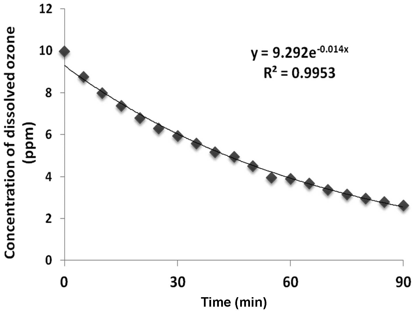
Changes in the concentration of dissolved ozone at
27°C are shown in Fig. 1. The
concentrations of dissolved ozone decreased constantly and
lineally. We also found that the half-life of the dissolved ozone
was 36.8±2.7 min (27°C, n=4).
Protective effect of ozonated water on
LPS-induced acute inflammation
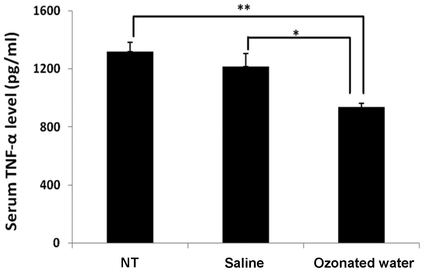
The TNF-α levels are shown in Fig. 2. In the ozonated water-treated
group, the level of TNF-α (935±87 pg/ml) in the serum was
significantly lower than the levels in the NT (935±87 pg/ml) and
saline-treated (935±87 pg/ml) groups (P<0.01 vs. NT and
P<0.05 vs. saline group).
Effect of ozonated water on superoxide
dismutase (SOD) serum activity
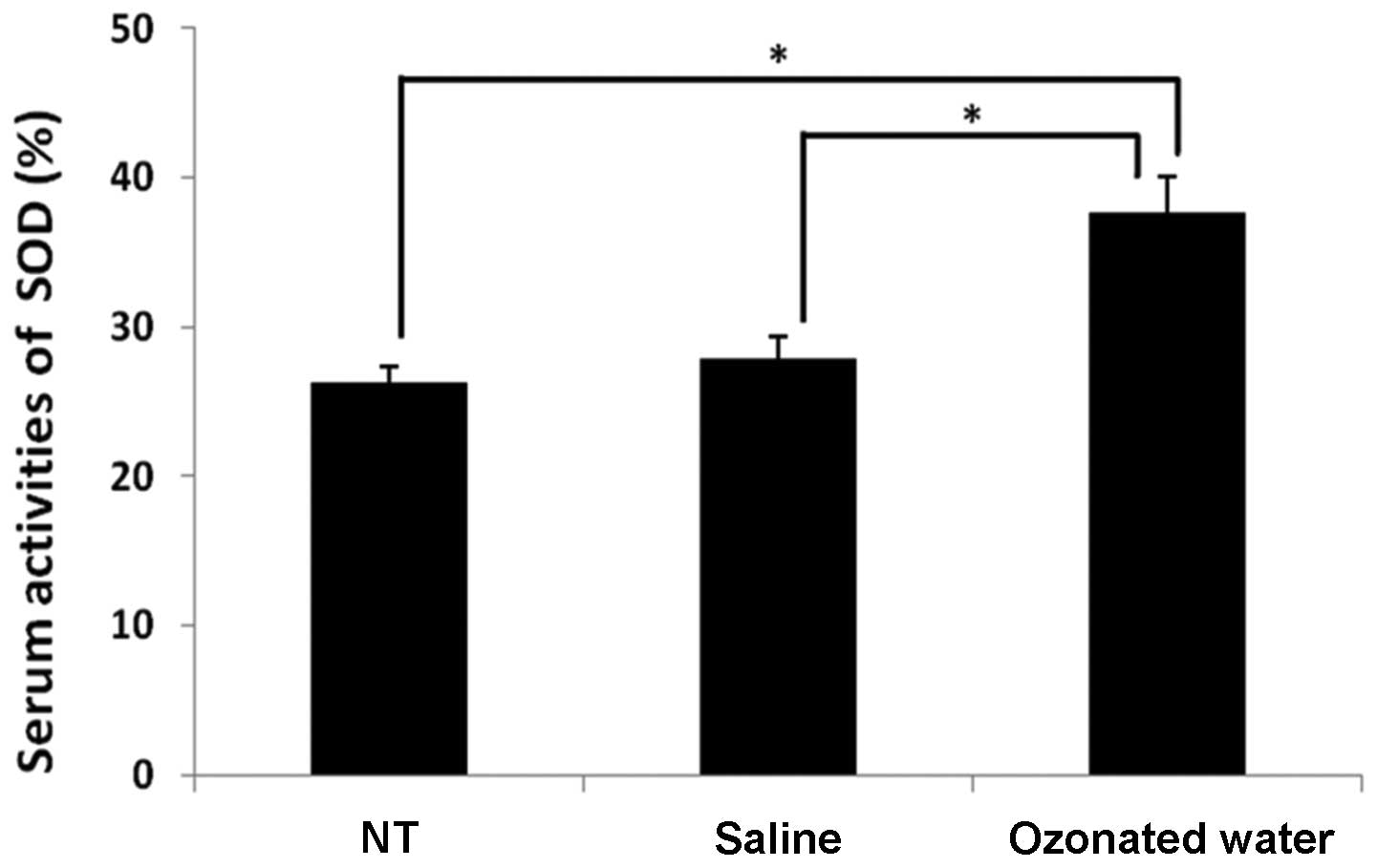
The serum SOD activity is shown in Fig. 3. In the ozonated water-treated
group, SOD activity (38±2%) was significantly higher than that in
the NT (26±1%) and saline-treated (28±1%) groups (P<0.05, vs.
the NT and saline groups).
Discussion
Oxidative damage to vital cellular molecules and
structures such as DNA, lipids, proteins and membranes is induced
by ROS (23). The mitochondrial
electron transport chain is a major source of intracellular ROS and
is susceptible to damage initiated by ROS (24). ROS potentially induces cellular
antioxidant defense enzymes, including SOD (25). Previously, it has been shown that
exposure to LPS increases circulating neutrophils (26) leading to systemic oxidative stress
(27). Tissue neutrophils,
macrophages and monocytes secrete inflammatory cytokines including
TNF-α (28,29). Previous reports indicate
O3 has strong antioxidant activities (4,5). In
the present study, the serum level of TNF-α and SOD serum activity
were significantly decreased and increased, respectively, in the
ozonated water-treated compared to the other groups. Inflammatory
cytokines, including TNF-α, are major mediators of inflammation in
peritonitis (30). Since
O3 therapy induces oxidation in the body, the activities
of antioxidant enzymes such as SOD are increased subsequent to
O3 treatment (5,31). In the experimental septic rat model,
the therapeutic effect of O3 treatment in the rectum was
confirmed (32). Our results show
the ozonated water also exhibit antioxidative and anti-inflammatory
activities. Previous studies indicate ROS was produced following
endotoxin shock-activated nuclear factor (NF)-κB and increased the
production of inflammatory cytokines (33–35).
Overexpression of SOD suppresses TNF-α production in human breast
cancer cells (36). Our findings
may indicate that ozonated water increased the activation of SOD,
which decreased ROS levels. However, additional studies should be
performed to gain an understanding of the mechanism involved in the
anti-inflammatory effects of ozonated water.
In conclusion, our results have shown that ozonated
water exerts critical anti-inflammatory effects. In addition,
ozonated water is useful as a therapeutic option for acute
inflammation.
References
|
1
|
Burns DT: Early problems in the analysis
and the determination of ozone. Fresenius J Anal Chem. 357:178–183.
1997. View Article : Google Scholar
|
|
2
|
Bocci V, Zanardi I and Travagli V: Has
oxygen-ozonetherapy a future in medicine? J Exp Integr Med. 1:5–11.
2011. View Article : Google Scholar
|
|
3
|
Bocci V: Ozone as Janus: this
controversial gas can be either toxic or medically useful.
Mediators Inflamm. 13:3–11. 2004. View Article : Google Scholar : PubMed/NCBI
|
|
4
|
Bocci V: Is it true that ozone is always
toxic? The end of a dogma. Toxicol Appl Pharmacol. 216:493–504.
2006. View Article : Google Scholar : PubMed/NCBI
|
|
5
|
Bocci V: Does ozone therapy normalize the
cellular redox balance? Implications for therapy of human
immunodeficiency virus infection and several other diseases. Med
Hypotheses. 46:150–154. 1996. View Article : Google Scholar
|
|
6
|
Lippmann M: Health effects of ozone. A
critical review. JAPCA. 39:672–695. 1989. View Article : Google Scholar : PubMed/NCBI
|
|
7
|
Devlin RB, McDonnell WF, Mann R, Becker S,
House DE, Schreinemachers D and Koren HS: Exposure of humans to
ambient levels of ozone for 6.6 hours causes cellular and
biochemical changes in the lung. Am J Respir Cell Mol Biol.
4:72–81. 1991. View Article : Google Scholar : PubMed/NCBI
|
|
8
|
Aris RM, Christian D, Hearne PQ, Kerr K,
Finkbeiner WE and Balmes JR: Ozone-induced airway inflammation in
human subjects as determined by airway lavage and biopsy. Am Rev
Respir Dis. 148:1363–1372. 1993. View Article : Google Scholar : PubMed/NCBI
|
|
9
|
Krishna MT, Madden J, Teran LM, Biscione
GL, Lau LC, Withers NJ, Sandstrom T, Mudway I, Kelly FJ, Walls A,
et al: Effects of 0.2 ppm ozone on biomarkers of inflammation in
bronchoalveolar lavage fluid and bronchial mucosa of healthy
subjects. Eur Respir J. 11:1294–1300. 1998. View Article : Google Scholar : PubMed/NCBI
|
|
10
|
Broeckaert F, Arsalane K, Hermans C,
Bergamaschi E, Brustolin A, Mutti A and Bernard A: Lung epithelial
damage at low concentrations of ambient ozone. Lancet. 353:900–901.
1999. View Article : Google Scholar : PubMed/NCBI
|
|
11
|
Bell ML, McDermott A, Zeger SL, Samet JM
and Dominici F: Ozone and short-term mortality in 95 US urban
communities, 1987–2000. JAMA. 292:2372–2378. 2004.
|
|
12
|
Tager IB, Balmes J, Lurmann F, Ngo L,
Alcorn S and Kunzli N: Chronic exposure to ambient ozone and lung
function in young adults. Epidemiology. 16:751–759. 2005.
View Article : Google Scholar : PubMed/NCBI
|
|
13
|
Soulage C, Perrin D, Cottet-Emard JM,
Pequignot J, Dalmaz Y and Pequignot JM: Central and peripheral
changes in catecholamine biosynthesis and turnover in rats after a
short period of ozone exposure. Neurochem Int. 45:979–986. 2004.
View Article : Google Scholar : PubMed/NCBI
|
|
14
|
Ruidavets JB, Cournot M, Cassadou S,
Giroux M, Meybeck M and Ferrieres J: Ozone air pollution is
associated with acute myocardial infarction. Circulation.
111:563–569. 2005. View Article : Google Scholar : PubMed/NCBI
|
|
15
|
Bell ML, Dominici F and Samet JM: A
meta-analysis of time-series studies of ozone and mortality with
comparison to the national morbidity, mortality, and air pollution
study. Epidemiology. 16:436–445. 2005. View Article : Google Scholar : PubMed/NCBI
|
|
16
|
Ito K, De Leon SF and Lippmann M:
Associations between ozone and daily mortality: analysis and
meta-analysis. Epidemiology. 16:446–457. 2005. View Article : Google Scholar : PubMed/NCBI
|
|
17
|
Levy JL, Chemerynski SM and Sarnat JA:
Ozone exposure and mortality: an empiric bayes metaregression
analysis. Epidemiology. 16:458–468. 2005. View Article : Google Scholar : PubMed/NCBI
|
|
18
|
Bates DV: Ambient ozone and mortality.
Epidemiology. 16:427–429. 2005. View Article : Google Scholar : PubMed/NCBI
|
|
19
|
Goodman SN: The methodologic ozone effect.
Epidemiology. 16:430–435. 2005. View Article : Google Scholar : PubMed/NCBI
|
|
20
|
Scott DB and Lesher EC: Effect of ozone on
survival and permeability of Escherichia coli. J Bacteriol.
85:567–576. 1963.PubMed/NCBI
|
|
21
|
Restaino L, Frampton EW, Hemphill JB and
Palnikar P: Efficacy of ozonated water against various food-related
microorganisms. Appl Environ Microbiol. 61:3471–3475.
1995.PubMed/NCBI
|
|
22
|
Ozmen V, Thomas WO, Healy JT, Fish JM,
Chambers R, Tacchi E, Nichols RL, Flint LM and Ferrara JJ:
Irrigation of the abdominal cavity in the treatment of
experimentally induced microbial peritonitis: efficacy of ozonated
saline. Am Surg. 59:297–303. 1993.PubMed/NCBI
|
|
23
|
Cadet J, Bellon S, Douki T, Frelon S,
Gasparutto D, Muller E, Pouget JP, Ravanat JL, Romieu A and
Sauvaigo S: Radiation-induced DNA damage: formation, measurement,
and biochemical features. J Environ Pathol Toxicol Oncol. 23:33–43.
2004. View Article : Google Scholar : PubMed/NCBI
|
|
24
|
Murphy MP: How mitochondria produce
reactive oxygen species. Biochem J. 417:1–13. 2009. View Article : Google Scholar : PubMed/NCBI
|
|
25
|
Turrens JF: Mitochondrial formation of
reactive oxygen species. J Physiol. 552:335–344. 2003. View Article : Google Scholar : PubMed/NCBI
|
|
26
|
Zhang B, Su Y, Ai G, Wang Y, Wang T and
Wang F: Involvement of peroxiredoxin I in protecting cells from
radiation-induced death. J Radiat Res. 46:305–312. 2005. View Article : Google Scholar : PubMed/NCBI
|
|
27
|
Cheah FC, Jobe AH, Moss TJ, Newnham JP and
Kallapur SG: Oxidative stress in fetal lambs exposed to
intra-amniotic endotoxin in a chorioamnionitis model. Pediatr Res.
63:274–279. 2008. View Article : Google Scholar : PubMed/NCBI
|
|
28
|
Lin HI, Chu SJ, Wang D and Feng NH:
Pharmacological modulation of TNF production in macrophages. J
Microbiol Immunol Infect. 37:8–15. 2004.PubMed/NCBI
|
|
29
|
Zamora ZB, Borrego A, Lopez OY, Delgado R,
Gonzalez R, Menendez S, Hernandez F and Schulz S: Effects of ozone
oxidative preconditioning on TNF-alpha release and
antioxidant-prooxidant intracellular balance in mice during
endotoxic shock. Mediators Inflamm. 2005.16–22. 2005. View Article : Google Scholar
|
|
30
|
Beutler B and Cerami A: The biology of
cachectin/TNF - a primary mediator of the host response. Annu Rev
Immunol. 7:625–655. 1989. View Article : Google Scholar : PubMed/NCBI
|
|
31
|
Peralta C, Leon OS, Xaus C, Prats N, Jalil
EC, Planell ES, Puig-Parellada P, Gelpi E and Rosello-Catafau J:
Protective effect of ozone treatment on the injury associated with
hepatic ischemia-reperfusion: antioxidant-prooxidant balance. Free
Radic Res. 31:191–196. 1999. View Article : Google Scholar : PubMed/NCBI
|
|
32
|
Souza YM, Fontes B, Martins JO, Sannomiya
P, Brito GS, Younes RN and Rasslan S: Evaluation of the effects of
ozone therapy in the treatment of intra-abdominal infection in
rats. Clinics (Sao Paulo). 65:195–202. 2010. View Article : Google Scholar : PubMed/NCBI
|
|
33
|
Chandel NS, Trzyna WC, McClintock DS and
Schumacker PT: Role of oxidants in NF-kappa B activation and
TNF-alpha gene transcription induced by hypoxia and endotoxin. J
Immunol. 165:1013–1021. 2000. View Article : Google Scholar : PubMed/NCBI
|
|
34
|
Gilmore TD: The Rel/NF-kappaB signal
transduction pathway: introduction. Oncogene. 18:6842–6844. 1999.
View Article : Google Scholar : PubMed/NCBI
|
|
35
|
Nakamura H, Nakamura K and Yodoi J: Redox
regulation of cellular activation. Annu Rev Immunol. 15:351–369.
1997. View Article : Google Scholar : PubMed/NCBI
|
|
36
|
Manna SK, Zhang HJ, Yan T, Oberley LW and
Aggarwal BB: Overexpression of manganese superoxide dismutase
suppresses tumor necrosis factor-induced apoptosis and activation
of nuclear transcription factor-kappaB and activated protein-1. J
Biol Chem. 273:13245–13254. 1998. View Article : Google Scholar
|