Introduction
The benefits of spring waters in the treatment of
actual pathologies and/or in re-establishing the physiological
wellness of different organs and systems have been demonstrated
since the most ancient times, and specific indications have been
historically attributed to each spring.
However, the molecular mechanisms and the various
interactions responsible for the anti-inflammatory and regenerative
properties of spring waters remain largely unknown and require
investigation.
Our previous study demonstrated that an Italian
spring water (Comano-Trentino) can improve skin regeneration in an
animal experimental model, not only by increasing keratinocyte
proliferation and migration, but also by modulating the regenerated
collagen and elastic fibers in the dermis (1).
However, such biological properties may not be
entirely explained by the mineral composition only.
As the non-pathogenic bacterial populations have
demonstrated an active role in different biological processes, the
potential presence of non-pathogenic bacterial species within the
Comano spring water have been investigated in order to identify any
possible correlation between these bacterial populations and the
demonstrated biological properties of this water.
Materials and methods
General
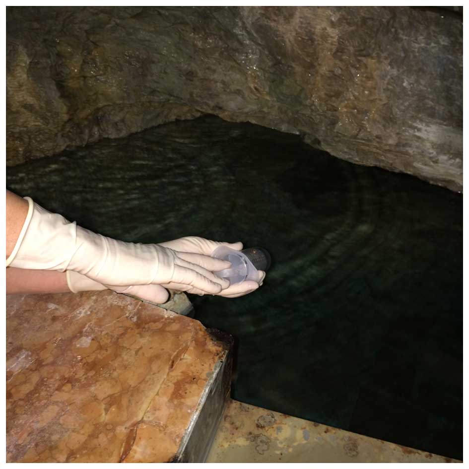
The Comano spring water was collected at the spring
with an aseptic procedure (Fig. 1) in
January, June and October 2014. A single operator wearing sterile
surgical gloves collected 3,000 ml of water each time with a
sterile 50-ml syringe. The samples were poured into 3 sterile
one-liter containers for microbiological analysis and stored at
4°C.
Sample processing, isolation and
identification of bacteria
Samples were transported to the Laboratory of
Bacteriology, Microbiology and Virology Department, San Matteo
Hospital Foundation, Research and Care Institute (Pavia, Italy), at
4°C and processed rapidly following collection.
Two pairs of BD BACTEC™ culture aerobic/anaerobic
vials were inoculated each with 10 ml water. Subsequently, the
samples were incubated in a BACTEC™ 9240 automated blood culture
system (BD Biosciences, Sparks, MD, USA), according to the
manufacturer's instructions, for 7 days.
Six 0.20-µm pore cellulose nitrate membranes
(Nalgene 0.2 Analytical filter Unit; Thermo Fisher Scientific,
Inc., Waltham, MA, USA) were used to filter 100 ml of water each.
Five membranes were subsequently placed on a different plating
medium (blood, chocolate, McConkey, mannitol salt or Sabouraud
dextrose agars) and incubated in aerobic conditions at 37°C for 3
days. One membrane was placed on Schaedler blood agar medium and
incubated in anaerobic conditions at 37°C for 6 days.
Similarly, a 1,000-ml water sample was filtered
through a 0.20-µm pore cellulose nitrate membrane and processed
according to the guidelines for Legionella detection in
water (2). In particular, buffered
charcoal yeast extract and glycine, vancomycin, polymyxin B and
cycloheximide plates were inoculated and incubated at 37°C with 5%
CO2 for 14 days.
Isolated organisms were biochemically identified
with API NE (BioMerieux SA, Marcy l'Etoile, France) or using the
Phoenix 100™ (BD Biosciences) automated system.
Results
Positive identification of
cultures
BD BACTEC™ culture aerobic/anaerobic vials became
positive after 3 days.
Biochemical identification of the
bacterial cultures
The biochemical identification of the cultured
bacteria provided different results at different times. The Phoenix
100™ (BD Biosciences) automated system identified Citrobacter
youngae and Pantoea agglomerans in January 2014,
Pseudomonas stutzeri and Streptococcus mitis in June
2014 and no colonies in October 2014. The microorganisms isolated
with API NE (BioMerieux sa) were Aeromonas hydrophila (CB 6
UFC/100 ml), Chromobacterium violaceum (CB 2 UFC/100 ml) and
Empedobacter brevis (CB 3 UFC/100 ml) in January 2014,
Brevundimonas vesicularis (CB 7 UFC/100 ml) and
Pseudomonas putida (CB 4 UFC/100 ml) in June 2014, and
Aeromonas hydrophila (CB 3 UFC/100 ml) and Pseudomonas
putida (CB 8 UFC/100 ml) in October 2014.
The isolates at different times per identification
system are summarized in Table I. A
classification of the isolates is provided in Table II.
 | Table I.Isolates at different times per
identification system. |
Table I.
Isolates at different times per
identification system.
|
| Identification
system |
|---|
|
|
|
|---|
| Dates | Phoenix 100™ | API NE |
|---|
| January 2014 | Citrobacter
youngae | Aeromonas
hydrophila (CB 6 UFC/100 ml) |
|
| Pantoea
agglomerans | Chromobacterium
violaceum (CB 2 UFC/100 ml) |
|
|
| Empedobacter
brevis (CB 3 UFC/100 ml) |
| June 2014 | Pseudomonas
stutzeri | Brevundimonas
vesicularis (CB 7 UFC/100 ml) |
|
| Streptococcus
mitis | Pseudomonas
putida (CB 4 UFC/100 ml) |
| October 2014 | Negative
culture | Aeromonas
hydrophila (CB 3 UFC/100 ml) |
|
|
| Pseudomonas
putida (CB 8 UFC/100 ml) |
| Table II.Classification of isolates. |
Table II.
Classification of isolates.
| Isolates | Family | Genus | Species |
|---|
| Aeromonas
hydrophila | Aeromonadaceae | Aeromonas | H.
hydrophila |
| Brevundimonas
vesicularis |
Caulobacteriaceae | Brevundimonas | B.
vesicularis |
| Chromobacterium
violaceum | Neisseriaceae |
Chromobacterium | C.
violaceum |
| Citrobacter
youngae |
Enterobacteriaceae | Citrobacter | C.
youngae |
| Empedobacter
brevis |
Flavobacteriaceae | Empedobacter | E.
brevis |
| Pantoea
agglomerans |
Enterobacteriaceae | Pantoea | P.
agglomerans |
| Pseudomonas
putida |
Pseudomonadaceae | Pseudomonas | P.
putida |
| Pseudomonas
stutzeri |
Pseudomonadaceae | Pseudomonas | P.
stutzeri |
| Streptococcus
mitis |
Streptococcaceae | Streptococcus | S.
mitis |
Discussion
The Comano spring water is a hypotonic,
bicarbonate-calcium-magnesium mineral water that is rich in
fluoride, and has a neutral pH and a low-buffer capacity. The
ECOOPERA S.C. laboratory (Gardolo, TN, Italy; ACCREDIA certified
no. 0252) regularly certifies this water as bacteriologically pure,
the latter definition meaning that it does not contain pathogenic
microorganisms nor microorganisms indicating fecal or other
contamination (3).
A total of 9 different strains were isolated from
the Comano spring water: Aeromonas hydrophila,
Brevundimonas vesicularis, Chromobacterium violaceum,
Citrobacter youngae, Empedobacter brevis, Pantoea
agglomerans, Pseudomonas putida, Pseudomonas
stutzeri and Streptococcus mitis.
Aeromonas hydrophila is the most prominent of
the 6 species of Aeromonas (4).
It is a heterotrophic Gram-negative bacterium mainly identified in
areas with a warm climate. This bacterium can be found in fresh or
brackish water and can survive in aerobic and anaerobic
environments. Aeromonas hydrophila represents a constant
component of the microbiota in fresh reservoirs where, together
with other microorganisms, it acts as a natural biofilter and
promotes water self-purification. It is present in normal
microflora of hydrobionts inhabiting fresh reservoirs (5). Recently, Aeromonas hydrophila was
isolated in samples from Moroccan Atlantic Ocean water (6), where antifungal and antibacterial
activity was demonstrated.
Brevundimonas vesicularis is an environmental
Gram-negative bacillum that has demonstrated the capacity to
degrade sulfonated naphthalene-formaldehyde condensate compounds
isolated from textile industry-activated sludge wastewater
(7).
Chromobacterium violaceum is a facultative
anaerobe Gram-negative bacterium, considered to be non-pathogenic
for humans. This bacterium appears to control microbial infection
and decrease the risks of resistance development (8). Violacein, the pigment of
Chromobacterium violaceum, has potential medical
applications as a drug with different properties: Antitrypanocidal,
antileishmaniosis, antimycobacterial, antimalaric,
anti-ulcerogenic, anticancer and antioxidant (9). Chromobacterium violaceum was also
isolated in samples from Moroccan Atlantic Ocean water (6).
The Citrobacter youngae species are straight,
facultative anaerobic Gram-negative bacilli that are commonly found
in water, soil, food and in the intestinal tracts of animals and
humans. Certain strains belonging to the genus Citrobacter
have been reported to produce chitin/chitosan-like bioflocculants
from acetate (10). Furthermore,
degradation of the lipopolysaccharide of Citrobacter youngae
releases polysaccharides with structure rarely identified in
bacteria (11).
Empedobacter brevis, a Gram-negative aerobe
also known as Flavobacterium breve, is the only member of
the Empedobacter genus. It produces an enzyme that catalyzes
the peptide-forming reaction producing l-alanyl-L-glutamine, a
dipeptide of significant industrial interest by virtue of its
widespread use in infusion therapy (12).
Pantoea agglomerans is a Gram-negative
bacterium that belongs to the family Enterobacteriaceae. It
is commonly isolated from plant surfaces, seeds, fruit (such as
mandarin oranges) and animal or human feces. Pantoea
agglomerans is primarily a plant epiphyte commonly found in
diverse ecological niches, including aquatic environments, soil or
sediments. Several strains of Pantoea agglomerans are sold
as commercial biological control agents against the fire blight
pathogen on apple and pear trees (13). The primary mode of action is
competitive exclusion, which involves the occupation of sites
otherwise colonized by the pathogens; however, according to the
literature, certain strains may also contribute with the production
of different antibiotic-like substances (herbicolins, pantocins,
putative phenazine and other unknown compounds) (14). This bacterium was also isolated in
samples from Moroccan Atlantic Ocean water (6).
Pseudomonas putida is a Gram-negative aerobic
saprotrophic soil bacterium. As it is able to degrade organic
solvents, such as toluene, and also convert styrene oil to
biodegradable plastic polyhydroxyalkanoates, it may be used to
degrade the polystyrene foam that was thought to be
non-biodegradable (15). Furthermore,
one of its engineered strains proved to be useful for in
situ bioremediation of soils co-contaminated with
organophosphorus and pyrethroid pesticides. In turn, Pseudomonas
putida induces plant growth and protects the plants from
pathogens. Τherefore, researchers use it in bioengineering research
to develop biopesticides and to the improve plant health (16).
Pseudomonas stutzeri, an almost universal
Gram-negative ammonia-oxidizing bacterium, exhibits abilities in
efficient heterotrophic nitrification and aerobic denitrification
(17), and in organophosphorus
pesticides degradation (18).
Therefore, it is considered a suitable candidate to simultaneously
remove nitrogen and phosphate in wastewater treatment. Similar to
Brevundimonas vesicularis, it also demonstrated the capacity
to degrade sulfonated naphthalene-formaldehyde condensate compounds
isolated from textile industry-activated sludge wastewater
(6).
Streptococcus mitis is a Gram-positive
facultative anaerobe coccus that is an abundant human oral
commensal and is reported as a potentially useful vector for
mucosal vaccination (19).
While in the past microorganisms were recognized
just as enemies of the human body, the trend has currently changed
as the position of microbiota has recently undergone a turning
point in the contemporary vision of medicine. Helminths,
saprophytic mycobacteria, bifidobacteria and
lactobacilli cause little, if any, harm and have been a part
of human microecology for millenia. Deficient exposure to these may
even explain the contemporary increase of immune disorders in a
modern, highly sanitized society (20).
The use of probiotics, prebiotics, helminths or
microbe-derived immunoregulatory substances has become a novel and
valuable approach to disease prevention (20), and emerging clinical studies indicate
that the supplementation and/or fecal microbial transplant
(21) can improve bowel health and
brain functions (22,23).
The skin microbiota is constituted by bacteria and
fungi with typical counts of 102−107
cells/cm2 in a diverse topography reflecting their
different niches (24,25).
Normal skin microorganisms are classified as either
resident (i.e. adhering predominantly to the skin and annexes,
maintaining viability and reproducibility) or transient (i.e.
deposited but not adhering to the skin surface, with little nor
sustained growth and reproduction). The skin ecosystem is a complex
environment, extending to sub-epidermal compartments (26), tending to withstand pathogen
colonization. Stability is maintained by interactions among
different microbial species and the host. The topical use of
probiotics has been reported to have a direct effect on the site of
application, as natural defence mechanisms are induced by the
competition with pathogens for nutrients, by the modulation of
mucosal immune functions and by the production of antimicrobial
metabolites (27–29).
Furthermore, epidermal keratinocytes have been
demonstrated to produce antimicrobial peptides (30–32).
A correlation between the well-known beneficial
effects on the skin and the resident non-pathogen bacterial
populations was demonstrated in certain spring waters.
From the culture of Aquaphilus dolomiae, a
non-spore forming bacterium belonging to the Neisseriaceae family
isolated from Avène thermal Water (France), an organic substance,
I-modulia, was obtained that is able to regulate keratinocyte
inflammatory and lymphocyte immune responses (33–36).
Similarly (37,38), the topical administration of a lysate
of Vitreoscilla filiformis, a Gram-negative aerobic
bacterium belonging to the Neisseriaceae family found in
LaRoche-Posay thermal water (France), has been demonstrated to
benefit the local skin immunity, possibly by the activation of
cutaneous regulatory T cells (39,40).
Our previous study on the properties of the Comano
water on experimental fresh wounds in an animal model demonstrated
a significant increase in the overall cell proliferation and a
corresponding reduction of the local inflammatory response
(1).
Such effects may not be entirely explained by the
mineral composition only, but may be correlated to the antifungal
and antimicrobial properties of some of the bacterial isolates as
well (40).
Although showing a rare potential virulence
(41–50), all the isolated bacterial strains
demonstrate peculiar and favorable metabolic attitudes in
controlling environmental pollution.
Skin regeneration is a complex process involving the
close and coordinated interaction of different cell strains, as
keratinocytes, fibroblasts and immune-system cells in the
extracellular matrix environment.
The modulation of the wound proliferative process is
likely to be influenced by the local microbiota as well, and this
hypothesis has not been investigated previously. Therefore, it
appears reasonable to conceive this study with the aim to provide a
more comprehensive understanding of the regenerative properties of
the Comano spring water.
Although the present study provided only preliminary
data, some of the non-pathogenic bacterial populations that were
identified in the Comano spring water are likely to produce
molecular mediators with a role in the wound healing process that,
thus far, remain unknown. Numerous other unknown bacterial species,
comprehensively termed DNA-rich ‘dark matter’, are likely to
contribute to the Comano water regenerative properties as well.
In conclusion, the therapeutical effects of certain
spring waters are currently being proven as correlated not only to
their peculiar mineral composition, but also to the complex
activity of their resident, non-pathogenic bacterial populations
(33–40).
Therefore, the non-pathogenic bacterial populations
of the Comano spring water are likely to be credited for its
demonstrated regenerative properties (1).
Such evidence may direct the introduction of novel
research opportunities for regeneration to the role of
microbiota.
References
|
1
|
Faga A, Nicoletti G, Gregotti C, Finotti
V, Nitto A and Gioglio L: Effects of thermal water on skin
regeneration. Int J Mol Med. 29:732–740. 2012.PubMed/NCBI
|
|
2
|
International Organization for
Standardization (ISO). Water quality-detection and enumeration of
Legionella: Direct membrane filtration method for waters with low
bacterial counts. ISO 11731–11732. 2004.http://www.legionellaonline.it/Accessed. May
26–2015
|
|
3
|
Éditeur officiel du Québec. Regulation
respecting bottled water: Food Products Act. http://www2.publicationsduquebec.gouv.qc.ca/dynamicSearch/telecharge.php?type=3&file=/P_29/P29R2_A.HTMAccessed.
August 1–2015
|
|
4
|
Horneman AJ and Ali A: Aeromonas. Manual
of Clinical Microbiology. Versalovic J, Carroll KC, Funke G,
Jorgensen JH, Landry ML and Warnock DW: (10th). ASM Press.
(Washington, DC). 658–665. 2011.
|
|
5
|
Kompanets EV, Isaeva NM and Balakhnin IA:
Bacteria of the genus Aeromonas and their role in
aquaculture. Mikrobiol Zh. 54:89–99. 1992.(In Russian). PubMed/NCBI
|
|
6
|
El Amraoui B, El Amraoui M, Cohen N and
Fassouane A: Antifungal and antibacterial activity of marine
microorganisms. Ann Pharm Fr. 72:107–111. 2014. View Article : Google Scholar : PubMed/NCBI
|
|
7
|
Cheriaa J, Mosrati R, Ladhari N and
Bakhrouf A: Acclimated biomass that degrades Sulfonated Naphthalene
Formaldehyde Condensate. Pak J Biol Sci. 11:1588–1593. 2008.
View Article : Google Scholar : PubMed/NCBI
|
|
8
|
Durán M, Faljoni-Alario A and Durán N:
Chromobacterium violaceum and its important metabolites -
review. Folia Microbiol (Praha). 55:535–547. 2010. View Article : Google Scholar : PubMed/NCBI
|
|
9
|
Hoshino T: Violacein and related
tryptophan metabolites produced by Chromobacterium
violaceum: Biosynthetic mechanism and pathway for construction
of violacein core. Appl Microbiol Biotechnol. 91:1463–1475. 2011.
View Article : Google Scholar : PubMed/NCBI
|
|
10
|
Kimura K, Inoue T, Kato D, Negoro S, Ike M
and Takeo M: Distribution of chitin/chitosan-like
bioflocculant-producing potential in the genus Citrobacter. Appl
Microbiol Biotechnol. 97:9569–9577. 2013. View Article : Google Scholar : PubMed/NCBI
|
|
11
|
Kocharova NA, Mieszała M, Zatonsky GV,
Staniszewska M, Shashkov AS, Gamian A and Knirel YA: Structure of
the O-polysaccharide of Citrobacter youngae O1 containing an
alpha-D-ribofuranosyl group. Carbohydr Res. 339:321–325. 2004.
View Article : Google Scholar : PubMed/NCBI
|
|
12
|
Yokozeki K and Hara S: A novel and
efficient enzymatic method for the production of peptides from
unprotected starting materials. J Biotechnol. 115:211–220. 2005.
View Article : Google Scholar : PubMed/NCBI
|
|
13
|
Stockwell VO, Johnson KB, Sugar D and
Loper JE: Antibiosis Contributes to Biological Control of Fire
Blight by Pantoea agglomerans Strain Eh252 in Orchards.
Phytopathology. 92:1202–1209. 2002. View Article : Google Scholar : PubMed/NCBI
|
|
14
|
Rezzonico F, Smits TH, Montesinos E, Frey
JE and Duffy B: Genotypic comparison of Pantoea agglomerans
plant and clinical strains. BMC Microbiol. 9:2042009. View Article : Google Scholar : PubMed/NCBI
|
|
15
|
Zuo Z, Gong T, Che Y, Liu R, Xu P, Jiang
H, Qiao C, Song C and Yang C: Engineering Pseudomonas putida KT2440
for simultaneous degradation of organophosphates and pyrethroids
and its application in bioremediation of soil. Biodegradation.
26:223–233. 2015. View Article : Google Scholar : PubMed/NCBI
|
|
16
|
Espinosa Urgel, Salido A and Ramos JL:
Genetic analysis of functions involved in adhesion of Pseudomonas
putida to seeds. J Bacteriol. 182:2363–2369. 2000. View Article : Google Scholar : PubMed/NCBI
|
|
17
|
Li C, Yang J, Wang X, Wang E, Li B, He R
and Yuan H: Removal of nitrogen by heterotrophic
nitrification-aerobic denitrification of a phosphate accumulating
bacterium Pseudomonas stutzeri YG-24. Bioresour Technol.
182:18–25. 2015. View Article : Google Scholar : PubMed/NCBI
|
|
18
|
Shi YH, Ren L, Jia Y and Yan YC: Genome
Sequence of Organophosphorus Pesticide-Degrading Bacterium
Pseudomonas stutzeri Strain YC-YH1. Genome Announc.
3:e00192–15. 2015. View Article : Google Scholar : PubMed/NCBI
|
|
19
|
Daifalla N, Cayabyab MJ, Xie E, Kim HB,
Tzipori S, Stashenko P, Duncan M and Campos-Neto A: Commensal
Streptococcus mitis is a unique vector for oral mucosal
vaccination. Microbes Infect. 17:237–242. 2015. View Article : Google Scholar : PubMed/NCBI
|
|
20
|
Guarner F, Bourdet-Sicard R, Brandtzaeg P,
Gill HS, McGuirk P, van Eden W, Versalovic J, Weinstock JV and Rook
GA: Mechanisms of disease: The hygiene hypothesis revisited. Nat
Clin Pract Gastroenterol Hepatol. 3:275–284. 2006. View Article : Google Scholar : PubMed/NCBI
|
|
21
|
Petrof EO, Gloor GB, Vanner SJ, Weese SJ,
Carter D, Daigneault MC, Brown EM, Schroeter K and Allen-Vercoe E:
Stool substitute transplant therapy for the eradication of
Clostridium difficile infection: ‘RePOOPulating’ the gut.
Microbiome. 1:32013. View Article : Google Scholar : PubMed/NCBI
|
|
22
|
Bested AC, Logan AC and Selhub EM:
Intestinal microbiota, probiotics and mental health: From
Metchnikoff to modern advances: Part III - convergence toward
clinical trials. Gut Pathog. 5:42013. View Article : Google Scholar : PubMed/NCBI
|
|
23
|
Selhub EM, Logan AC and Bested AC:
Fermented foods, microbiota, and mental health: Ancient practice
meets nutritional psychiatry. J Physiol Anthropol. 33:22014.
View Article : Google Scholar : PubMed/NCBI
|
|
24
|
Grice EA and Segre JA: The skin
microbiome. Nat Rev Microbiol. 9:244–253. 2011. View Article : Google Scholar : PubMed/NCBI
|
|
25
|
Huttenhower C, Gevers D, Knight R, et al:
Human Microbiome Project Consortium: Structure, function and
diversity of the healthy human microbiome. Nature. 486:207–214.
2012. View Article : Google Scholar : PubMed/NCBI
|
|
26
|
Nakatsuji T, Chiang HI, Jiang SB,
Nagarajan H, Zengler K and Gallo RL: The microbiome extends to
subepidermal compartments of normal skin. Nat Commun. 4:14312013.
View Article : Google Scholar : PubMed/NCBI
|
|
27
|
Krutmann J: Pre- and probiotics for human
skin. Clin Plast Surg. 39:59–64. 2012. View Article : Google Scholar : PubMed/NCBI
|
|
28
|
Al-Ghazzewi FH and Tester RF: Impact of
prebiotics and probiotics on skin health. Benef Microbes. 5:99–107.
2014. View Article : Google Scholar : PubMed/NCBI
|
|
29
|
Bockmühl D, Jassoy C, Nieveler S,
Scholtyssek R, Wadle A and Waldmann-Laue M: Prebiotic Cosmetics: An
Alternative to Antibacterial Products. Int J Cosmet Sci. 29:63–64.
2007. View Article : Google Scholar
|
|
30
|
Gallo RL, Murakami M, Ohtake T and Zaiou
M: Biology and clinical relevance of naturally occurring
antimicrobial peptides. J Allergy Clin Immunol. 110:823–831. 2002.
View Article : Google Scholar : PubMed/NCBI
|
|
31
|
de Jongh GJ, Zeeuwen PL, Kucharekova M,
Pfundt R, van der Valk PG, Blokx W, Dogan A, Hiemstra PS, van de
Kerkhof PC and Schalkwijk J: High expression levels of keratinocyte
antimicrobial proteins in psoriasis compared with atopic
dermatitis. J Invest Dermatol. 125:1163–1173. 2005. View Article : Google Scholar : PubMed/NCBI
|
|
32
|
Faga A, Pelfini C, Concia E and Bona F:
Variazioni della naturale attività antibatterica della cute: Un
metodo di valutazione di detergenti e/o antisettici. Chron Derm.
2:233–241. 1981.(In Italian).
|
|
33
|
Nocera T, Fabre P, Rossi AB and Mengeaud
V: Clinical development program of a new dermocosmetic range of
products containing I-modulia (Aquaphilus dolomiae extract)
in atopic dermatitis. J Am Acad Dermatol. 70:(suppl 1).
AB62014.
|
|
34
|
Patrizi A, Bacquey A, Fabre P, Schmitt AM,
Decoster CJ, Phulpin C, Theunis J and Mengeaud V: Clinical and
biometrologic evaluation of a novel emollient balm containing an
Aquaphilus dolomiae extract in 1- to 4-year-old children
suffering from atopic dermatitis: International, multicenter,
randomized versus control group study. J Am Acad Dermatol.
70:(suppl 1). AB622014. View Article : Google Scholar
|
|
35
|
Aries MF, Fabre P, Vaissière C, Delga H,
Leveque M, Castex-Rizzi N, Bessou-Touya S and Nguyen T:
Antiinflammatory and immunomodulatory effect of I-modulia, an
Aquaphilus dolomiae extract, on atopic dermatitis in vitro.
J Am Acad Dermatol. 70:(suppl 1). AB612014. View Article : Google Scholar
|
|
36
|
Aries MF, Fabre P, Duplan H, Hernandez
Pigeon H, Galliano MF, Castex-Rizzi N, Bessou-Touya S and Nguyen T:
I-modulia, an Aquaphilus dolomiae extract, stimulates innate immune
response through Toll-like receptor activation. J Am Acad Dermatol.
70:(suppl 1). AB632014. View Article : Google Scholar
|
|
37
|
Mahe YF, Perez MJ, Tacheau C, Fanchon C,
Martin R, Rousset F and Seite S: A new Vitreoscilla
filiformis extract grown on spa water-enriched medium activates
endogenous cutaneous antioxidant and antimicrobial defenses through
a potential Toll-like receptor 2/protein kinase C, zeta
transduction pathway. Clin Cosmet Investig Dermatol. 6:191–196.
2013.PubMed/NCBI
|
|
38
|
Gueniche A, Knaudt B, Schuck E, Volz T,
Bastien P, Martin R, Röcken M, Breton L and Biedermann T: Effects
of nonpathogenic gram-negative bacterium Vitreoscilla
filiformis lysate on atopic dermatitis: A prospective,
randomized, double-blind, placebo-controlled clinical study. Br J
Dermatol. 159:1357–1363. 2008. View Article : Google Scholar : PubMed/NCBI
|
|
39
|
Volz T, Skabytska Y, Guenova E, Chen KM,
Frick JS, Kirschning CJ, Kaesler S, Röcken M and Biedermann T:
Nonpathogenic bacteria alleviating atopic dermatitis inflammation
induce IL-10-producing dendritic cells and regulatory Tr1 cells. J
Invest Dermatol. 134:96–104. 2014. View Article : Google Scholar : PubMed/NCBI
|
|
40
|
Nakatsuji T and Gallo RL: Dermatological
therapy by topical application of non-pathogenic bacteria. J Invest
Dermatol. 134:11–14. 2014. View Article : Google Scholar : PubMed/NCBI
|
|
41
|
Agger WA, McCormick JD and Gurwith MJ:
Clinical and microbiological features of Aeromonas
hydrophila-associated diarrhea. J Clin Microbiol. 21:909–913.
1985.PubMed/NCBI
|
|
42
|
Minnaganti VR, Patel PJ, Iancu D, Schoch
PE and Cunha BA: Necrotizing fasciitis caused by Aeromonas
hydrophila. Heart Lung. 29:306–308. 2000. View Article : Google Scholar : PubMed/NCBI
|
|
43
|
Sneath PH, Whelan JP, Bhagwan Singh R and
Edwards D: Fatal infection by Chromobacterium violaceum.
Lancet. 265:276–277. 1953. View Article : Google Scholar : PubMed/NCBI
|
|
44
|
Janknecht P, Schneider CM and Ness T:
Outbreak of Empedobacter brevis endophthalmitis after cataract
extraction. Graefes Arch Clin Exp Ophthalmol. 240:291–295. 2002.
View Article : Google Scholar : PubMed/NCBI
|
|
45
|
Jain S, Bohra I, Mahajan R, Jain S and
Chugh TD: Pantoea agglomerans infection behaving like a
tumor after plant thorn injury: An unusual presentation. Indian J
Pathol Microbiol. 55:386–388. 2012. View Article : Google Scholar : PubMed/NCBI
|
|
46
|
Shih CC, Chen YC, Chang SC, Luh KT and
Hsieh WC: Bacteremia due to Citrobacter species:
Significance of primary intraabdominal infection. Clin Infect Dis.
23:543–549. 1996. View Article : Google Scholar : PubMed/NCBI
|
|
47
|
Shang ST, Chiu SK, Chan MC, Wang NC, Yang
YS, Lin JC and Chang FY: Invasive Brevundimonas vesicularis
bacteremia: Two case reports and review of the literature. J
Microbiol Immunol Infect. 45:468–472. 2012. View Article : Google Scholar : PubMed/NCBI
|
|
48
|
Von Graevenitz A and Weinstein J:
Pathogenic significance of Pseudomonas fluorescensPseudomonas
putida. Yale J Biol Med. 44:265–273. 1971.PubMed/NCBI
|
|
49
|
Noble RC and Overman SB: Pseudomonas
stutzeri infection. A review of hospital isolates and a review
of the literature. Diagn Microbiol Infect Dis. 19:51–56. 1994.
View Article : Google Scholar : PubMed/NCBI
|
|
50
|
Mitchell J: Streptococcus mitis:
Walking the line between commensalism and pathogenesis. Mol Oral
Microbiol. 26:89–98. 2011. View Article : Google Scholar : PubMed/NCBI
|