Introduction
Glial cell tumors form the most common group of
primary brain tumors, accounting for 40% of all central nervous
system (CNS) neoplasms (1). Despite
marked efforts to improve the clinical outcome of glioma patients,
very little progress has been made, particularly in patients with
high-grade gliomas (2). As in other
types of cancer, malignant progression of glial cell tumors is
accompanied by abnormal molecular changes (3). Various molecules are vital in the pro- or
anti-survival signaling pathways (4).
Therefore, the identification and development of chemotherapeutic
agents that selectively target molecular events associated to
malignant progression may be an effective strategy for the
treatment of glial cell tumors (5).
The molecular mechanisms of tumorigenesis and
malignant progression of tumor cells have been identified to be
correlated with the inactivation of tumor suppressor genes or the
overexpression of oncogenes. However, the mechanisms of
tumorigenesis and malignant progression of glial cell have remained
unclear. Survivin is a bifunctional member of the inhibitor of
apoptosis protein (IAP) family that counteracts cell death and
controls mitotic progression. The expression of survivin is among
the most tumor specific of all human genes. It is undetectable in
the majority of healthy adult tissues, but becomes prominently
expressed in nearly all types of human cancer. Tumors with
overexpression of survivin generally have a worse prognosis and are
associated with resistance to radiotherapy or chemotherapy
(6).
By contrast, B-cell lymphoma 2 (Bcl-2) is one of a
family of interacting proteins involved in the regulation of
controlled cell death, apoptosis. Bcl-2 appears to inhibit
apoptosis by preventing cells from responding to stimuli to
initiate apoptosis. Apoptosis is a recognized feature of malignant
glioma, however, the association with Bcl-2 expression remains
unclear. Furthermore, it is unclear whether Bcl-2 expression is
associated with the pathological grade of glial cell tumors. In a
previous study, survivin expression was significantly associated
with Bcl-2 expression and anti-apoptotic properties, which were
strongly correlated with poor clinical outcome following resection
in gastric and colorectal cancers (7).
The aim of the present study was to analyze survivin
and Bcl-2 expression in glial cell tumors, and investigate the
correlation of survivin and Bcl-2 expression with pathological
malignancy and anti-apoptotic properties.
Materials and methods
Patient population
Tumor specimens from 58 patients with glial cell
tumors were prospectively collected from consecutive patients who
had undergone curative resection between July 2003 and December
2004 at the Department of Neurosurgery, Hanyang University Guri
Hospital (Guri-si, South Korea). Donors provided informed consent.
The pathological types were categorized according to the World
Health Organization classification (8). Clinical data were reviewed for each
medical record and follow-up information was retrieved from medical
records, including neuroradiological images. All tumor specimens
were immediately frozen following surgical removal and stored in a
liquid nitrogen tank (at −80°C) until performance of experimental
procedures. The study protocol was approved by the Institutional
Review Board of Hanyang University Medical Center.
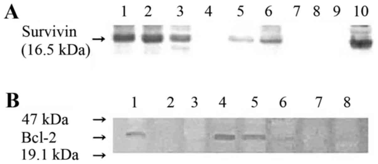
Western blot analysis
Tumor specimens were washed three times for 15 min
each time in phosphate-buffered saline (PBS) and lysed using a
homogenization buffer containing 0.1 M sodium phosphate buffer (pH
6.1), 1 mM ethylenediaminetetraacetic acid (EDTA), 1 mM
dithiothreitol, 0.1 mM phenylmethylsulfonyl fluoride and 1 mM
benzamidine (1%). Following protein quantification using the DC
Protein Assay (Bio-Rad Laboratories, Inc., Hercules, CA, USA), 15
µg protein was loaded into each lane and subjected to SDS-PAGE on
10% acrylamide gel. Proteins were transferred overnight onto
polyvinylidene difluoride membranes (Bio-Rad Laboratories, Inc.) at
a constant voltage (40 V). Membranes were saturated for 1 h using
5% dry milk in Tris-buffered saline with Tween-20 [10 mM
Tris-buffer (pH 7.5), 100 mM NaCl and 0.1% Tween-20] and incubated
overnight with 1 µg/ml anti-survivin antibody (cat. no. NB500-238;
Novus Biologicals LLC, Littleton, CO, USA) or Bcl-2 monoclonal
antibody (cat. no. sc-7382; Santa Cruz Biotechnology, Inc., Dallas,
TX, USA) at 4°C. The membranes were then incubated for 1 h with a
mouse anti-mouse immunoglobulin G antibody conjugated to alkaline
phosphatase (cat. no. CN55455; Zymed Laboratories, Inc., San
Francisco, CA, USA). Detection was then performed using an ECL
detection system (GE Healthcare Life Sciences, Arlington Height,
IL, USA). The blots were washed in PBS for 2 min, and the bands
were visualized using ECL plus (GE Healthcare Life Sciences) and
autoradiography (Fig. 1).
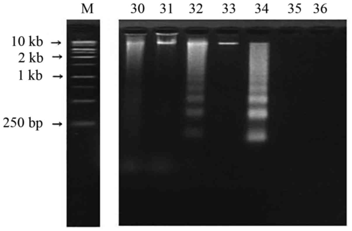
DNA fragmentation analysis
Tumor specimens were washed with PBS (pH 7.4) three
times and homogenized with lysis buffer (10 mM Tris-HCl, 100 mM
EDTA and 0.5% SDS; pH 8.0). The homogenized samples were
centrifuged at 15,000 × g for 5 min at 4°C and 50 µg of the
supernatants were placed in fresh tubes. During the time course
experiments, the experimental cells and control cells (normal glial
cells) were terminated at the same time. Samples were dissolved in
20 µl sample buffer (50 mM Tris-HCl, 0.05% SDS and 10 mM EDTA; pH
8.0) and incubated with 10 mg/ml heat-treated RNase A
(Sigma-Aldrich Corporation; Merck KGaA, Darmstadt, Germany) at 56°C
for 1 h. Proteinase K (10 mg/ml; Promega Corporation, Madison, WI,
USA) was added to each sample and incubated for 1 h at 37°C. The
reaction was stopped by increasing the temperature to 70°C for 10
min. Five microliters of loading buffer (20% glycerol, 20 mM EDTA
and 0.15% bromphenol blue) were added and the samples were analyzed
on 2% agarose gels in Tris/boric acid/EDTA buffer (89 mM Tris base,
89 mM boric acid and 2.5 mM EDTA; pH 8.0) containing 50 ng/ml
ethidium bromide. Electrophoresis was performed at 100 V for 50 min
and DNA fragmentations were visualized by ultraviolet
transillumination (Fig. 2). An
anti-apoptotic rate was defined as the percentage of patients that
did not exhibit apoptosis.
Statistical analysis
Statistical analysis was performed using SPSS 10
software (SPSS, Inc., Chicago, IL, USA). The difference between
survivin and Bcl-2 expression levels according to the pathological
type of brain tumor was analyzed using the Kruskal-Wallis test. To
determine whether there was an association between anti-apoptosis
and survivin or Bcl-2 expression levels, or between the
co-expression of survivin and Bcl-2, a χ2 test was
performed. P<0.05 was considered to indicate a statistically
significant difference.
Results
Demographic data and tumor
pathology
The patient groups comprised of 32 males and 26
females with a median age of 40 years (range, 6–68 years). The
tumor specimens consisted of 45 astrocytic tumors (comprising 15
astrocytomas, 7 anaplastic astrocytomas and 23 glioblastomas), 6
oligodendroglial tumors (including 2 oligodendrogliomas and 4
anaplastic oligodendrogliomas) and 7 ependymal tumors (4
ependymomas and 3 anaplastic ependymomas).
Expression levels of survivin and
Bcl-2, and apoptosis in tumor specimens
Survivin and Bcl-2 expression levels were analyzed
by western blotting in 58 brain tumor specimens. As demonstrated in
Fig. 1, survivin and Bcl-2 were
detected in the specimens from the majority of tumor types. The
positive expression of survivin was exhibited in 35 (60.3%) of the
58 patients. A high expression level of survivin was observed in
glioblastomas. The frequency of survivin expression in the tumor
samples was significantly correlated with the pathological grade of
glial cell tumors (P<0.05; Table
I). Positive Bcl-2 protein expression was observed in 25
(43.1%) of 58 patients, although its expression was not correlated
with the pathological grade. Survivin and Bcl-2 were co-expressed
in 15 (25.9%) patients.
 | Table I.Expression levels of survivin and
Bcl-2 protein, and apoptosis in 58 patients with glial cell
tumors. |
Table I.
Expression levels of survivin and
Bcl-2 protein, and apoptosis in 58 patients with glial cell
tumors.
|
| Expression rate, n
(%) |
|
|---|
|
|
|
|
|---|
| Pathological
type | Survivin | Bcl-2 | Apoptosis n (%) |
|---|
| Astrocytic tumor,
n=45 |
|
|
|
|
Astrocytoma, n=15 | 7 (46.7) | 9 (60.0) | 4 (26.6) |
|
Anaplastic astrocytoma,
n=7 | 4 (57.1) | 1 (14.3) | 2 (28.5) |
|
Glioblastoma, n=23 | 17
(73.3)a | 7 (30.4) | 7 (30.4) |
| Oligodendroglial
tumor, n=6 |
|
|
|
|
Oligodendroglioma, n=2 | 2 (100.0) | 2 (100.0) | 0 (0.0) |
|
Anaplastic oligodendroglioma,
n=4 | 2 (50.0) | 3 (75.0) | 1 (25.0) |
| Ependymal tumor,
n=7 |
|
|
|
|
Ependymoma, n=4 | 1 (25.0) | 2 (50.0) | 0 (0.0) |
|
Anaplastic ependymoma n=3 | 2 (66.7) | 1 (33.3) | 0 (0.0) |
| Total,
n=58 | 35 (60.3) | 25 (43.1) | 14 (24.1) |
Apoptosis in tumor specimens was
investigated using DNA fragmentation
A typical laddering pattern of DNA fragmentation is
presented in Fig. 2. Apoptosis was
confirmed in 14 (24.1%) of the 58 glial cell tumor samples obtained
from patients (Table I).
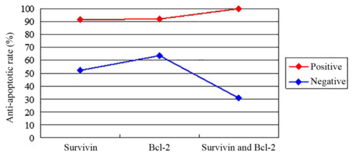
Association between survivin and Bcl-2
expression levels, and anti-apoptosis
To investigate the anti-apoptotic rate and its
association with survivin and/or Bcl-2 expression in glial cell
tumor specimens, the anti-apoptotic rate was compared between the
survivin-positive, Bcl-2-positive and co-expression groups.
Anti-apoptosis effects were detected in 32 (91.4%) of 35 patients
for survivin-positive tumors, while anti-apoptosis effects for
Bcl-2-positive tumors were found in 23 (92%) of 25 patients. An
anti-apoptosis effect was observed in all cases of co-expression of
survivin- and Bcl-2-positive tumors. A statistically significant
association was observed between an anti-apoptosis effect and the
expression of survivin or Bcl-2, and the co-expression of survivin
and Bcl-2 (P<0.05; Table II).
However, no difference in the frequency of anti-apoptosis was
identified between the three groups, including patients expressing
survivin, Bcl-2, or patients co-expressing survivin and Bcl-2
(P>0.05). The anti-apoptotic rate between the positive and
negative expression groups of survivin, Bcl-2 or survivin and Bcl-2
was identified to be significantly different (P=0.003; Fig. 3).
| Table II.Association between apoptosis, and
survivin and Bcl-2 expression levels in 58 patients with glial cell
tumors. |
Table II.
Association between apoptosis, and
survivin and Bcl-2 expression levels in 58 patients with glial cell
tumors.
|
| Apoptosis, n (%) |
|
|---|
|
|
|
|
|---|
| Expression | (+) | (−) | P-valuea |
|---|
| Survivin
expression |
|
| 0.001 |
| Positive,
n=35 | 3 (8.6) | 32 (91.4) |
|
| Negative,
n=23 | 11 (47.8) | 12 (52.2) |
|
| Bcl-2 expression |
|
| 0.01 |
| Positive,
n=25 | 2 (8.0) | 23 (92.0) |
|
| Negative,
n=3 | 12 (36.4) | 21 (63.6) |
|
| Survivin and Bcl-2
expression |
|
| 0.000 |
| Positive,
n=15 | 0 (0.0) | 15 (100.0) |
|
| Negative,
n=13 | 9 (69.2) | 4 (30.8) |
|
Discussion
Survivin is a unique member of the IAP family. It is
of special interest in oncology, as it is strongly expressed in
embryonic and fetal organs, undetectable in the majority of healthy
adult tissues, and markedly overexpressed in various types of human
cancer (9,10). In the present study, survivin was
expressed in 35 of 58 glial cell tumor patient samples.
Furthermore, survivin expression was identified to be correlated
with pathological grades of astrocytic tumors and anti-apoptosis in
glial cell tumors. In addition, the co-expression of survivin and
Bcl-2 exerted a potent anti-apoptotic influence, although, did not
exert a synergistic effect on the anti-apoptotic properties of
glial cell tumors. The current results indicate that proteins
involved in anti-apoptosis are actively expressed in glial cell
tumors. Proteins, such as survivin and Bcl-2 may, therefore,
promote the malignant progression of glial cell tumors.
Novel anti-apoptotic proteins that signal through
caspase-dependent and -independent mechanisms have previously been
described. Six members of this IAP family have been introduced in
humans: HIAP1, HIAP2, XIAP, NIAP, survivin and livin (11). Survivin is a member of the IAP family
that directly inhibits caspase-3 and −7 activity and regulates the
G2/M phase of the cell cycle. Its gene is encoded at chromosome
17q25. Furthermore, survivin links to microtubules of the mitotic
spindle (12). Interruption of
survivin-microtubule interactions resulted in the loss of
anti-apoptotic properties and increased caspase-3 activity during
mitosis. The over-expression of survivin in cancer may eliminate
apoptotic checkpoints and allow aberrant progression of transformed
cells through mitosis.
Expression of survivin has been reported in various
types of human cancer, including lung, stomach, colon, pancreas,
breast and prostate, as well as neuroblastomas, melanomas, and
lymphomas (7,13). Survivin is normally expressed during
development, but it is undetectable in healthy adult tissues and
prominently overexpressed in human cancers. However, the timing of
its overexpression during tumorigenesis and malignant progression
remains unknown. Survivin was identified to be expressed in primary
brain tumors, particularly in glioblastomas, meningiomas and
schwannomas (14,15). The present results demonstrate that
survivin was expressed at high levels in high-grade glial cell
tumors. A previous study indicated that survivin expression in
human cancers was associated with a more aggressive and invasive
clinical phenotype, and correlated with the inhibition of apoptosis
(7). IAPs may have greater
anti-apoptotic properties than any other family of apoptotic
inhibitors, including Bcl-2.
The expression of survivin has been significantly
correlated with the malignant grade of astrocytic tumors and a
worse clinical outcome (16).
Furthermore, patients who had been diagnosed with glioblastomas,
with over-expression of survivin, have been observed to have a
significantly shorter survival time (17). Therefore, it is suggested that survivin
may be involved in tumorigenesis and malignant progression of glial
cell tumors (18). Therefore, survivin
may be a useful indicator for predicting the clinical outcome of
patients and may be an ideal target for novel chemotherapeutic
agents. Consequently, survivin inhibition may manage glial cell
tumors without affecting normal brain tissue (19). In the current study, the association
between survivin expression and pathological malignancy of
astrocytic tumors was also consistent with previous findings
(16). However, the association
between survivin expression and the pathological malignancy of
oligodendrogliomas or ependymomas has not been confirmed.
Various previous studies have shown a correlation
between survivin expression and radiation resistance in certain
human cancers. Chakravarti et al (17) reported that survivin improves
double-strand DNA break repair in tumor cells, inhibiting
radiation-induced cell death. Sasaki et al (15) demonstrated that suppression of survivin
by small interfering ribonucleic acid improved the radiosensitivity
of tumor cells. Based on these results, anti-survivin therapeutic
strategies may enhance the radiation response and be correlated
with favorable clinical outcomes (20).
Bcl-2 regulates cytochrome c release from
mitochondria and suppresses upstream initiation of the caspase
activation cascade. Bcl-2 acts to extend cell survival by blocking
apoptosis and may, therefore, influence pathological malignancy
(21). In the current study, Bcl-2
expression was not correlated with a malignant grade of astrocytic
cell tumors. Numerous authors previously reported that cytoplasmic
Bcl-2 expression levels were higher in low-grade astrocytomas than
in high-grade astrocytomas and were not associated with the
pathological malignancy of astrocytic cell tumors.
Survivin and Bcl-2 possess potent anti-apoptotic
properties. Survivin reportedly affects caspase-3 or −7 via various
signaling pathways of the Bcl-2 family. In addition, Bcl-2
expression was identified to be positively correlated with survivin
expression in epithelial ovarian cancer (22). However, survivin expression was not
correlated with Bcl-2 expression in glial cell tumors (16). Due to these varying results, the
anti-apoptotic effect of survivin remains controversial and is not
clearly understood. Low survivin expression levels in certain human
cancer cells were associated with apoptotic properties and
correlated with Bcl-2 expression (23). Conversely, survivin expression in
bladder tumors was not associated with high expression levels of
Bcl-2. This result was not surprising, due to the fact that
survivin and Bcl-2 do not share completely common mechanisms of
transcriptional activation (11).
However, the expression levels of survivin and Bcl-2 in melanomas
may provide two independent mechanisms of apoptosis inhibition
leading to malignant progression of cancer cells (24).
Survivin and Bcl-2 expression in breast cancer cells
is significantly involved in apoptosis inhibition, which
potentially contributes to malignant progression of cancer cells
and resistance to chemotherapy or radiotherapy. Survivin expression
was significantly associated with Bcl-2 expression and
anti-apoptotic properties, which were strongly correlated with a
worse clinical outcome following resection in gastric and
colorectal cancers (7). Expression of
the survivin gene alone or the survivin gene plus other
anti-apoptotic genes, such as Bcl-2 may have more noticeable
anti-apoptotic properties in breast carcinomas (13). However, in the current study, no
synergistic anti-apoptotic effects were noted in the co-expression
group using glial cell tumor samples. Differences in the results
may arise from the small number of glial cell tumors, which may be
unrepresentative due to the tumor heterogeneity.
A major limitation to verifying the hypothesis of
the current study is the small number of glial cell tumor samples.
Additionally, anti-apoptosis in the same pathology of glial cell
tumors was not quantitatively evaluated. Thus, studies of other
molecular markers, such as other IAP members, and investigations
into the response to treatments, such as radiotherapy and
chemotherapy, are required. Furthermore, the correlation between
molecular markers and the clinical outcome of glial cell tumors
requires investigation. Larger prospective studies using the same
pathology of glial cell tumors must be performed to determine the
association between apoptosis, and survivin and Bcl-2 expression
levels.
Previous studies have indicated that survivin is an
ideal target for cancer treatment, either directly or by affecting
endothelial cell viability (25). A
previously reported study on survivin antisense treatment
demonstrated that survivin caused regression in vascular capillary
formation (26). Thus, survivin
antisense treatment has anti-angiogenic effects and anti-tumor cell
effects, and may present as an attractive target for glial cell
tumor treatment.
In conclusion, the current results indicate that
survivin or Bcl-2 expression exert potent anti-apoptotic effects in
glial cell tumors. In addition, survivin expression in astrocytic
tumors was identified to be significantly associated with
pathological malignancy. Survivin or Bcl-2 may be attractive
targets for use in cancer therapy to target tumor cells while
leaving the normal cells unaffected. Thus, suppression of survivin
or Bcl-2 expression may be an effective treatment modality against
glial cell tumors.
References
|
1
|
Ahmed R, Oborski MJ, Hwang M, Lieberman FS
and Mountz JM: Malignant gliomas: Current perspectives in
diagnosis, treatment, and early response assessment using advanced
quantitative imaging methods. Cancer Manag Res. 6:149–170.
2014.PubMed/NCBI
|
|
2
|
Corwin D, Holdsworth C, Rockne RC, Trister
AD, Mrugala MM, Rockhill JK, Stewart RD, Phillips M and Swanson KR:
Toward patient-specific, biologically optimized radiation therapy
plans for the treatment of glioblastoma. PLoS One. 8:e791152013.
View Article : Google Scholar : PubMed/NCBI
|
|
3
|
Kanamori M, Higa T, Sonoda Y, Murakami S,
Dodo M, Kitamura H, Taguchi K, Shibata T, Watanabe M, Suzuki H, et
al: Activation of the NRF2 pathway and its impact on the prognosis
of anaplastic glioma patients. Neuro-oncol. 17:555–565. 2015.
View Article : Google Scholar : PubMed/NCBI
|
|
4
|
Hu B, Emdad L, Bacolod MD, Kegelman TP,
Shen XN, Alzubi MA, Das SK, Sarkar D and Fisher PB: Astrocyte
elevated gene-1 interacts with Akt isoform 2 to control glioma
growth, survival, and pathogenesis. Cancer Res. 74:7321–7332. 2014.
View Article : Google Scholar : PubMed/NCBI
|
|
5
|
Würth R, Pattarozzi A, Gatti M, Bajetto A,
Corsaro A, Parodi A, Sirito R, Massollo M, Marini C, Zona G, et al:
Metformin selectively affects human glioblastoma tumor-initiating
cell viability: A role for metformin-induced inhibition of Akt.
Cell Cycle. 12:145–156. 2013. View
Article : Google Scholar : PubMed/NCBI
|
|
6
|
Yamamoto T and Tanigawa N: The role of
survivin as a new target of diagnosis and treatment in human
cancer. Med Electron Microsc. 34:207–212. 2001. View Article : Google Scholar : PubMed/NCBI
|
|
7
|
Kawasaki H, Altieri DC, Lu CD, Toyoda M,
Tenjo T and Tanigawa N: Inhibition of apoptosis by survivin
predicts shorter survival rates in colorectal cancer. Cancer Res.
58:5071–5074. 1998.PubMed/NCBI
|
|
8
|
Louis DN, Ohgaki H, Wiestler OD and
Cavenee WK: World Health Organization histological classification
of tumours of the central nervous system. International Agency for
Research on Cancer; Lyon: 2007
|
|
9
|
Altieri DC: The molecular basis and
potential role of survivin in cancer diagnosis and therapy. Trends
Mol Med. 7:542–547. 2001. View Article : Google Scholar : PubMed/NCBI
|
|
10
|
Altieri DC: Validating survivin as a
cancer therapeutic target. Nat Rev Cancer. 3:46–54. 2003.
View Article : Google Scholar : PubMed/NCBI
|
|
11
|
Ashhab Y, Alian A, Polliack A, Panet A and
Ben Yehuda D: Two splicing variants of a new inhibitor of apoptosis
gene with different biological properties and tissue distribution
pattern. FEBS Lett. 495:56–60. 2001. View Article : Google Scholar : PubMed/NCBI
|
|
12
|
O'Connor DS, Grossman D, Plescia J, Li F,
Zhang H, Villa A, Tognin S, Marchisio PC and Altieri DC: Regulation
of apoptosis at cell division by p34cdc2 phosphorylation of
survivin. Proc Natl Acad Sci USA. 97:13103–13107. 2000. View Article : Google Scholar : PubMed/NCBI
|
|
13
|
Tanaka K, Iwamoto S, Gon G, Nohara T,
Iwamoto M and Tanigawa N: Expression of survivin and its
relationship to loss of apoptosis in breast carcinomas. Clin Cancer
Res. 6:127–134. 2000.PubMed/NCBI
|
|
14
|
Kleinschmidt-DeMasters BK, Heinz D,
McCarthy PJ, Bobak JB, Lillehei KO, Shroyer AL and Shroyer KR:
Survivin in glioblastomas. Protein and messenger RNA expression and
comparison with telomerase levels. Arch Pathol Lab Med.
127:826–833. 2003.PubMed/NCBI
|
|
15
|
Sasaki T, Lopes MBS, Hankins GR and Helm
GA: Expression of survivin, an inhibitor of apoptosis protein, in
tumors of the nervous system. Acta Neuropathol. 104:105–109. 2002.
View Article : Google Scholar : PubMed/NCBI
|
|
16
|
Kajiwara Y, Yamasaki F, Hama S, Yahara K,
Yoshioka H, Sugiyama K, Arita K and Kurisu K: Expression of
survivin in astrocytic tumors: Correlation with malignant grade and
prognosis. Cancer. 97:1077–1083. 2003. View Article : Google Scholar : PubMed/NCBI
|
|
17
|
Chakravarti A, Noll E, Black PM,
Finkelstein DF, Finkelstein DM, Dyson NJ and Loeffler JS:
Quantitatively determined survivin expression levels are of
prognostic value in human gliomas. J Clin Oncol. 20:1063–1068.
2002. View Article : Google Scholar : PubMed/NCBI
|
|
18
|
Xie D, Zeng YX, Wang HJ, Wen JM, Tao Y,
Sham JS and Guan XY: Expression of cytoplasmic and nuclear Survivin
in primary and secondary human glioblastoma. Br J Cancer.
94:108–114. 2006. View Article : Google Scholar : PubMed/NCBI
|
|
19
|
Altieri DC: Survivin, cancer networks and
pathway-directed drug discovery. Nat Rev Cancer. 8:61–70. 2008.
View Article : Google Scholar : PubMed/NCBI
|
|
20
|
Kanwar RK, Cheung CH, Chang JY and Kanwar
JR: Recent advances in anti-survivin treatments for cancer. Curr
Med Chem. 17:1509–1515. 2010. View Article : Google Scholar : PubMed/NCBI
|
|
21
|
Plati J, Bucur O and Khosravi-Far R:
Apoptotic cell signaling in cancer progression and therapy. Integr
Biol. 3:279–296. 2011. View Article : Google Scholar
|
|
22
|
Zhang SL, Zhao CQ, Lin B, Li Y and Gao H:
Expression of survivin gene and its relation with the expression of
bcl-2 and bax protein in epithelial ovarian cancer. Zhonghua Fu
Chan Ke Za Zhi. 38:203–206. 2003.(In Chinese). PubMed/NCBI
|
|
23
|
Rödel F, Hoffmann J, Grabenbauer GG,
Papadopoulos T, Weiss C, Günther K, Schick C, Sauer R and Rödel C:
High survivin expression is associated with reduced apoptosis in
rectal cancer and may predict disease-free survival after
preoperative radiochemotherapy and surgical resection. Strahlenther
Onkol. 178:426–435. 2002. View Article : Google Scholar : PubMed/NCBI
|
|
24
|
Gradilone A, Gazzaniga P, Ribuffo D,
Scarpa S, Cigna E, Vasaturo F, Bottoni U, Innocenzi D, Calvieri S,
Scuderi N, et al: Survivin, bcl-2, bax, and bcl-X gene expression
in sentinel lymph nodes from melanoma patients. J Clin Oncol.
21:306–312. 2003. View Article : Google Scholar : PubMed/NCBI
|
|
25
|
O'Connor DS, Schechner JS, Adida C, Mesri
M, Rothermel AL, Li F, Nath AK, Pober JS and Altieri DC: Control of
apoptosis during angiogenesis by survivin expression in endothelial
cells. Am J Pathol. 156:393–398. 2000. View Article : Google Scholar : PubMed/NCBI
|
|
26
|
Mesri M, Morales-Ruiz M, Ackermann EJ,
Bennett CF, Pober JS, Sessa WC and Altieri DC: Suppression of
vascular endothelial growth factor-mediated endothelial cell
protection by survivin targeting. Am J Pathol. 158:1757–1765. 2001.
View Article : Google Scholar : PubMed/NCBI
|