Spandidos Publications Logo
  • About
    • About Spandidos
    • Aims and Scopes
    • Abstracting and Indexing
    • Editorial Policies
    • Reprints and Permissions
    • Job Opportunities
    • Terms and Conditions
    • Contact
  • Journals
    • All Journals
    • Oncology Letters
      • Oncology Letters
      • Information for Authors
      • Editorial Policies
      • Editorial Board
      • Aims and Scope
      • Abstracting and Indexing
      • Bibliographic Information
      • Archive
    • International Journal of Oncology
      • International Journal of Oncology
      • Information for Authors
      • Editorial Policies
      • Editorial Board
      • Aims and Scope
      • Abstracting and Indexing
      • Bibliographic Information
      • Archive
    • Molecular and Clinical Oncology
      • Molecular and Clinical Oncology
      • Information for Authors
      • Editorial Policies
      • Editorial Board
      • Aims and Scope
      • Abstracting and Indexing
      • Bibliographic Information
      • Archive
    • Experimental and Therapeutic Medicine
      • Experimental and Therapeutic Medicine
      • Information for Authors
      • Editorial Policies
      • Editorial Board
      • Aims and Scope
      • Abstracting and Indexing
      • Bibliographic Information
      • Archive
    • International Journal of Molecular Medicine
      • International Journal of Molecular Medicine
      • Information for Authors
      • Editorial Policies
      • Editorial Board
      • Aims and Scope
      • Abstracting and Indexing
      • Bibliographic Information
      • Archive
    • Biomedical Reports
      • Biomedical Reports
      • Information for Authors
      • Editorial Policies
      • Editorial Board
      • Aims and Scope
      • Abstracting and Indexing
      • Bibliographic Information
      • Archive
    • Oncology Reports
      • Oncology Reports
      • Information for Authors
      • Editorial Policies
      • Editorial Board
      • Aims and Scope
      • Abstracting and Indexing
      • Bibliographic Information
      • Archive
    • Molecular Medicine Reports
      • Molecular Medicine Reports
      • Information for Authors
      • Editorial Policies
      • Editorial Board
      • Aims and Scope
      • Abstracting and Indexing
      • Bibliographic Information
      • Archive
    • World Academy of Sciences Journal
      • World Academy of Sciences Journal
      • Information for Authors
      • Editorial Policies
      • Editorial Board
      • Aims and Scope
      • Abstracting and Indexing
      • Bibliographic Information
      • Archive
    • International Journal of Functional Nutrition
      • International Journal of Functional Nutrition
      • Information for Authors
      • Editorial Policies
      • Editorial Board
      • Aims and Scope
      • Abstracting and Indexing
      • Bibliographic Information
      • Archive
    • International Journal of Epigenetics
      • International Journal of Epigenetics
      • Information for Authors
      • Editorial Policies
      • Editorial Board
      • Aims and Scope
      • Abstracting and Indexing
      • Bibliographic Information
      • Archive
    • Medicine International
      • Medicine International
      • Information for Authors
      • Editorial Policies
      • Editorial Board
      • Aims and Scope
      • Abstracting and Indexing
      • Bibliographic Information
      • Archive
  • Articles
  • Information
    • Information for Authors
    • Information for Reviewers
    • Information for Librarians
    • Information for Advertisers
    • Conferences
  • Language Editing
Spandidos Publications Logo
  • About
    • About Spandidos
    • Aims and Scopes
    • Abstracting and Indexing
    • Editorial Policies
    • Reprints and Permissions
    • Job Opportunities
    • Terms and Conditions
    • Contact
  • Journals
    • All Journals
    • Biomedical Reports
      • Information for Authors
      • Editorial Policies
      • Editorial Board
      • Aims and Scope
      • Abstracting and Indexing
      • Bibliographic Information
      • Archive
    • Experimental and Therapeutic Medicine
      • Information for Authors
      • Editorial Policies
      • Editorial Board
      • Aims and Scope
      • Abstracting and Indexing
      • Bibliographic Information
      • Archive
    • International Journal of Epigenetics
      • Information for Authors
      • Editorial Policies
      • Editorial Board
      • Aims and Scope
      • Abstracting and Indexing
      • Bibliographic Information
      • Archive
    • International Journal of Functional Nutrition
      • Information for Authors
      • Editorial Policies
      • Editorial Board
      • Aims and Scope
      • Abstracting and Indexing
      • Bibliographic Information
      • Archive
    • International Journal of Molecular Medicine
      • Information for Authors
      • Editorial Policies
      • Editorial Board
      • Aims and Scope
      • Abstracting and Indexing
      • Bibliographic Information
      • Archive
    • International Journal of Oncology
      • Information for Authors
      • Editorial Policies
      • Editorial Board
      • Aims and Scope
      • Abstracting and Indexing
      • Bibliographic Information
      • Archive
    • Medicine International
      • Information for Authors
      • Editorial Policies
      • Editorial Board
      • Aims and Scope
      • Abstracting and Indexing
      • Bibliographic Information
      • Archive
    • Molecular and Clinical Oncology
      • Information for Authors
      • Editorial Policies
      • Editorial Board
      • Aims and Scope
      • Abstracting and Indexing
      • Bibliographic Information
      • Archive
    • Molecular Medicine Reports
      • Information for Authors
      • Editorial Policies
      • Editorial Board
      • Aims and Scope
      • Abstracting and Indexing
      • Bibliographic Information
      • Archive
    • Oncology Letters
      • Information for Authors
      • Editorial Policies
      • Editorial Board
      • Aims and Scope
      • Abstracting and Indexing
      • Bibliographic Information
      • Archive
    • Oncology Reports
      • Information for Authors
      • Editorial Policies
      • Editorial Board
      • Aims and Scope
      • Abstracting and Indexing
      • Bibliographic Information
      • Archive
    • World Academy of Sciences Journal
      • Information for Authors
      • Editorial Policies
      • Editorial Board
      • Aims and Scope
      • Abstracting and Indexing
      • Bibliographic Information
      • Archive
  • Articles
  • Information
    • For Authors
    • For Reviewers
    • For Librarians
    • For Advertisers
    • Conferences
  • Language Editing
Login Register Submit
  • This site uses cookies
  • You can change your cookie settings at any time by following the instructions in our Cookie Policy. To find out more, you may read our Privacy Policy.

    I agree
Search articles by DOI, keyword, author or affiliation
Search
Advanced Search
presentation
Biomedical Reports
Join Editorial Board Propose a Special Issue
Print ISSN: 2049-9434 Online ISSN: 2049-9442
Journal Cover
June-2019 Volume 10 Issue 6

Full Size Image

Sign up for eToc alerts
Recommend to Library

Journals

International Journal of Molecular Medicine

International Journal of Molecular Medicine

International Journal of Molecular Medicine is an international journal devoted to molecular mechanisms of human disease.

International Journal of Oncology

International Journal of Oncology

International Journal of Oncology is an international journal devoted to oncology research and cancer treatment.

Molecular Medicine Reports

Molecular Medicine Reports

Covers molecular medicine topics such as pharmacology, pathology, genetics, neuroscience, infectious diseases, molecular cardiology, and molecular surgery.

Oncology Reports

Oncology Reports

Oncology Reports is an international journal devoted to fundamental and applied research in Oncology.

Experimental and Therapeutic Medicine

Experimental and Therapeutic Medicine

Experimental and Therapeutic Medicine is an international journal devoted to laboratory and clinical medicine.

Oncology Letters

Oncology Letters

Oncology Letters is an international journal devoted to Experimental and Clinical Oncology.

Biomedical Reports

Biomedical Reports

Explores a wide range of biological and medical fields, including pharmacology, genetics, microbiology, neuroscience, and molecular cardiology.

Molecular and Clinical Oncology

Molecular and Clinical Oncology

International journal addressing all aspects of oncology research, from tumorigenesis and oncogenes to chemotherapy and metastasis.

World Academy of Sciences Journal

World Academy of Sciences Journal

Multidisciplinary open-access journal spanning biochemistry, genetics, neuroscience, environmental health, and synthetic biology.

International Journal of Functional Nutrition

International Journal of Functional Nutrition

Open-access journal combining biochemistry, pharmacology, immunology, and genetics to advance health through functional nutrition.

International Journal of Epigenetics

International Journal of Epigenetics

Publishes open-access research on using epigenetics to advance understanding and treatment of human disease.

Medicine International

Medicine International

An International Open Access Journal Devoted to General Medicine.

Journal Cover
June-2019 Volume 10 Issue 6

Full Size Image

Sign up for eToc alerts
Recommend to Library

  • Article
  • Citations
    • Cite This Article
    • Download Citation
    • Create Citation Alert
    • Remove Citation Alert
    • Cited By
  • Similar Articles
    • Related Articles (in Spandidos Publications)
    • Similar Articles (Google Scholar)
    • Similar Articles (PubMed)
  • Download PDF
  • Download XML
  • View XML
Case Report

Giant peritoneal loose body: A case report

  • Authors:
    • Shanshan Guo
    • Haijuan Yuan
    • Yingying Xu
    • Ping Chen
    • Liang Zong
  • View Affiliations / Copyright

    Affiliations: Department of Gastrointestinal Surgery, Northern Jiangsu People's Hospital, Clinical Medical College, Yangzhou University, Yangzhou, Jiangsu 225001, P.R. China
  • Pages: 351-353
    |
    Published online on: April 18, 2019
       https://doi.org/10.3892/br.2019.1208
  • Expand metrics +
Metrics: Total Views: 0 (Spandidos Publications: | PMC Statistics: )
Metrics: Total PDF Downloads: 0 (Spandidos Publications: | PMC Statistics: )
Cited By (CrossRef): 0 citations Loading Articles...

This article is mentioned in:


Abstract

Peritoneal loose body (PLB) is a rare finding and it is usually incidentally discovered during laparotomy, examination or autopsy, as it is usually asymptomatic or presents with non‑specific symptoms. In particular, giant PLBs, measuring >50 mm in maximum diameter, are even more rare, and only a few cases of laparoscopic extraction of these giant bodies have been reported in the literature to date. We herein describe our experience with the diagnosis and treatment of a giant PLB in a 49‑year‑old male patient who was admitted with complaints of intermittent pain the in lower abdomen 3 months earlier. An abdominal computed tomography scan revealed a 5.5x5.0‑cm midline mass with central high density. The diagnosis was confirmed by open surgery and postoperative pathological examination. The postoperative recovery of the patient was uneventful and he was discharged from the hospital 7 days after surgery.

Introduction

Peritoneal loose bodies (PLBs) are quite rare and usually present as small, white or pale gray, egg-shaped objects with a smooth glistening surface. They are usually located freely in the peritoneal cavity (1) and rarely cause symptoms. PLBs are usually 5-20 mm in diameter and are diagnosed via laparotomy or autopsy by accident (2). Matsubara et al (3) reviewed 20 reported PLB cases and determined that PLB is more common in males, with a male:female ratio of 17:3. Although the pathogenesis of PLB remains unclear, it is widely accepted that they arise from infarcted appendices epiploicae, which then go through several sequential processes, including saponification, calcification and fibrosis (4). PLBs then increase in size by accumulating albumin from exudative peritoneal fluid (4). In the pathological examination of PBLs, hyperplastic fibrillar collagen with partial microcalcifications are usually used as criteria for diagnosis (5,6). In most cases, patients present with non-specific symptoms and do not require treatment. However, in cases of giant PLBs, surgical intervention is important for treatment, as giant PLBs are more likely to cause acute or life-threatening symptoms (7). Furthermore, other diseases may not be fully excluded following radiological imaging alone so surgical exploration may be necessary for the definitive diagnosis and treatment of PLBs (8).

Case report

A 49-year-old man was admitted to Northern Jiangsu People's Hospital in March 2016 with a complaint of intermittent pain in the lower abdomen 3 months earlier. The patient had no fever, nausea or vomiting. The physical examination was unremarkable. The medical history was significant for acute appendicitis ~4 years earlier. At that time, only conservative treatment with antibiotics was administered instead of an appendectomy.

The laboratory tests revealed high levels of γ-glutamyl-transpeptidase (80 U/l; normal range, 0-50 U/l), direct bilirubin and alanine aminotransferase, and increased levels of carcinoembryonic antigen (CEA); infectious markers included positive hepatitis B virus (HBV) markers, namely HB surface 
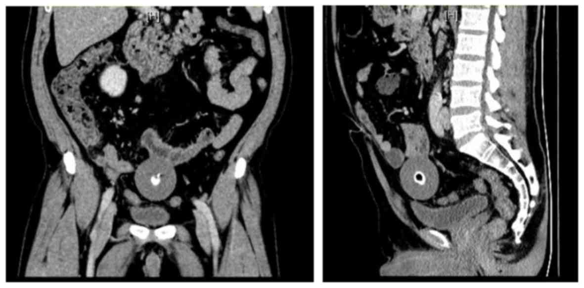
antigen, anti-HBe antibody and HB core antibody. An abdominal computed tomography (CT) scan revealed a 5.5x5.0-cm midline mass with central high density (Fig. 1). In addition, the CT examination revealed a fatty liver. Abdominal 
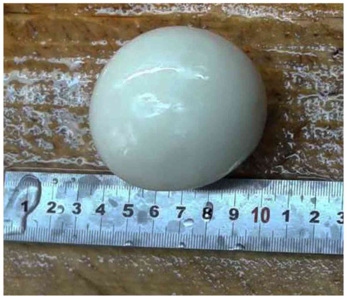
open surgery was performed to remove the mass, and the postoperative pathological examination revealed that it was composed of hyperplastic fibrillar collagen with partial microcalcifications.

Figure 1.

Anteroposterior (left) and lateral (right) computed tomography images of the patient.

The postoperative recovery of the patient was uneventful and he was discharged from the hospital 7 days after surgery. The intermittent pain in the lower abdomen resolved immediately after removal of the mass. After a follow-up period of two years, the patient had no abnormal complaints.

Discussion

PLBs are a rare occurrence, and only a few cases of giant PLBs (>5 cm in diameter) have been reported in the literature to date. PLBs are usually incidentally discovered during surgery or examination. The majority of PLBs are asymptomatic, but a small proportion of giant PLBs may cause symptoms, such as abdominal pain, bowel obstruction, urinary retention or urinary frequency (9). Matsubara et al reviewed 20 cases of giant PLBs reported in the literature and reported that PLBs were more common in men, with a male:female incidence of 17:3(3). The majority of the patients were aged >50 years. In addition, most cases shared a distinct histological characteristic, namely calcified necrosis of fat tissue with hypocellular fibrolamellar tissue and numerous microcalcifications.

Clinically, PLBs must be differentiated from other tumors, such as mesenteric tumors, leiomyoma and teratoma. Abdominal CT examination may be helpful, as it demonstrates the characteristic features of PLBs, which are well-defined oval or round soft tissue masses with central calcification, usually located in the abdomen, with a distinct fat plane around the mass separating it from adjacent organs (10). Since PLBs are freely mobile, additional scanning in the prone position or a follow-up CT examination can demonstrate the change in location. Moreover, on magnetic resonance imaging (MRI) examination, PLB appears as a well-defined, low-intensity mass on both T1- and T2-weighted images (11). The MRI signal is similar to that of muscle, and a central high-intensity area may be seen on T1-weighted images. PLBs do not exhibit any enhancement, which is useful in differential diagnosis, as leiomyoma and teratoma exhibit contrast enhancement. Performing CT and MRI is crucial for identifying the characteristic features of PLB and establishing an accurate diagnosis.

The pathogenesis of PLBs remains unclear; however, the general consensus is that PLBs are derived from the epiploic appendices via sequential torsion, infarction, saponification and calcification. Sussman and Murdock described a rare case of a PLB in a 62-year-old patient in 2015(9). As Sussman and Murdock (9) mentioned, the formation of the PLB was attributed to the torsion and detachment of an epiploic appendage, with subsequent transformation into a fibrotic mass. With continuous peritoneal exudate deposition on the surface, PLBs may slowly grow to a larger size. However, although there are a number of cases with severe abdominal or pelvic inflammation, only few result in the formation of a PLB, suggesting that some specific condition may be required. In the present case, CT imaging revealed a fatty liver, and the laboratory tests revealed a compromised liver function and increased CEA levels. It may be hypothesized that the compromised liver function may have increased the volume of peritoneal exudate, thereby facilitating PLB formation.

Of note, the formation of the PLB resembles that of a pearl, which originates from an external stimulus, such as a parasite, inducing a defense mechanism in mollusks, which then create a pearl sac to seal it off (Fig. 2).

Figure 2.

Macroscopic appearance of the giant peritoneal loose body.

Acknowledgements

Not applicable.

Funding

The present study was supported by the Jiangsu Natural Science Foundation (grant no. BK20180274).

Availability of data and materials

All the information relevant to the present study are available from the corresponding author on reasonable request.

Authors' contributions

SG drafted the manuscript and examined the patient; PC assisted in the preparation of the manuscript and examined the patient; LZ critically reviewed the manuscript and examined the patient. All authors have read and approved the final version of this manuscript.

Ethics approval and consent to participate

Not applicable.

Patient consent for publication

Written informed consent was obtained from the patient for the publication of the case details and any associated images.

Competing interests

The authors declare that they have no competing interests to disclose.

References

1 

Takabe K, Greenberg JI and Blair SL: Giant peritoneal loose bodies. J Gastrointest Surg. 10:465–468. 2006.PubMed/NCBI View Article : Google Scholar

2 

Zhang H, Ling YZ, Cui MM, Xia ZX, Feng Y and Chen CS: Giant peritoneal loose body in the pelvic cavity confirmed by laparoscopic exploration: A case report and review of the literature. World J Surg Oncol. 13(118)2015.PubMed/NCBI View Article : Google Scholar

3 

Matsubara K, Takakura Y, Urushihara T, Nishisaka T and Itamoto T: Laparoscopic extraction of a giant peritoneal loose body: Case report and review of literature. Int J Surg Case Rep. 39:188–191. 2017.PubMed/NCBI View Article : Google Scholar

4 

Sewkani A, Jain A, Maudar K and Varshney S: ‘Boiled egg’ in the peritoneal cavity-a giant peritoneal loose body in a 64-year-old man: A case report. J Med Case Reports. 5(297)2011.PubMed/NCBI View Article : Google Scholar

5 

Asabe K, Maekawa T, Yamashita Y and Shirakusa T: Endoscopic extraction of a peritoneal loose body: A case report of an infant. Pediatr Surg Int. 21:388–389. 2005.PubMed/NCBI View Article : Google Scholar

6 

Van Zyl C, Davis R, Hurter D and Van Der Westhuizen G: Giant peritoneal loose bodies. S Afr J Rad. 19:730–733. 2015. View Article : Google Scholar

7 

Elsner A, Walensi M, Fuenfschilling M, Rosenberg R and Mechera R: Symptomatic giant peritoneal loose body in the pelvic cavity: A case report. Int J Surg Case Rep. 21:32–35. 2016.PubMed/NCBI View Article : Google Scholar

8 

Obaid M and Gehani S: Deciding to Remove or Leave a Peritoneal Loose Body: A Case Report and Review of Literature. Am J Case Rep. 19:854–857. 2018.PubMed/NCBI View Article : Google Scholar

9 

Sussman R and Murdock J: Images in clinical medicine. Peritoneal loose body. N Engl J Med. 372(1359)2015.PubMed/NCBI View Article : Google Scholar

10 

Gayer G and Petrovitch I: CT diagnosis of a large peritoneal loose body: A case report and review of the literature. Br J Radiol. 84:e83–e85. 2011.PubMed/NCBI View Article : Google Scholar

11 

Takayama S, Sakamoto M and Takeyama H: Clinical challenges and images in GI. Image 1: huge peritoneal loose body in the pelvic cavity. Gastroenterology. 136(404): 730. 2009.PubMed/NCBI View Article : Google Scholar

Related Articles

  • Abstract
  • View
  • Download
  • Twitter
Copy and paste a formatted citation
Spandidos Publications style
Guo S, Yuan H, Xu Y, Chen P and Zong L: Giant peritoneal loose body: A case report. Biomed Rep 10: 351-353, 2019.
APA
Guo, S., Yuan, H., Xu, Y., Chen, P., & Zong, L. (2019). Giant peritoneal loose body: A case report. Biomedical Reports, 10, 351-353. https://doi.org/10.3892/br.2019.1208
MLA
Guo, S., Yuan, H., Xu, Y., Chen, P., Zong, L."Giant peritoneal loose body: A case report". Biomedical Reports 10.6 (2019): 351-353.
Chicago
Guo, S., Yuan, H., Xu, Y., Chen, P., Zong, L."Giant peritoneal loose body: A case report". Biomedical Reports 10, no. 6 (2019): 351-353. https://doi.org/10.3892/br.2019.1208
Copy and paste a formatted citation
x
Spandidos Publications style
Guo S, Yuan H, Xu Y, Chen P and Zong L: Giant peritoneal loose body: A case report. Biomed Rep 10: 351-353, 2019.
APA
Guo, S., Yuan, H., Xu, Y., Chen, P., & Zong, L. (2019). Giant peritoneal loose body: A case report. Biomedical Reports, 10, 351-353. https://doi.org/10.3892/br.2019.1208
MLA
Guo, S., Yuan, H., Xu, Y., Chen, P., Zong, L."Giant peritoneal loose body: A case report". Biomedical Reports 10.6 (2019): 351-353.
Chicago
Guo, S., Yuan, H., Xu, Y., Chen, P., Zong, L."Giant peritoneal loose body: A case report". Biomedical Reports 10, no. 6 (2019): 351-353. https://doi.org/10.3892/br.2019.1208
Follow us
  • Twitter
  • LinkedIn
  • Facebook
About
  • Spandidos Publications
  • Careers
  • Cookie Policy
  • Privacy Policy
How can we help?
  • Help
  • Live Chat
  • Contact
  • Email to our Support Team