Introduction
Diffuse large B-cell lymphoma (DLBCL) is the most
common type of non-Hodgkin's lymphoma (NHL), accounting for 31% of
all cases of NHL in western countries and >40% of NHL cases in
Asia (1-3). With
the currently available options for chemoimmunotherapy and systemic
disease control, the 5-year survival rate of patients with DLBCL
has improved (60-90%) (4,5). Secondary involvement of the central
nervous system (CNS) in patients with DLBCL is a relatively
uncommon manifestation, encountered in only 5-7% of cases (6-9);
however, its incidence is higher in patients with certain high-risk
clinical characteristics at the time of diagnosis (10). These risk factors include a high
International Prognostic Index score (11); involvement of more than two extranodal
sites, retroperitoneal lymph node involvement, elevated lactate
dehydrogenase level, or DLBCL originating from high-risk locations,
such as the bone marrow, paranasal sinuses, testis, breast, adrenal
gland and kidney (8,12-16).
The outcome following CNS relapse is poor, with the overall
survival shortened to <6 months (17). Intrathecal (IT) and intravenous
high-dose (HD) methotrexate are common methods of CNS prophylaxis
(11). Given the low CNS relapse rate
in DLBCL, and evaluating the benefits against the adverse effects,
the application of CNS prophylaxis for DLBCL is not widely
implemented (12,18). As it is preferable that CNS
prophylaxis is administered during primary chemotherapy, the
identification of patients with DLBCL who are at high risk for
subsequent CNS recurrence at the time of diagnosis is crucial.
There is currently no consensus regarding a diagnostic algorithm
for CNS involvement in DLBCL. Neurological symptoms, CNS imaging,
stereotactic biopsy and cerebrospinal fluid (CSF) cytology are the
current methods commonly used for diagnosis and evaluating patients
at high risk of, or with suspected, CNS involvement (19). CSF examination includes cytology, flow
cytometric analysis and biochemical biomarkers. CSF cytology is a
specific diagnostic approach, but it can only detect malignant
lymphoid cells in 40% of patients with suspected CNS dissemination
(20). Flow cytometric analysis of
the CSF has already demonstrated increased sensitivity (21); however, since the introduction of
rituximab, the majority of CNS relapse events are parenchymal
(65-70%), and CSF flow cytometry is of limited diagnostic value in
such cases (22-24).
Biochemical biomarker examination of the CSF exhibits higher
sensitivity compared with CSF cytology (58-85%), but only a
moderate improvement in specificity (85%) (25). Therefore, it is necessary to develop a
convenient and accurate method for evaluating the risk of CNS
involvement at diagnosis in order to implement adequate CNS
prophylaxis.
Quantitative global proteomics is an advanced
approach to the accurate characterization of proteins in complex
biological systems, which is applied to identify unbiased
biomarkers or key proteins associated with specific physiological
and pathological states (26). The
advantages of label-free quantification liquid chromatography-mass
spectrometry (LC/MS) analysis are as follows: First, the cost,
procedure and artificial expenses of labeling samples are
eliminated. Second, it has the capacity to quantify a large number
of proteins per LC/MS measurement (27). By using proteomics, numerous studies
have identified potential CSF biomarkers of neurological diseases,
including amyotrophic lateral sclerosis, cerebral malaria and
tuberculous meningitis (28-30).
Quantitative proteomics of CSF samples from serial lumbar punctures
during induction in patients with acute lymphoblastic leukemia have
found potential predictive markers of CNS thrombosis (31). However, to the best of our knowledge,
global proteomic profiling of CSF from patients with DLBCL has not
been reported to date. Therefore, in the present study, a
high-throughput label-free quantitative proteomic analysis was
performed to identify proteins present in the CSF of patients with
DLBCL with CNS recurrence compared with those in healthy controls,
in order to identify potential CSF biomarkers for patients at high
risk of developing CNS recurrence.
Materials and methods
Subjects
The subjects included four patients diagnosed with
DLBCL at the West China Medical Center of Sichuan University
(Chengdu, China), and six healthy control subjects recruited at the
Physical Examination Center of West China Hospital, Sichuan
University from January 2016 to January 2017. The patients with
DLBCL were evaluated for CNS recurrence based on the clinical
characteristics at the time of diagnosis. The healthy control
subjects were defined as individuals without active DLBCL or any
neurological complaints. Informed consent was obtained from all the
participants and the study protocol was approved by the Medical
Ethics Committee of West China Hospital, Sichuan University.
Sample preparation
The CSF was collected in plastic tubes containing
Trasylol (Bayer Diagnostics) to prevent proteolysis. Following
clinical analysis, the CSF samples were centrifuged at 3,000 x g at
4˚C for 5 min. All supernatant, aside from the last 0.5 ml, was
transferred to a new container and then immediately frozen at -20˚C
and stored at -80˚C until further use. A total of 30 µl CSF were
solubilized in cold RIPA buffer [150 mm NaCl, 50 mm Tris-HCl (pH
7.61), NP-40, 1% deoxycholic acid] with phosphatase and protease
inhibitors on ice for 10 min. The samples were then centrifuged at
20,000 x g at 4˚C for 5 min and quantified using a Bradford protein
assay. Buffer containing 100 mM NH4HCO3 was
added to equivalent proteins at 100 µg for trypsin digestion. The
protein samples were then treated with 5 mM DL-dithioreitol (DTT,
Sigma-Aldrich; Merck KGaA) and incubated for 60 min at 37˚C. To
alkylate the cysteines, iodoacetamide (IAM; Sigma-Aldrich; Merck
KGaA) was added to a final concentration of 15 mM, followed by
incubation in the dark at room temperature for 45 min. A total of
30 mM L-cysteine (Promega Corporation) was required for blocking
redundant IAM. The protein samples were digested with trypsin
(Promega Corporation) overnight at 37˚C at a protein: Trypsin ratio
of 50:1. The digestion reaction was terminated by heating the
samples to 90˚C to inactivate the enzyme. Finally, C18 ZipTip
(Merck KGaA) was used for desalination of the in-solution digested
samples.
MS analysis
Prior to being analyzed by LC-MS/MS, coupling an
Easy nLC1000 nanoflow HPLC system to the Q-Exactive
quadrupole-orbitrap mass spectrometer (Thermo Fisher Scientific,
Inc.), all samples were lyophilized and resuspended in buffer A [2%
acetonitrile (ACN) + 0.1% formic acid (FA)]. A two-column setup was
used. Both the trap column (100 µm x 2 cm) and analytical column
(75 µm x 12 cm) were packed in-house with Magic C18 AQ resin (200A,
5 µm; Michrom Bioresources). The composition (v/v) of the LC buffer
was as follows; buffer A: 97.9% water, 2% ACN and 0.1% FA; and
buffer B: 95% ACN, 4.9% water and 0.1% FA. The mobile phases were
initially 4% B for 3 min, reaching 22% B between 3 and 43 min at a
flow rate of 400 nl/min. An increase to 30% B over the next 8 min
was at 300 nl/min. An increase to 95% B occurred between 52 and 60
min and lasted for the final 5 min. The mass spectrometer was set
to perform data-dependent acquisition in positive ion mode. Full MS
spectra were acquired at a resolution of 70,000 over a mass range
of 350-1,800 m/z. The automatic gain control (AGC) value was set to
3x10⁶ with maximum fill times of 20 ms. For the MS/MS
scans, the 20 most intense parent ions were selected with a 1.6 m/z
mass window and fragmented with a normalized collision energy of
27%. The MS/MS spectra were recorded at a resolution of 17,500,
with the AGC value target set to 1x10⁶ and a maximum
fill time of 64 ms. Parent ions with a single charge or with
unassigned charge states were not selected for fragmentation, and
the intensity threshold for selection was set to
3.1x10⁶. Dynamic exclusion with a time window of 30 sec
was applied.
Data analysis
The raw files acquired on the Q-Exactive plus were
subjected to data analysis using MaxQuant software (version 1.3,
https://maxquant.org/). The searches were
performed against the SwissProt human database (updated on 05/2012,
86,758 sequences, https://www.uniprot.org/statistics/Swiss-Prot). The
following settings were selected for analysis: Cysteine
carbamidomethylation was set as a fixed modification. Oxidation of
methionine and acetylation of the protein N-terminal were set as
variable modifications. Precursor peptide mass deviation was set to
10 ppm and fragment ion mass deviation was set to 0.02 Da. The
maximum number of missed trypsin cleavages was set to two in the
searches. The peptide false discovery rate was calculated with
searches against the corresponding reverse database and set to
0.01. Label-free quantification was performed in MaxQuant, as
described previously (32). Peptides
with the same mass but different oxidation states were considered
to be the same peptide in all data analyses, in case the oxidation
was due to sample manipulation.
Proteomics analysis
The heat map of Global protein expression profiles
was carried out by R version. The enrichment functional analysis
for Gene Ontology (GO) terms and KEGG pathways of the identified
proteins was performed using David 6.7 (https://david-d.ncifcrf.gov/). The protein-protein
interacts network (PPIN) of the proteins selected in this study was
constructed by the String database (https://string-db.org/). Then the hub gene network
modules from the PPIN were done by MCODE (Cytoscape 3.6.0,
https://cytoscape.org/).
Statistical analysis
Statistical analyses were performed with GraphPad
Prism 7.0 (GraphPad Software, San Diego, CA, USA) and the data were
expressed as mean ± standard deviations (SD). P<0.05 was
considered statistically significant. Multiple comparisons of
different groups were performed using paired t-test.
Results
Subject characteristics
CSF samples were obtained from the four patients
with DLBCL with CNS involvement and six healthy controls. The DLBCL
group included two men and two women. The mean age of the patients
was 45 years (range, 20.25-62.50 years). The healthy control group
included three men and three women, with a mean age of 40.5 years
(range, 26.50-55.25 years). There were no statistically significant
differences between the two groups in terms of age or sex
(P>0.05). The demographic details of the patients with DLBCL are
summarized in Table SI.
Quantitative proteomics of CSF
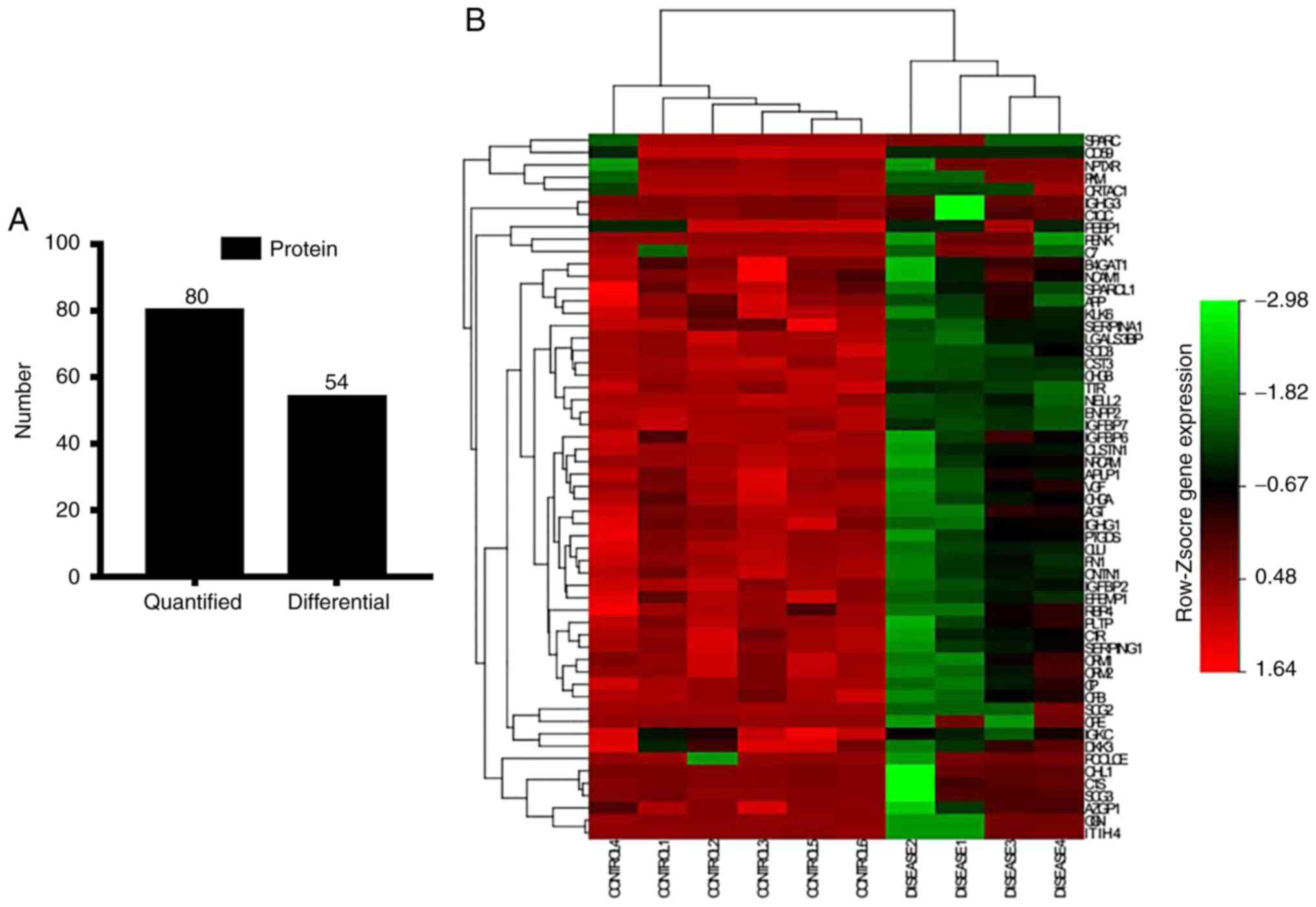
A total of 80 proteins from the four patients and
six healthy control subjects were identified using label-free
LC-MS/MS, and only 53 differentially expressed proteins with >1
log2 fold change and P<0.05 between the two groups
were selected for further analysis (Fig.
1A; Table I). Heat maps of all
proteins selected are shown in Fig.
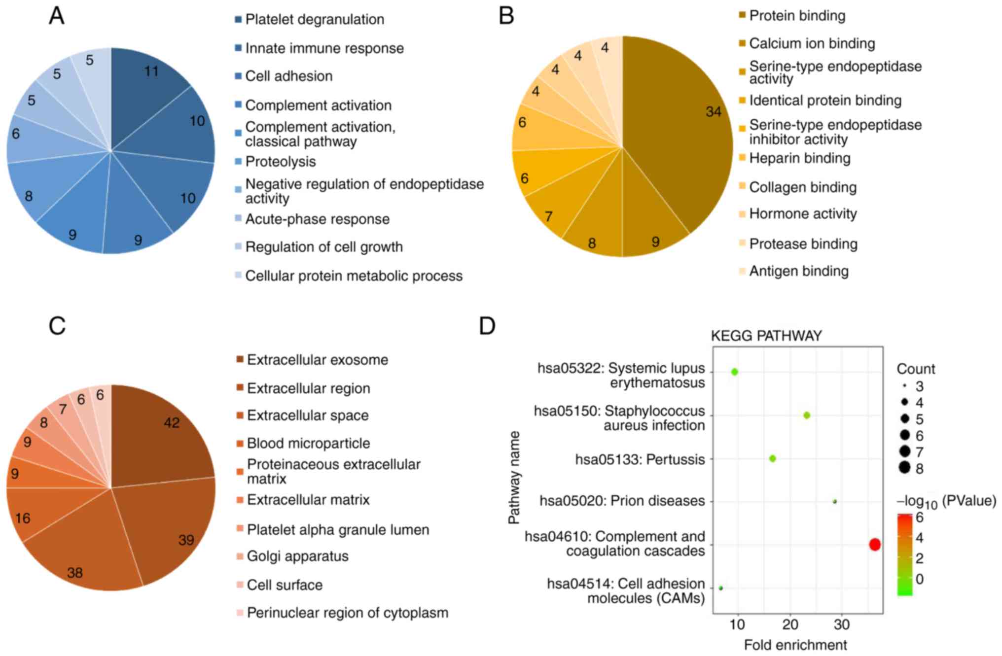
1B. The Database for Annotation, Visualization and Integrated
Discovery (DAVID) was utilized for the functional annotation of the
53 differential proteins. According to the Gene Ontology database,
certain proteins can be attributed to multiple cellular components,
functions and/or processes. As shown in Fig. 2A, the ‘biological processes’ analysis
revealed significant enrichment in platelet degranulation, innate
immune response and cell adhesion. The analysis of ‘molecular
function’ indicated that the differential CSF proteins were mainly
associated with protein binding, calcium ion binding and
serine-type endopeptidase activity (Fig.
2B). The ‘cellular component’ analysis (Fig. 2C) revealed that extracellular exosome,
extracellular space and blood microparticle were the most
over-represented terms (P<0.001). Kyoto Encyclopedia of Genes
and Genomes pathway analysis revealed three canonical pathways:
Complement and coagulation cascades, prion diseases and
Staphylococcus aureus infection, which provided insight into
the function of the 53 differential proteins in the CSF (Fig. 2D).
 | Table IDifferential proteins identified by
label-free LC-MS/MS ordered by log2FC. |
Table I
Differential proteins identified by
label-free LC-MS/MS ordered by log2FC.
| Protein ID | Gene name | Protein name | Log2FC |
|---|
| Q13822 | ENPP2 | Ectonucleotide
pyrophosphatase/phosphodiesterase family member 2 | 5.32 |
| Q16270 | IGFBP7 | Insulin-like growth
factor-binding protein 7 | 4.97 |
| P08294 | SOD3 | Extracellular
superoxide dismutase | 4.29 |
| O94985 | CLSTN1 | Calsyntenin-1 | 4.18 |
| P09486 | SPARC | Secreted protein
acidic and rich in cysteine | 4.13 |
| P10643 | C7 | Complement
component C7 | 4.11 |
| P01210 | PENK | Proenkephalin | 3.95 |
| P00736 | C1R | Complement C1r | 3.94 |
| Q08380 | LGALS3BP | Galectin-3-binding
protein | 3.92 |
| Q8WXD2 | SCG3 |
Secretogranin-3 | 3.82 |
| P02747 | C1QC | Complement C1q
subcomponent subunit C | 3.69 |
| P14618 | PKM | Pyruvate
kinase | 3.41 |
| P16870 | CPE | Carboxypeptidase
E | 3.33 |
| P00751 | CFB | Complement factor
B | 3.29 |
| P05155 | SERPING1 | Plasma protease C1
inhibitor | 3.19 |
| P09871 | C1S | Complement C1s
subcomponent | 3.14 |
| P18065 | IGFBP2 | Insulin-like growth
factor-binding protein 2 | 3.12 |
| P01034 | CST3 | Cystatin-C | 3.11 |
| P00450 | CP | Ceruloplasmin | 3.10 |
| P02751 | FN1 | Fibronectin | 3.10 |
| P05060 | CHGB | Secretogranin-1;
CCB peptide | 3.08 |
| Q92823 | NRCAM | Neuronal cell
adhesion molecule | 3.08 |
| Q15113 | PCOLCE | Procollagen C
endopeptidase enhancer 1 | 3.03 |
| Q12860 | CNTN1 | Contactin-1 | 2.95 |
| P02766 | TTR | Transthyretin | 2.93 |
| Q99435 | NELL2 | Neural epidermal
growth factor-like like 2 | 2.92 |
| P19652 | ORM2 | Alpha-1-acid
glycoprotein 2 | 2.85 |
| P02763 | ORM1 | Alpha-1-acid
glycoprotein 1 | 2.84 |
| P10909 | CLU | Clusterin | 2.77 |
| O95502 | NPTXR | Neuronal pentraxin
receptor | 2.64 |
| P51693 | APLP1 | Amyloid-like
protein 1; C30 | 2.60 |
| Q12805 | EFEMP1 | EGF-containing
fibulin-like extracellular matrix protein 1 | 2.58 |
| P10645 | CHGA | Chromogranin-A | 2.46 |
| P20774 | OGN | Mimecan | 2.44 |
| P05067 | APP | Amyloid beta A4
protein | 2.33 |
| O15240 | VGF | Neurosecretory
protein VGF | 2.32 |
| P55058 | PLTP | Phospholipid
transfer protein | 2.32 |
| Q14624 | ITIH4 | Inter-alpha-trypsin
inhibitor heavy chain H4 | 2.31 |
| Q92876 | KLK6 | Kallikrein-6 | 2.30 |
| P01857 | IGHG1 | Ig gamma-1 chain C
region | 2.29 |
| P24592 | IGFBP6 | Insulin-like growth
factor-binding protein 6 | 2.27 |
| O00533 | CHL1 | Neural cell
adhesion molecule L1-like protein | 2.24 |
| P01019 | AGT |
Angiotensinogen | 2.19 |
| P01860 | IGHG3 | Ig gamma-3 chain C
region | 2.10 |
| Q9NQ79 | CRTAC1 | Cartilage acidic
protein 1 | 1.87 |
| P02753 | RBP4 | Retinol-binding
protein 4 | 1.84 |
| P01009 | SERPINA1 |
Alpha-1-antitrypsin | 1.79 |
| O43505 | B4GAT1 |
Beta-1,4-glucuronyltransferase 1 | 1.68 |
| P25311 | AZGP1 |
Zinc-alpha-2-glycoprotein | 1.52 |
| Q14515 | SPARCL1 | SPARC-like protein
1 | 1.50 |
| P13591 | NCAM1 | Neural cell
adhesion molecule 1 | 1.34 |
| Q9UBP4 | DKK3 | Dickkopf-related
protein 3 | 1.27 |
| P01834 | IGKC | Ig kappa chain C
region | 1.14 |
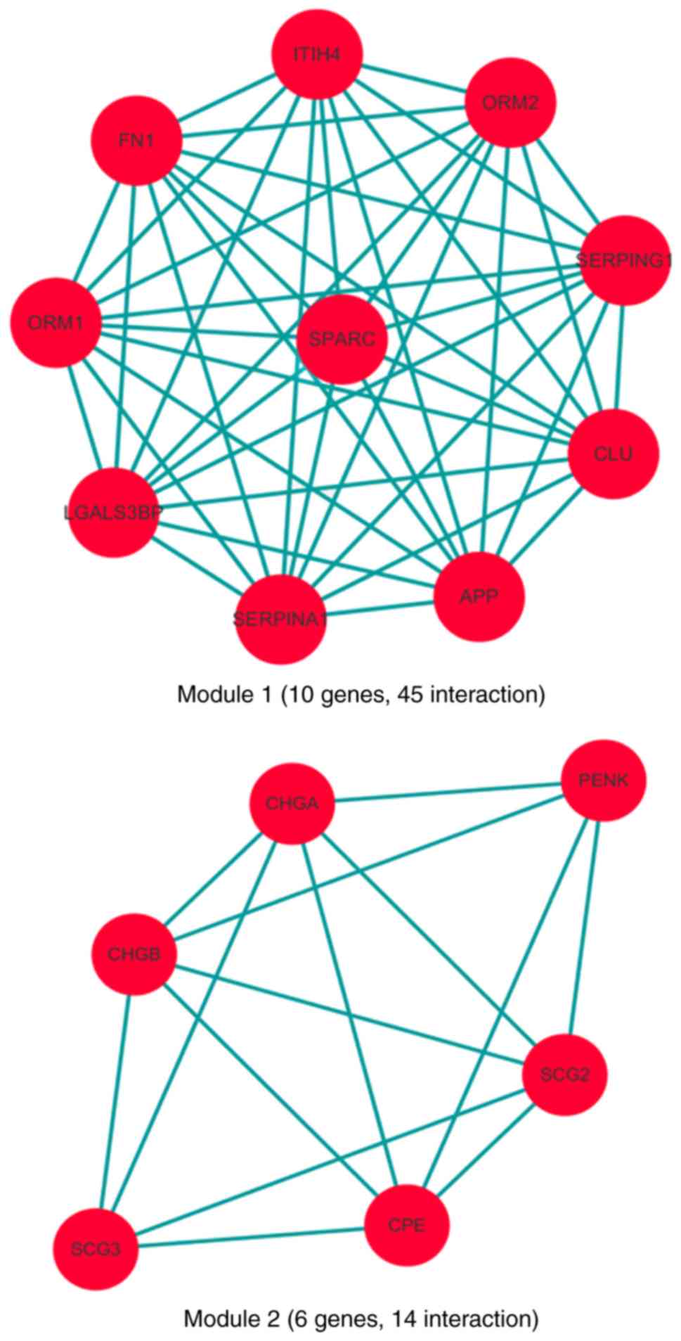
Protein-protein interaction (PPI)
network (PPIN) construction
Based on the Search Tool for the Retrieval of
Interacting Genes/Proteins (STRING) database, a PPIN of 53 proteins
was constructed (Fig. 3). As shown in
Fig. 4, two hub gene network modules
were obtained from the PPIN analyzed by MCODE. There were 10 genes
with 45 interactions in module 1 (MCODE score 8.182), and six genes
with 14 interactions in module 2 (MCODE score 4). In order to
further understand the function of the obtained hub modules, they
were analyzed using the DAVID database (Table II). The first module was mainly
enriched in platelet degranulation, and the complement and
coagulation cascades. The second module was mainly related to
neuropeptide signaling pathway and secretion. Furthermore, secreted
protein acidic and rich in cysteine (SPARC) was found to be at the
center of network module 1 with the highest fold change
(log2FC: 4.13). In network module 2, proenkephalin
(PENK) was the protein with the highest fold change
(log2FC: 4.13). These findings indicated that the
decreased expression of SPARC and PENK in the CSF was the most
sensitive, and these proteins may serve as early-phase biomarkers
to evaluate the risk of CNS involvement in DLBCL.
| Table IIFunctional enrichment analysis of the
hub modules. |
Table II
Functional enrichment analysis of the
hub modules.
| Module | Database | GO and pathway | Genes (n) | P-value |
|---|
| 1 | GO term-BP | Platelet
degranulation | 9 | 0.15 |
| | | Acute-phase
response | 5 |
<0.01b |
| | | Negative regulation
of endopeptidase activity | 4 |
<0.01b |
| | | Cell adhesion | 3 | 0.02a |
| | KEGG | Complement and
coagulation cascades | 2 | 0.03a |
| 2 | GO term-BP | Neuropeptide
signaling pathway | 2 | 0.02a |
| | | Secretion | 3 | 0.02a |
| | | G-protein coupled
receptor signaling pathway | 3 | 0.03a |
Discussion
In the present study, high-throughput quantitative
proteomic analysis was applied to analyze the expression of
proteins in the CSF of patients with DLBCL. Four CSF samples from
patients with DLBCL with CNS involvement were compared with six CSF
samples from healthy control subjects. Among the 53 differentially
expressed proteins identified, the most significantly altered
proteins, namely SPARC and PENK, appear to hold promise as a
diagnostic biomarker to evaluate the risk of CNS involvement in
DLBCL, as they exhibited the highest fold change and cross-talk
with other proteins found to be altered in the CSF of patients with
DLBCL.
SPARC is a 43-kDa glycoprotein, also referred to as
BM-40 or osteonectin (33). SPARC is
a multifunctional protein that can modulate extracellular matrix
assembly, integrin activity and growth factor signaling (34-37).
In cancer, SPARC has different functions, depending on the tissue
and cell type. In certain types of cancer, such as melanoma and
glioma, SPARC is associated with a highly aggressive tumor
phenotype (38). However, in
neuroblastomas, and ovarian, colorectal and primitive
neuroectodermal tumors, SPARC-induced changes in the tumor
microenvironment can suppress tumor progression (38,39). It
was previously reported that patients with DLBCL with any
SPARC-positive cells in the microenvironment had a significantly
longer overall survival, and patients with high SPARC-positivity in
the microenvironment also had a significantly longer event-free
survival (40). These findings
suggested that SPARC-positive stromal cells in the microenvironment
of DLBCL may act as tumor suppressors. In the present study, the
expression of SPARC was also found to be significantly
downregulated in the CSF of patients with DLBCL with CNS
involvement, which was in accordance with the suppressive function
of SPARC in DLBCL. The mechanism underlying the suppressive effect
of SPARC on the progression of DLBCL and improved patient survival
rates remains unclear. Possible explanations include the decreased
production of necessary growth factors, alterations of the
extracellular matrix preventing tumor cell interactions, and
decreased integrin production by tumor cells, resulting in altered
extracellular matrix interactions (40).
PENK is a nuclear protein responsive to growth
arrest and differentiation signals, and is required for the
induction of apoptosis (41). It is
reported that the expression of PENK is downregulated by two
proto-oncogenes, Fos and Jun (42).
Previous studies have reported that PENK is downregulated in
prostate cancer and glioblastoma; PENK was also reported to be
aberrantly methylated in colorectal, bladder and pancreatic cancer
(43-48).
PENK has been shown to stimulate stress-activated apoptosis,
particularly under treatment with chemotherapeutic drugs, in colon
cancer. In the present study, PENK was found to be downregulated in
the CSF of patients with DLBCL with CNS involvement. These findings
suggest that the tumors may attenuate apoptosis by downregulating
the protein expression of PENK.
Taken together, these data indicate that the
decreased expression of certain proteins in the CSF of patients
with DLBCL with CNS involvement is closely associated with the
antitumor and/or anti-invasion process. Therefore, detecting the
concentration of proteins, including SPARC and PENK, offers a
potential method for identifying patients with DLBCL at risk of
subsequent CNS involvement. In the present study, the small sample
size is a limitation, but it also provides future direction to
collect additional samples for further investigation.
In conclusion, through high-throughput label-free
quantitative proteomic analysis of the CSF from patients with DLBCL
and healthy control subjects, 53 differentially expressed proteins
and two gene (protein) hub network modules were identified in
total. Protein biomarkers, including SPARC and PENK, which were
found to be closely associated with DLBCL invasion, were expressed
at low levels in the CSF of patients with early-stage DLBCL.
Therefore, they may be valuable biomarkers for assessing the risk
of CNS involvement in DLBCL at initial diagnosis. Considering the
potential limitations of the present study in terms of design,
technique and analytical strategy, additional investigations with
larger cohorts of patients are required to confirm the robustness
of these findings.
Supplementary Material
Subject demographics.
Acknowledgements
Not applicable.
Funding
No funding was received.
Availability of data and materials
The datasets used and/or analyzed during the current
study are available from the corresponding author on reasonable
request.
Authors' contributions
XBL and XLM participated in the design of the
studies. XBL and FM collected data from participants. HZ performed
the statistical analysis; XBL and SZ participated in writing the
manuscript and performed the literature search. All the authors
have read and approved the final version of this manuscript for
publication.
Ethics approval and consent to
participate
All procedures performed in studies were in
accordance with the ethical standards of the Medical Ethics
Committee of West China Hospital, Sichuan University (2016.285).
All participants underwent an informed consent process.
Patient consent for publication
Not applicable.
Competing interests
The authors declare that they have no competing
interests.
References
|
1
|
A clinical evaluation of the International
Lymphoma Study Group classification of non-Hodgkin's lymphoma. The
Non-Hodgkin's Lymphoma Classification Project. Blood 89: 3909-3918,
1997.
|
|
2
|
Chen W, Wang H, Chen H, Liu S, Lu H, Kong
D, Huang X, Kong Q and Lu Z: Clinical significance and detection of
microRNA-21 in serum of patients with diffuse large B-cell lymphoma
in Chinese population. Eur J Haematol. 92:407–412. 2014.PubMed/NCBI View Article : Google Scholar
|
|
3
|
Martelli M, Ferreri AJ, Agostinelli C, Di
Rocco A, Pfreundschuh M and Pileri SA: Diffuse large B-cell
lymphoma. Crit Rev Oncol Hematol. 87:146–171. 2013.PubMed/NCBI View Article : Google Scholar
|
|
4
|
Feugier P, Van Hoof A, Sebban C,
Solal-Celigny P, Bouabdallah R, Fermé C, Christian B, Lepage E,
Tilly H, Morschhauser F, et al: Long-term results of the R-CHOP
study in the treatment of elderly patients with diffuse large
B-cell lymphoma: A study by the Groupe d'Etude des Lymphomes de
l'Adulte. J Clin Oncol. 23:4117–4126. 2005.PubMed/NCBI View Article : Google Scholar
|
|
5
|
Pfreundschuh M: Therapy of diffuse large
B-cell lymphomas. Eur J Cancer. 1 (Suppl 45):S386–S387. 2009.
|
|
6
|
Feugier P, Virion JM, Tilly H, Haioun C,
Marit G, Macro M, Bordessoule D, Recher C, Blanc M, Molina T, et
al: Incidence and risk factors for central nervous system
occurrence in elderly patients with diffuse large-B-cell lymphoma:
Influence of rituximab. Ann Oncol. 15:129–133. 2004.PubMed/NCBI View Article : Google Scholar
|
|
7
|
Herrlinger U, Glantz M, Schlegel U,
Gisselbrecht C and Cavalli F: Should intra-cerebrospinal fluid
prophylaxis be part of initial therapy for patients with
non-Hodgkin lymphoma: What we know, and how we can find out more.
Semin Oncol. 36 (4 Suppl 2):S25–S34. 2009.PubMed/NCBI View Article : Google Scholar
|
|
8
|
van Besien K, Ha CS, Murphy S, McLaughlin
P, Rodriguez A, Amin K, Forman A, Romaguera J, Hagemeister F,
Younes A, et al: Risk factors, treatment, and outcome of central
nervous system recurrence in adults with intermediate-grade and
immunoblastic lymphoma. Blood. 91:1178–1184. 1998.PubMed/NCBI
|
|
9
|
Zinzani PL, Magagnoli M, Frezza G, Prologo
G, Gherlinzoni F, Bendandi M, Albertini P, Babini L, D'Alessandro R
and Tura S: Isolated central nervous system relapse in aggressive
non-Hodgkin's lymphoma: The Bologna experience. Leuk Lymphoma.
32:571–576. 1999.PubMed/NCBI View Article : Google Scholar
|
|
10
|
Hollender A, Kvaloy S, Nome O, Skovlund E,
Lote K and Holte H: Central nervous system involvement following
diagnosis of non-Hodgkin's lymphoma: A risk model. Ann Oncol.
13:1099–1107. 2002.PubMed/NCBI View Article : Google Scholar
|
|
11
|
Zahid MF, Khan N, Hashmi SK, Kizilbash SH
and Barta SK: Central nervous system prophylaxis in diffuse large
B-cell lymphoma. Eur J Haematol. 97:108–120. 2016.PubMed/NCBI View Article : Google Scholar
|
|
12
|
Haioun C, Besson C, Lepage E, Thieblemont
C, Simon D, Rose C, Tilly H, Sonet A, Lederlin P, Attal M, et al:
Incidence and risk factors of central nervous system relapse in
histologically aggressive non-Hodgkin's lymphoma uniformly treated
and receiving intrathecal central nervous system prophylaxis: A
GELA study on 974 patients. Groupe d'Etudes des Lymphomes de
l'Adulte. Ann Oncol. 11:685–690. 2000.PubMed/NCBI View Article : Google Scholar
|
|
13
|
Barosi G, Carella A, Lazzarino M,
Marchetti M, Martelli M, Rambaldi A, Tarella C, Vitolo U, Zinzani
PL, Tura S, et al: Management of nodal diffuse large B-cell
lymphomas: Practice guidelines from the italian society of
hematology, the italian society of experimental hematology and the
italian group for bone marrow transplantation. Haematologica.
91:96–103. 2006.PubMed/NCBI
|
|
14
|
Boehme V, Zeynalova S, Kloess M, Loeffler
M, Kaiser U, Pfreundschuh M and Schmitz N: German High-Grade
Non-Hodgkin's Lymphoma Study Group (DSHNHL): Incidence and risk
factors of central nervous system recurrence in aggressive
lymphoma-a survey of 1693 patients treated in protocols of the
German High-Grade Non-Hodgkin's Lymphoma Study Group (DSHNHL). Ann
Oncol. 18:149–157. 2007.PubMed/NCBI View Article : Google Scholar
|
|
15
|
Hill QA and Owen RG: CNS prophylaxis in
lymphoma: Who to target and what therapy to use. Blood Rev.
20:319–332. 2006.PubMed/NCBI View Article : Google Scholar
|
|
16
|
Liang R, Chiu E and Loke SL: Secondary
central nervous system involvement by non-Hodgkin's lymphoma: The
risk factors. Hematol Oncol. 8:141–145. 1990.PubMed/NCBI
|
|
17
|
Fletcher CD and Kahl BS: Central nervous
system involvement in diffuse large B-cell lymphoma: An analysis of
risks and prevention strategies in the post-rituximab era. Leuk
Lymphoma. 55:2228–2240. 2014.PubMed/NCBI View Article : Google Scholar
|
|
18
|
Holte H, Leppä S, Björkholm M, Fluge O,
Jyrkkiö S, Delabie J, Sundström C, Karjalainen-Lindsberg ML,
Erlanson M, Kolstad A, et al: Dose-densified chemoimmunotherapy
followed by systemic central nervous system prophylaxis for younger
high-risk diffuse large B-cell/follicular grade 3 lymphoma
patients: Results of a phase II Nordic Lymphoma Group study. Ann
Oncol. 24:1385–1392. 2013.PubMed/NCBI View Article : Google Scholar
|
|
19
|
Penalver FJ, Sancho JM, de la Fuente A,
Olave MT, Martín A, Panizo C, Pérez E, Salar A and Orfao A: Spanish
Lymphoma Group (GELTAMO): Guidelines for diagnosis, prevention and
management of central nervous system involvement in diffuse large
B-cell lymphoma patients by the Spanish Lymphoma Group (GELTAMO).
Haematologica. 102:235–245. 2017.PubMed/NCBI View Article : Google Scholar
|
|
20
|
Glass JP, Melamed M, Chernik NL and Posner
JB: Malignant cells in cerebrospinal fluid (CSF): The meaning of a
positive CSF cytology. Neurology. 29:1369–1375. 1979.PubMed/NCBI View Article : Google Scholar
|
|
21
|
Hegde U, Filie A, Little RF, Janik JE,
Grant N, Steinberg SM, Dunleavy K, Jaffe ES, Abati A,
Stetler-Stevenson M and Wilson WH: High incidence of occult
leptomeningeal disease detected by flow cytometry in newly
diagnosed aggressive B-cell lymphomas at risk for central nervous
system involvement: The role of flow cytometry versus cytology.
Blood. 105:496–502. 2005.PubMed/NCBI View Article : Google Scholar
|
|
22
|
McMillan A, Ardeshna KM, Cwynarski K,
Lyttelton M, McKay P and Montoto S: British Committee for Standards
in Haematology: Guideline on the prevention of secondary central
nervous system lymphoma: British committee for standards in
haematology. Br J Haematol. 163:168–181. 2013.PubMed/NCBI View Article : Google Scholar
|
|
23
|
Villa D, Connors JM, Shenkier TN, Gascoyne
RD, Sehn LH and Savage KJ: Incidence and risk factors for central
nervous system relapse in patients with diffuse large B-cell
lymphoma: The impact of the addition of rituximab to CHOP
chemotherapy. Ann Oncol. 21:1046–1052. 2010.PubMed/NCBI View Article : Google Scholar
|
|
24
|
Shimazu Y, Notohara K and Ueda Y: Diffuse
large B-cell lymphoma with central nervous system relapse:
Prognosis and risk factors according to retrospective analysis from
a single-center experience. Int J Hematol. 89:577–583.
2009.PubMed/NCBI View Article : Google Scholar
|
|
25
|
Scott BJ, Douglas VC, Tihan T, Rubenstein
JL and Josephson SA: A systematic approach to the diagnosis of
suspected central nervous system lymphoma. JAMA Neurol. 70:311–319.
2013.PubMed/NCBI View Article : Google Scholar
|
|
26
|
Shen X, Young R, Canty JM and Qu J:
Quantitative proteomics in cardiovascular research: Global and
targeted strategies. Proteomics Clin Appl. 8:488–505.
2014.PubMed/NCBI View Article : Google Scholar
|
|
27
|
Weisser H, Nahnsen S, Grossmann J, Nilse
L, Quandt A, Brauer H, Sturm M, Kenar E, Kohlbacher O, Aebersold R
and Malmström L: An automated pipeline for high-throughput
label-free quantitative proteomics. J Proteome Res. 12:1628–1644.
2013.PubMed/NCBI View Article : Google Scholar
|
|
28
|
Gitau EN, Kokwaro GO, Karanja H, Newton CR
and Ward SA: Plasma and cerebrospinal proteomes from children with
cerebral malaria differ from those of children with other
encephalopathies. J Infect Dis. 208:1494–1503. 2013. View Article : Google Scholar
|
|
29
|
Ou Q, Liu X and Cheng X: An iTRAQ approach
to quantitative proteome analysis of cerebrospinal fluid from
patients with tuberculous meningitis. Biosci Trends. 7:186–192.
2013.PubMed/NCBI View Article : Google Scholar
|
|
30
|
Pasinetti GM, Ungar LH, Lange DJ, Yemul S,
Deng H, Yuan X, Brown RH, Cudkowicz ME, Newhall K, Peskind E, et
al: Identification of potential CSF biomarkers in ALS. Neurology.
66:1218–1222. 2006.PubMed/NCBI View Article : Google Scholar
|
|
31
|
Priola GM, Foster MW, Deal AM, Richardson
BM, Thompson JW and Blatt J: Cerebrospinal fluid proteomics in
children during induction for acute lymphoblastic leukemia: A pilot
study. Pediatr Blood Cancer. 62:1190–1194. 2015.PubMed/NCBI View Article : Google Scholar
|
|
32
|
Rardin MJ, Newman JC, Held JM, Cusack MP,
Sorensen DJ, Li B, Schilling B, Mooney SD, Kahn CR, Verdin E and
Gibson BW: Label-free quantitative proteomics of the lysine
acetylome in mitochondria identifies substrates of SIRT3 in
metabolic pathways. Proc Natl Acad Sci USA. 110:6601–6606.
2013.PubMed/NCBI View Article : Google Scholar
|
|
33
|
Chlenski A and Cohn SL: Modulation of
matrix remodeling by SPARC in neoplastic progression. Semin Cell
Dev Biol. 21:55–65. 2010.PubMed/NCBI View Article : Google Scholar
|
|
34
|
Bradshaw AD, Puolakkainen P, Dasgupta J,
Davidson JM, Wight TN and Helene Sage E: SPARC-null mice display
abnormalities in the dermis characterized by decreased collagen
fibril diameter and reduced tensile strength. J Invest Dermatol.
120:949–955. 2003.PubMed/NCBI View Article : Google Scholar
|
|
35
|
Raines EW, Lane TF, Iruela-Arispe ML, Ross
R and Sage EH: The extracellular glycoprotein SPARC interacts with
platelet-derived growth factor (PDGF)-AB and -BB and inhibits the
binding of PDGF to its receptors. Proc Natl Acad Sci USA.
89:1281–1285. 1992.PubMed/NCBI View Article : Google Scholar
|
|
36
|
Sangaletti S, Stoppacciaro A, Guiducci C,
Torrisi MR and Colombo MP: Leukocyte, rather than tumor-produced
SPARC, determines stroma and collagen type IV deposition in mammary
carcinoma. J Exp Med. 198:1475–1485. 2003.PubMed/NCBI View Article : Google Scholar
|
|
37
|
Weaver MS, Workman G and Sage EH: The
copper binding domain of SPARC mediates cell survival in vitro via
interaction with integrin beta1 and activation of integrin-linked
kinase. J Biol Chem. 283:22826–22837. 2008.PubMed/NCBI View Article : Google Scholar
|
|
38
|
Tai IT and Tang MJ: SPARC in cancer
biology: Its role in cancer progression and potential for therapy.
Drug Resist Updat. 11:231–246. 2008.PubMed/NCBI View Article : Google Scholar
|
|
39
|
Bhoopathi P, Chetty C, Gujrati M, Dinh DH,
Rao JS and Lakka S: Cathepsin B facilitates autophagy-mediated
apoptosis in SPARC overexpressed primitive neuroectodermal tumor
cells. Cell Death Differ. 17:1529–1539. 2010.PubMed/NCBI View Article : Google Scholar
|
|
40
|
Meyer PN, Fu K, Greiner T, Smith L,
Delabie J, Gascoyne R, Ott G, Rosenwald A, Braziel R, Campo E, et
al: The stromal cell marker SPARC predicts for survival in patients
with diffuse large B-cell lymphoma treated with rituximab. Am J
Clin Pathol. 135:54–61. 2011. View Article : Google Scholar
|
|
41
|
Bottger A and Spruce BA: Proenkephalin is
a nuclear protein responsive to growth arrest and differentiation
signals. J Cell Biol. 130:1251–1262. 1995.PubMed/NCBI View Article : Google Scholar
|
|
42
|
Sonnenberg JL, Rauscher FJ III, Morgan JI
and Curran T: Regulation of proenkephalin by Fos and Jun. Science.
246:1622–1625. 1989.PubMed/NCBI View Article : Google Scholar
|
|
43
|
Ueki T, Toyota M, Skinner H, Walter KM,
Yeo CJ, Issa JP, Hruban RH and Goggins M: Identification and
characterization of differentially methylated CpG islands in
pancreatic carcinoma. Cancer Res. 61:8540–8546. 2001.PubMed/NCBI
|
|
44
|
Fukushima N, Sato N, Ueki T, Rosty C,
Walter KM, Wilentz RE, Yeo CJ, Hruban RH and Goggins M: Aberrant
methylation of preproenkephalin and p16 genes in pancreatic
intraepithelial neoplasia and pancreatic ductal adenocarcinoma. Am
J Pathol. 160:1573–1581. 2002.PubMed/NCBI View Article : Google Scholar
|
|
45
|
Goo YA, Goodlett DR, Pascal LE,
Worthington KD, Vessella RL, True LD and Liu AY: Stromal mesenchyme
cell genes of the human prostate and bladder. BMC Urol.
5(17)2005.PubMed/NCBI View Article : Google Scholar
|
|
46
|
Tan AC, Jimeno A, Lin SH, Wheelhouse J,
Chan F, Solomon A, Rajeshkumar NV, Rubio-Viqueira B and Hidalgo M:
Characterizing DNA methylation patterns in pancreatic cancer
genome. Mol Oncol. 3:425–438. 2009.PubMed/NCBI View Article : Google Scholar
|
|
47
|
Roperch JP, Incitti R, Forbin S, Bard F,
Mansour H, Mesli F, Baumgaertner I, Brunetti F and Sobhani I:
Aberrant methylation of NPY, PENK, and WIF1 as a promising marker
for blood-based diagnosis of colorectal cancer. BMC Cancer.
13(566)2013.PubMed/NCBI View Article : Google Scholar
|
|
48
|
Lee EJ, Rath P, Liu J, Ryu D, Pei L,
Noonepalle SK, Shull AY, Feng Q, Litofsky NS, Miller DC, et al:
Identification of global DNA methylation signatures in
glioblastoma-derived cancer stem cells. J Genet Genomics.
42:355–371. 2015.PubMed/NCBI View Article : Google Scholar
|