Introduction
Diabetic retinopathy (DR), one of the leading causes
of blindness worldwide, results from chronic hyperglycemia in
patients with diabetes mellitus (DM) (1). Proliferative diabetic retinopathy
(PDR), the advanced stage of DR, is characterized by severe retinal
ischemia and highly upregulated expression of VEGF, resulting in
the development of neovascularization in ocular tissues, vitreous
hemorrhage and inflammatory fibrovascular membranes (2). Hyperglycemia also induces
non-enzymatic glycation of proteins, lipids and nucleic acids, via
a chemical process known as the Maillard reaction, and this
promotes the formation of advanced glycation end-products (AGE)
(3). AGE acts directly or
indirectly via the receptor for AGE (RAGE) as a pathogenic cue in
inflammation, angiogenesis and cell proliferation (4,5), and
there is considerable evidence of increased serum AGE levels in DM
patients with diabetic complications (6,7). In
the pathogenesis of DR, AGE contributes to the expression of
pro-angiogenic factors such as VEGF-A and TNF-α by means of NF-κB
activation induced by RAGE-AGE interaction and the decline in the
number of retinal pericytes caused by reactive oxygen species
(ROS)-derived apoptosis (8,9). Furthermore, AGE-modified albumin shows
pro-inflammatory effects through the induction of leukocyte
adhesion in the retinal microvasculature, resulting in breakdown of
the blood-retinal barrier (10).
αB-crystallin, also known as heat shock protein B5
(HSPB5), belongs to the crystallin family of proteins. It has
pleiotropic roles not only in cellular protection and induction of
cell proliferation, but as a molecular chaperone for VEGF-A
(11,12). Blood αB-crystallin levels are
reported to increase in inflammatory conditions such as multiple
sclerosis and in the experimental autoimmune encephalomyelitis
(EAE) mouse model (13), in which
the increase may be correlated with the reduction of symptoms
including paralysis (14). In
ocular tissues, Kannan et al (15) revealed that αB-crystallin can exert
cell-protective effects by inhibiting oxidative stress and
endoplasmic reticulum stress in retinal pigment epithelium cells.
Conversely, angiogenesis was inhibited by deletion of αB-crystallin
through VEGF-A degradation in models of intraocular angiogenesis
(16). Colocalization of
αB-crystallin and VEGF in the endothelial cells of retinal tissues
obtained from PDR patients has also been observed, suggesting that
αB-crystallin is a key molecule of angiogenesis and inflammation
(17).
Previous studies showed that AGE are likely to
regulate αB-crystallin expression. In fact, in our previous study,
it was demonstrated that αB-crystallin protein is downregulated
while αA-crystallin protein is upregulated in wild-type murine
posterior eyecups exposed to AGE protein (18). Moreover, αB-crystallin protein
expression is noted and AGE accumulates in the retinal tissues of
human cadaver diabetic eyes without retinopathy (18,19).
Other reports showed that AGE concentrations are elevated in the
vitreous fluid and aqueous humor of DR patients compared with
age-matched controls (20,21), and the AGE concentration was higher
in PDR patients' blood than in the control group (22,23).
Conversely, the αB-crystallin protein concentration increased in
the vitreous fluid of PDR patients compared with eyes without DR
(24). Another report showed
increased intravitreal αB-crystallin levels in rhegmatogenous
retinal detachment patients (25).
Given this evidence, it is possible that αB-crystallin and AGE
proteins may be released from somatic cells, including retinal
pigment epithelium cells, and eventually be secreted into humoral
fluids such as vitreous fluids and sera. However, there are no
reports on concentrations of αB-crystallin and AGE in sera of the
same PDR patients. In this study, the correlation between
αB-crystallin and AGE in serum samples were explored, which were
prospectively collected from PDR patients.
Patients and methods
Study population
This study was performed according to the tenets of
the Declaration of Helsinki and approved by the institutional
review board of Hokkaido University Hospital (approval no.
019-0186; December 2019). Patients who came to the Department of
Ophthalmology of Hokkaido University Hospital (Hokkaido, Japan) and
underwent pars plana vitrectomy were enrolled after
providing written informed consent. Patients' sera were collected
between December 2019 and December 2020 prior to vitrectomy.
Grading of DR was performed according to the literature (26) by retina specialists in our hospital
who have >3 years of experience as ophthalmologists, and PDR was
defined as proliferative tissue caused by neovessels. Serum samples
and matching clinical data were obtained from 7 PDR patients (5
females and 2 males, referred to as the PDR group) and 8
non-diabetic patients with idiopathic macular diseases including an
epiretinal membrane and a macular hole (3 females and 5 males,
non-DM group). The random blood sugar level, estimated glomerular
filtration rate (eGFR), aspartate aminotransferase and alanine
transaminase were routinely measured in patients' blood. Patient
backgrounds and demographics are described in Table I. The median age of the seven
enrolled PDR patients and eight non-DM patients was 62 years (age
range, 34-78 years) and 72 years (age range, 59-83 years),
respectively.
 | Table IClinicopathological characteristics of
participating patients. |
Table I
Clinicopathological characteristics of
participating patients.
| Factor | Non-diabetes
mellitus | Proliferative
diabetic retinopathy | P-value |
|---|
| Number of
patients | 8 | 7 | |
| Ages,
yearsb | 72 (range 59-83) | 62 (range 34-78) | 0.118 |
| Sex, % | | | 0.315 |
|
Male | 63 | 29 | |
|
Female | 37 | 71 | |
| Blood sugar level,
mmol/lc | 5.92±0.29 | 9.21±1.20 | 0.011a |
| Estimated glomerular
filtration rate, ml/minc | 58.05±4.49 | 61.80±9.13 | 0.418 |
| Aspartate
aminotransferase, U/lc | 20.50±0.96 | 21.14±2.20 | 0.861 |
| Alanine transaminase,
U/lc | 22.25±3.86 | 20.86±3.64 | 0.728 |
ELISA
The protein levels of αB-crystallin and AGE were
determined using an αB-crystallin ELISA kit (cat. no. SKT-123,
StressMarq Biosciences Inc.) and an AGE assay kit (cat. no.
ab238539, Abcam) according to the manufacturers' instructions.
αB-crystallin and AGE were measured by sandwich ELISA and
competitive ELISA, respectively. The optical density was determined
at 450 nm using a microplate reader (Sunrise absorbance reader,
Tecan Group, Ltd.). For AGE measurement, the standard curve was
calibrated using AGE-BSA (µg/ml) and the serum AGE concentration is
expressed in terms of AGE-BSA.
Statistical analysis
All results are presented as the mean ± SEM. A
Mann-Whitney U test was used to assess differences in serum
concentrations between PDR and non-DM. Spearman's rank correlation
analysis was used to analyze the correlation between serum AGE and
αB-crystallin concentrations. A Fisher's exact test was used to
examine if there were any sex-based differences. P<0.05 was
considered to indicate a statistically significant difference. A
Smirnov-Grubbs test was used to exclude outlier data in the
determination of the normal ranges. Correlation analysis of
αB-crystallin and AGE was performed after excluding any
outliers.
Results
Study population
There was no significant difference in age between
the two groups (P=0.082). None of the male-to-female ratio, levels
of hepatic enzymes, or eGFR showed significant differences between
the two groups. The random blood sugar level was significantly
higher in the PDR group (non-DM, 5.92±0.29 mmol/l; PDR, 9.21±1.20
mmol/l; P<0.05; Table I).
Therefore, the PDR patients enrolled in this study were
characterized by hyperglycemia without renal dysfunction.
Correlation between serum AGE and
αB-crystallin
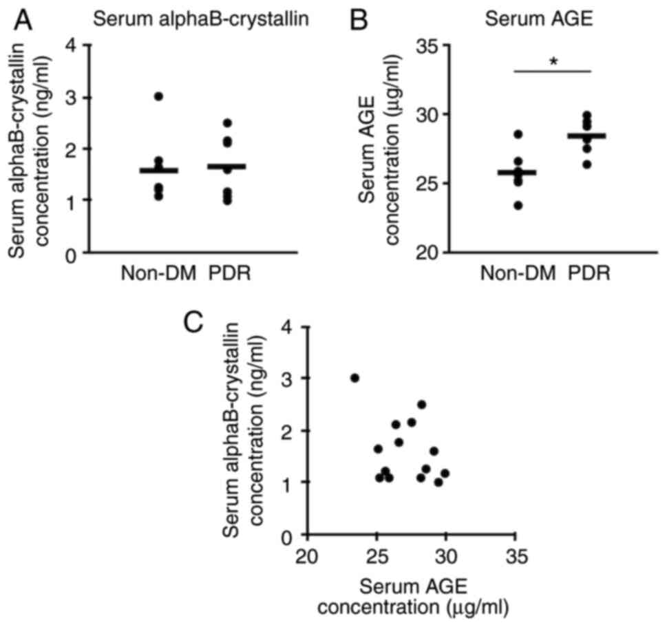
Serum levels of AGE were significantly higher in the
PDR group than in the non-DM group (PDR, 28.41±0.46 µg/ml; non-DM,
25.76±0.60 µg/ml; P=0.015). In contrast, serum αB-crystallin levels
did not differ significantly between the non-DM and PDR groups
(1.58±0.26 and 1.66±0.23 ng/ml, respectively; P=0.949; Fig. 1). There was no significant
correlation between serum αB-crystallin and AGE (ρ=-0.276,
P=0.340). The existence of outlier data from one patient was
confirmed by the Smirnov-Grubbs test, and these were excluded from
this analysis; the serum αB-crystallin levels of this patient in
the non-DM group was extremely high (123.9 ng/ml). In fact, this
patient was treated with oral prednisolone for 2 decades, with a
total dose of ~10,000 mg, due to rheumatoid arthritis and
interstitial pneumonia, whereas no other patients had received
systemic corticosteroid therapy.
Discussion
The present study was a prospective observational
study with PDR patients' sera, and the results demonstrated
significantly higher AGE levels in the PDR group, which is
consistent with the report of a case controlled study (22). Blood-retinal barrier breakdown
occurs in patients with DR, and AGE induces microvascular
hyperpermeability in vascular endothelial cells via RAGE (10,27).
Prolonged hyperglycemia causes AGE accumulation in various tissues,
and it has been reported that intravitreal AGE elevation is
correlated with serum AGE and hemoglobin A1c (HbA1c) levels in
diabetic patients (28,29). HbA1c is one of the non-enzymatic
glycation products as well as AGE; however, AGE reflects
longer-term cumulative diabetic exposure as they are harder to
degrade than HbA1c (30). AGE
immunoreactivity was strongly noted in cadaver diabetic retinal
tissues including central retinal arteries/veins (19). Therefore, the AGE concentration is
likely to be higher in sera of PDR patients with a long-term
DM.
Our previous report revealed that αB-crystallin is
expressed in neovessels within PDR membranes (17), where αB-crystallin is phosphorylated
on serine 59 through upregulation of phosphorylated p38
mitogen-activated protein kinase (31). αB-crystallin showed cytosolic
localization (15), but it could be
secreted through an unconventional secretion pathway via exosomes
following phosphorylation on serine 59(32). Although there are no reports on the
relationship between AGE and αB-crystallin concentrations in
vitreous fluid, it was reported that αB-crystallin protein
concentrations increased in vitreous fluids of PDR patients
compared with those without DR in proportion to VEGF concentrations
(24). Therefore, it was
hypothesized that αB-crystallin may also be secreted into the sera
together with AGE in PDR patients. However, the current prospective
study found neither αB-crystallin elevation in the PDR group nor a
significant correlation between serum αB-crystallin and AGE
concentrations. Further studies are needed to elucidate the
mechanisms underlying extracellular secretion of αB-crystallin from
intraocular tissues, such as from the retina of diabetic
patients.
It should be noted that the serum αB-crystallin
levels were ~80X higher than in the non-DM group in one patient
with systemic inflammatory diseases who had been treated with oral
corticosteroids. Among patients with multiple sclerosis, a chronic
neuroinflammatory disease characterized by demyelination of the
central nerve system, blood αB-crystallin levels were higher than
those of healthy controls (13).
Furthermore, it has been shown that proinflammatory cytokines
including IL-1β and TNF-α promote αB-crystallin mRNA upregulation
in a glioblastoma cell line (33).
As indicated in the literature, inflammatory conditions can induce
αB-crystallin expression both intra- and extracellularly, and this
is involved in the protection of targeted cells from apoptosis.
While inflammation can affect αB-crystallin expression, it has been
shown that dexamethasone increases the amount of αB-crystallin
protein in glomerular podocytes (34), which indicates that therapeutic
glucocorticoid is also capable of elevating αB-crystallin
expression. Additional research using DR models is needed to
further elucidate how glucocorticoid treatment changes the
expression levels of αB-crystallin.
The present study has some limitations. First,
patients without ophthalmic diseases and diabetic and non-diabetic
patients who have not undergone vitrectomy were not enrolled in
this study. Therefore, further studies are required to confirm
whether serum levels of αB-crystallin or AGE differ between
subjects without any ocular diseases and with idiopathic macular
diseases. Next, although the number of patients examined was
limited, this study prospectively enrolled only patients who
consented to participation in the IRB-approved study based on
written informed consent during designated study periods.
Furthermore, the novel coronavirus disease (COVID-19) pandemic
spread in our province in Japan from March 2020, in the middle of
the recruitment of participants. In fact, it has been reported that
surgical activity was significantly lower during the COVID-19
pandemic than before (35). Thus,
we encountered similar social challenges in Japan, and this severe
pandemic prevented the collection of a larger number of samples.
Further studies with a larger cohort will be needed to confirm the
results.
In summary, the current prospective study revealed
that serum AGE levels were significantly higher in diabetic
patients, while serum αB-crystallin levels in diabetic patients did
not differ from those of non-diabetic subjects.
Acknowledgements
Not applicable.
Funding
Funding: This study was supported in part by MEXT KAKENHI, Japan
(grant no. JP18K09394).
Availability of data and materials
The datasets used and/or analyzed during the present
study are available from the corresponding author on reasonable
request.
Authors' contributions
TY contributed to data curation, analysis,
experimentation and writing the manuscript. SK designed the study,
collected the samples and edited the manuscript. MM and SI
interpreted the data and edited the manuscript. TY and SK confirmed
the authenticity of all the raw data. All authors have read and
approved the final manuscript.
Ethics approval and consent to
participate
This study was performed according to the tenets of
the Declaration of Helsinki and approved by the institutional
review board of Hokkaido University Hospital (Hokkaido, Japan;
approval no. s019-0186; December 2019). All patients provided
written informed consent.
Patient consent for publication
Not applicable.
Competing interests
The authors declare that they have no competing
interests.
References
|
1
|
Jampol LM, Glassman AR and Sun J:
Evaluation and care of patients with diabetic retinopathy. N Engl J
Med. 382:1629–1637. 2020.PubMed/NCBI View Article : Google Scholar
|
|
2
|
Kase S, Saito W, Ohno S and Ishida S:
Proliferative diabetic retinopathy with lymphocyte-rich epiretinal
membrane associated with poor visual prognosis. Invest Ophthalmol
Vis Sci. 50:5909–5912. 2009.PubMed/NCBI View Article : Google Scholar
|
|
3
|
Chaudhuri J, Bains Y, Guha S, Kahn A, Hall
D, Bose N, Gugliucci A and Kapahi P: The role of advanced glycation
end products in aging and metabolic diseases: Bridging association
and causality. Cell Metab. 28:337–352. 2018.PubMed/NCBI View Article : Google Scholar
|
|
4
|
Zong H, Ward M, Madden A, Yong PH, Limb
GA, Curtis TM and Stitt AW: Hyperglycaemia-induced pro-inflammatory
responses by retinal Müller glia are regulated by the receptor for
advanced glycation end-products (RAGE). Diabetologia. 53:2656–2666.
2010.PubMed/NCBI View Article : Google Scholar
|
|
5
|
Chen L, Cui Y, Li B, Weng J, Wang W, Zhang
S, Huang X, Guo X and Huang Q: Advanced glycation end products
induce immature angiogenesis in in vivo and ex vivo mouse models.
Am J Physiol Heart Circ Physiol. 318:H519–H533. 2020.PubMed/NCBI View Article : Google Scholar
|
|
6
|
Makita Z, Radoff S, Rayfield EJ, Yang Z,
Skolnik E, Delaney V, Friedman EA, Cerami A and Vlassara H:
Advanced glycosylation end products in patients with diabetic
nephropathy. N Engl J Med. 325:836–842. 1991.PubMed/NCBI View Article : Google Scholar
|
|
7
|
Monnier VM, Sell DR and Genuth S:
Glycation products as markers and predictors of the progression of
diabetic complications. Ann N Y Acad Sci. 1043:567–581.
2005.PubMed/NCBI View Article : Google Scholar
|
|
8
|
Stitt AW: AGEs and diabetic retinopathy.
Invest Ophthalmol Vis Sci. 51:4867–4874. 2010.PubMed/NCBI View Article : Google Scholar
|
|
9
|
Xu J, Chen LJ, Yu J, Wang HJ, Zhang F, Liu
Q and Wu J: Involvement of advanced glycation end products in the
pathogenesis of diabetic retinopathy. Cell Physiol Biochem.
48:705–717. 2018.PubMed/NCBI View Article : Google Scholar
|
|
10
|
Moore TC, Moore JE, Kaji Y, Frizzell N,
Usui T, Poulaki V, Campbell IL, Stitt AW, Gardiner TA, Archer DB
and Adamis AP: The role of advanced glycation end products in
retinal microvascular leukostasis. Invest Ophthalmol Vis Sci.
44:4457–4464. 2003.PubMed/NCBI View Article : Google Scholar
|
|
11
|
Ghosh JG, Shenoy AK Jr and Clark JI:
Interactions between important regulatory proteins and human alphaB
crystallin. Biochemistry. 46:6308–6317. 2007.PubMed/NCBI View Article : Google Scholar
|
|
12
|
Mymrikov EV, Seit-Nebi AS and Gusev NB:
Large potentials of small heat shock proteins. Physiol Rev.
91:1123–1159. 2011.PubMed/NCBI View Article : Google Scholar
|
|
13
|
Rothbard JB, Kurnellas MP, Brownell S,
Adams CM, Su L, Axtell RC, Chen R, Fathman CG, Robinson WH and
Steinman L: Therapeutic effects of systemic administration of
chaperone αB-crystallin associated with binding proinflammatory
plasma proteins. J Biol Chem. 287:9708–9721. 2012.PubMed/NCBI View Article : Google Scholar
|
|
14
|
Ousman SS, Tomooka BH, van Noort JM,
Wawrousek EF, O'Connor KC, Hafler DA, Sobel RA, Robinson WH and
Steinman L: Protective and therapeutic role for alphaB-crystallin
in autoimmune demyelination. Nature. 448:474–479. 2007.PubMed/NCBI View Article : Google Scholar
|
|
15
|
Kannan R, Sreekumar PG and Hinton DR:
Alpha crystallins in the retinal pigment epithelium and
implications for the pathogenesis and treatment of age-related
macular degeneration. Biochim Biophys Acta. 1860:258–268.
2016.PubMed/NCBI View Article : Google Scholar
|
|
16
|
Kase S, He S, Sonoda S, Kitamura M, Spee
C, Wawrousek E, Ryan SJ, Kannan R and Hinton DR: AlphaB-crystallin
regulation of angiogenesis by modulation of VEGF. Blood.
115:3398–3406. 2010.PubMed/NCBI View Article : Google Scholar
|
|
17
|
Dong Z, Kase S, Ando R, Fukuhara J, Saito
W, Kanda A, Murata M, Noda K and Ishida S: Alphab-crystallin
expression in epiretinal membrane of human proliferative diabetic
retinopathy. Retina. 32:1190–1196. 2012.PubMed/NCBI View Article : Google Scholar
|
|
18
|
Kase S, Ishida S and Rao NA: Increased
expression of αA-crystallin in human diabetic eye. Int J Mol Med.
28:505–511. 2011.PubMed/NCBI View Article : Google Scholar
|
|
19
|
Kase S, Ishida S and Rao NA:
Immunolocalization of advanced glycation end products in human
diabetic eyes: An immunohistochemical study. J Diabetes Mellit.
1:57–62. 2011.
|
|
20
|
Endo M, Yanagisawa K, Tsuchida K, Okamoto
T, Matsushita T, Higuchi M, Matsuda A, Takeuchi M, Makita Z and
Koike T: Increased levels of vascular endothelial growth factor and
advanced glycation end products in aqueous humor of patients with
diabetic retinopathy. Horm Metab Res. 33:317–322. 2001.PubMed/NCBI View Article : Google Scholar
|
|
21
|
Stitt AW, Moore JE, Sharkey JA, Murphy G,
Simpson DA, Bucala R, Vlassara H and Archer DB: Advanced glycation
end products in vitreous: Structural and functional implications
for diabetic vitreopathy. Invest Ophthalmol Vis Sci. 39:2517–2523.
1998.PubMed/NCBI
|
|
22
|
Boehm BO, Schilling S, Rosinger S, Lang
GE, Lang GK, Kientsch-Engel R and Stahl P: Elevated serum levels of
N(epsilon)-carboxymethyl-lysine, an advanced glycation end product,
are associated with proliferative diabetic retinopathy and macular
oedema. Diabetologia. 47:1376–1379. 2004.PubMed/NCBI View Article : Google Scholar
|
|
23
|
Hase K, Kanda A, Noda K and Ishida S:
Increased plasma galectin-1 correlates with advanced glycation end
products and interleukin-1β in patients with proliferative diabetic
retinopathy. Int J Ophthalmol. 12:692–694. 2019.PubMed/NCBI View Article : Google Scholar
|
|
24
|
Chen W, Lu Q, Lu L and Guan H: Increased
levels of alphaB-crystallin in vitreous fluid of patients with
proliferative diabetic retinopathy and correlation with vascular
endothelial growth factor. Clin Exp Ophthalmol. 45:379–384.
2017.PubMed/NCBI View Article : Google Scholar
|
|
25
|
Baba T, Oshitari T and Yamamoto S: Level
of vitreous alpha-B crystallin in eyes with rhegmatogenous retinal
detachment. Graefes Arch Clin Exp Ophthalmol. 253:1251–1254.
2015.PubMed/NCBI View Article : Google Scholar
|
|
26
|
Klein R, Klein BE, Magli YL, Brothers RJ,
Meuer SM, Moss SE and Davis MD: An alternative method of grading
diabetic retinopathy. Ophthalmology. 93:1183–1187. 1986.PubMed/NCBI View Article : Google Scholar
|
|
27
|
Zhang W, Xu Q, Wu J, Zhou X, Weng J, Xu J,
Wang W, Huang Q and Guo X: Role of src in vascular
hyperpermeability induced by advanced glycation end products. Sci
Rep. 5(14090)2015.PubMed/NCBI View Article : Google Scholar
|
|
28
|
Choudhuri S, Dutta D, Sen A, Chowdhury IH,
Mitra B, Mondal LK, Saha A, Bhadhuri G and Bhattacharya B: Role of
N-ε-carboxy methyl lysine, advanced glycation end products and
reactive oxygen species for the development of nonproliferative and
proliferative retinopathy in type 2 diabetes mellitus. Mol Vis.
19:100–113. 2013.PubMed/NCBI
|
|
29
|
Loho T, Venna V, Setiabudy RD, Sukartini
N, Immanuel S, Kumalawati J, Victor AA and Waspadji S: Correlation
between vitreous advanced glycation end products, and D-dimer with
blood HbA1c levels in proliferative diabetic retinopathy. Acta Med
Indones. 50:132–137. 2018.PubMed/NCBI
|
|
30
|
Sanchis P, Rivera R, Fortuny R, Rio C,
Mas-Gelabert M, Gonzalez-Freire M, Grases F and Masmiquel L: Role
of advanced glycation end products on aortic calcification in
patients with type 2 diabetes mellitus. J Clin Med.
9(1751)2020.PubMed/NCBI View Article : Google Scholar
|
|
31
|
Dong Y, Dong Z, Kase S, Ando R, Fukuhara
J, Kinoshita S, Inafuku S, Tagawa Y, Ishizuka ET, Saito W, et al:
Phosphorylation of alphaB-crystallin in epiretinal membrane of
human proliferative diabetic retinopathy. Int J Ophthalmol.
9:1100–1105. 2016.PubMed/NCBI View Article : Google Scholar
|
|
32
|
D'Agostino M, Scerra G, Cannata Serio M,
Caporaso MG, Bonatti S and Renna M: Unconventional secretion of
α-Crystallin B requires the Autophagic pathway and is controlled by
phosphorylation of its serine 59 residue. Sci Rep.
9(16892)2019.PubMed/NCBI View Article : Google Scholar
|
|
33
|
Kore RA and Abraham EC: Inflammatory
cytokines, interleukin-1 beta and tumor necrosis factor-alpha,
upregulated in glioblastoma multiforme, raise the levels of CRYAB
in exosomes secreted by U373 glioma cells. Biochem Biophys Res
Commun. 453:326–331. 2014.PubMed/NCBI View Article : Google Scholar
|
|
34
|
Ransom RF, Vega-Warner V, Smoyer WE and
Klein J: Differential proteomic analysis of proteins induced by
glucocorticoids in cultured murine podocytes. Kidney Int.
67:1275–1285. 2005.PubMed/NCBI View Article : Google Scholar
|
|
35
|
Cano-Valderrama O, Morales X, Ferrigni CJ,
Martin-Antona E, Turrado V, Garcia A, Cuñarro-López Y,
Zarain-Obrador L, Duran-Poveda M, Balibrea JM and Torres AJ:
Reduction in emergency surgery activity during COVID-19 pandemic in
three Spanish hospitals. Br J Surg. 107(e239)2020.PubMed/NCBI View Article : Google Scholar
|