Introduction
Antiangiogenic drugs have an antitumor effect
through suppressing neoangiogenesis in tumors and reducing
vascularization, which leads to inhibition of the proliferation of
tumor cells and their metastasis. The vascular endothelial growth
factor (VEGF) A isoform is secreted by tumors to stimulate the
proliferation, migration and survival of endothelial cells by
binding and activating VEGF receptors expressed on endothelial
cells (1). Anti-VEGF drugs are
aimed at blocking VEGF itself or its receptors (VEGFR). Indications
for the use of antiangiogenic drugs are metastatic forms of various
malignant tumors, including the following: Colorectal, ovarian,
breast, stomach and non-small-cell lung cancer (2,3). The
most common adverse effects of anti-VEGF therapy are hypertension,
proteinuria and thrombotic microangiopathy (4-6).
The result of these pathological processes can be a decrease in the
glomerular filtration rate up to the development of acute kidney
injury, even in patients with intravitreal administration (7,8). In
some cases, therapy with anti-VEGF drugs can lead to gradual but
irreversible changes in renal function, up to end-stage renal
failure (9). Assessment of the risk
of deterioration of renal function and factors that can increase
the nephrotoxicity of anti-VEGF drugs and their compensation can
improve renal prognosis (10). The
identification of biomarkers that will allow the recognition of
renal interstitial ischemia and glomerular damage in the early
stages can help reduce the risk of renal dysfunction when
optimizing treatment and concomitant therapy. Currently,
neutrophilic gelatinase-associated lipocalin (NGAL) and KIM-1 are
known early markers present in the urine for detection of acute
renal injury in ischemic and toxic tubular injury, for example, as
a result of cisplatin therapy (11-14).
NGAL, also known as lipocalin 2, is the best-studied biomarker for
acute kidney injury (9-11).
NGAL expression is significantly increased in the kidney, namely,
in the distal segments of the nephron, especially in the thick
ascending part of Henle's loop and collecting ducts after exposure
to a damaging factor, such as ischemic or toxic insults (15-17).
NGAL participates in the suppression of tubular cell apoptosis
(18). However, an increase in NGAL
can also predict the development of chronic kidney disease, up to
end-stage renal failure (19).
KIM-1 is a proximal tubule transmembrane protein
that is virtually absent in the urine under normal conditions
(20). For ischemic or direct toxic
damage to the proximal tubule, the ectodomain is cleaved by matrix
metalloproteases (MMPs), and the soluble form of KIM-1 is shed into
the urine (21,22). Urine KIM-1 levels are associated
with KIM-1 protein expression in experimental and clinical renal
disease (23,24). KIM-1 and NGAL are considered
biomarkers of tubular injury in the development of acute renal
injury associated with the administration of nephrotoxic drugs,
such as cisplatin (12,25). To date, there are no predictive
biomarkers of kidney dysfunction for patients receiving
antiangiogenic drugs. The aim of this study was to evaluate the
possibility of using NGAL, KIM-1, hypoxia inducible factor-1α
(HIF-1α) and nephrin in urine as early biomarkers of nephrotoxicity
following treatment with anti-VEGF drugs.
Materials and methods
Study subjects
The study included 50 patients who received
chemotherapy with antiangiogenic drugs (bevacizumab, aflibercept,
or ramucirumab) either as monotherapy or in combination regimens
that did not result in nephrotoxicity. The median age of the
patients was 46 [interquartile range (IQR) 34-57] years, with an
age range of 24-80 years. The study included 22 men (44%) and 28
(56%) women.
Of the 50 patients, 17 (34%) received monotherapy
with antiangiogenic drugs, and in the other 33 (66%),
antiangiogenic drugs were used as part of combined treatment
regimens (5-fluorouracil + irinotecan; irinotecan, capecitabine,
paclitaxel or eribulin). A total of 11 patients (22%) received
bevacizumab, 29 (58%) received ramucirumab, and 10 (20%) received
aflibercept.
The present study was approved by Local Ethics
Committee A.S. Loginov Moscow Clinical Scientific Center of Moscow
Healthcare Department (approval no. 2/2020, 17th Feb 2020). All
subjects provided informed written consent before participation,
and the study conformed with the guidelines described in the
Declaration of Helsinki (26).
Patients received the recommended doses of anti-VEGF
drugs: Aflibercept 4 mg/kg every 2 weeks, bevacizumab 5 mg/kg every
2 weeks, 10 mg/kg every 3 weeks, ramucirumab 8 mg/kg every 2 weeks.
The study included patients with cancers of different sites:
Colorectal cancer 52% (n=26), ovarian cancer 20% (n=10), breast
cancer 20% (n=10) and stomach cancer 8% (n=4). Patient
characteristics are presented in Table
I. The exclusion criteria for the appointment of therapy was a
decrease in GFR to <60 ml/min/1.73 m2 according to
the CKD-EPI formula (27), other
chronic diseases or kidney and urinary tract tumors, heart failure,
uncontrolled arterial hypertension, decompensated diabetes
mellitus, extensive atherosclerosis with renal arteries damage or
prolonged hospitalization. Acute kidney injury was defined
according to the KDIGO-Clinical Practice Guideline for AKI
(28).
 | Table ICharacteristics of the recruited
cohort. |
Table I
Characteristics of the recruited
cohort.
|
Characteristics | Value |
|---|
| Age, median
(range) | 46 (24-80) |
| Sex, n (%) | |
|
Female | 28(56) |
|
Male | 22(44) |
| Diagnosis, n
(%) | |
|
Gastric
cancer | 4(8) |
|
Colon
cancer | 14(28) |
|
Sigmoid
cancer | 6(12) |
|
Rectal
cancer | 6(12) |
|
Breast
cancer | 10(20) |
|
Ovarian
cancer | 10(20) |
| Body mass index,
median (range) | 27.3
(16.4-40.3) |
| Smoking, n (%) | |
|
Smokers | 9(18) |
|
Non-smokers | 41(82) |
| Concomitant
diseases, n (%) | |
|
Cerebral
vascular accident | 1(2) |
|
Myocardial
infarction | 2(4) |
|
Ischemic
heart disease | 14(28) |
|
Arterial
hypertension | 15(30) |
|
Diabetes
mellitus | 6(12) |
| Chemotherapy, n
(%) | |
|
Monotherapy | 17(34) |
|
Polychemotherapy | 33(66) |
|
5-fluorouracil
+ irinotecan | 14(28) |
|
Irinotecan | 7(14) |
|
Capecitabine | 7(14) |
|
Paclitaxel | 3(6) |
|
Eribulin | 2(4) |
| Anti-VEGF-drug, n
(%) | |
|
Aflibercept | 10(20) |
|
Bevacizumab | 29(58) |
|
Ramucirumab | 11(22) |
| Antihypertensive, n
(%) | 15(30) |
|
Angiotensin
converting enzyme inhibitors | 5(10) |
|
Angiotensin
receptor blockers | 7(14) |
|
β-blockers | 1(2) |
|
Diuretics | 2(4) |
|
No
treatment | 35(70) |
|
Anticoagulants,
n (%) | 13(26) |
|
Low
molecular heparin | 6(12) |
|
Хa
inhibitors | 7(14) |
|
No
treatment | 37(74) |
Among the clinical characteristics, we assessed sex,
age, body mass index, the presence of arterial hypertension before
treatment, levels of systolic and diastolic blood pressure, the
type of antiangiogenic drug (bevacizumab, aflibercept,
ramucirumab), the use of anticoagulants (low molecular weight
heparin, Xa inhibitors), and nonsteroidal anti-inflammatory drugs.
Among the laboratory parameters, the hemoglobin level, the number
of platelets and schistocytes, D-dimer, serum lactate dehydrogenase
(LDH) levels, serum creatinine levels and the calculated estimated
(e)GFR according to the CKD-EPI formula, and the levels of 24 h
albuminuria were assessed. These parameters were entered into the
database before treatment and during the course of administration
at the end of weeks 1, 2, 4 and 8.
ELISA for NGAL, KIM-1, nephrin and
HIF-1α quantification
A total of 10 ml morning urine was collected into
dry plastic sterile tubes. The urine was centrifuged to remove
cellular and crystalline sediment at room temperature for 15 min at
a speed of 1,027 x g. The resulting supernatant was transferred to
Eppendorf tubes for subsequent freezing. Frozen samples were stored
at -20˚C until required. Urine samples for research were taken
before the start of treatment and over time at the following
points: At the end of 1, 2, 4 and 8 weeks from the start of
therapy. The concentrations of biomarkers in urine samples were
determined by ELISA using specific kits: NGAL Human ELISA
(BioVendor, cat. no. RD191102200R), Human KIM-1 ELISA Kit (cat. no.
ELH-TIM1, RayBio), Human HIF-1α ELISA Kit (cat. no. ELH-HIF1a,
RayBio) and Human ELISA Kit for Nephrin (Cloud Clone, cat. no.
SEA937Hu), according to the manufacturer's protocol. Each
measurement of standard and experimental samples was performed in
duplicate. Urine levels of the biomarkers were standardized to
urine creatinine levels.
Statistical analysis
Statistical analysis was performed using Jamovi
version 2.2.2 (Jamovi team). Data are presented as the median and
IQR. When analyzing the sample, in connection with the abnormal
distribution of indicators, the nonparametric Friedman's test was
used for statistical processing. To determine the significance of
the increase in the levels of biomarkers, a Friedman test with a
post hoc pairwise comparison using the Durbin-Conover test was
performed. To show that there were no differences between subgroups
of patients treated with different chemotherapeutic regimens, a
Kruskal-Wallis test with a Bonferroni post hoc test was used.
As an endpoint, the risk of nephrotoxicity was
assessed as a decrease in the glomerular filtration rate to <60
ml/min/1.73 m2 at 8 weeks of treatment with
antiangiogenic drugs was used.
A receiver operating characteristic curve was used
for analysis of urinary biomarkers after 1 and 2 weeks.
The results after a 9-month follow-up were also
assessed. To assess risk factors for nephrotoxicity, logistic
regression analysis was performed with the inclusion of the
following factors: Age, sex, body mass index, smoking status, the
presence of concomitant cardiovascular diseases (myocardial
infarction, coronary artery disease, or cerebrovascular accident),
arterial hypertension status, presence of diabetes mellitus, and
the type of antiangiogenic drug, type of cancer, eGFR before
treatment, achievement of blood pressure target ≤130/80 mmHg, as
well as the concentration of biomarkers in the urine before
treatment and after the 1st and 2nd weeks of therapy. The odds
ratio (OR) was estimated with a 95% CI (confidence interval).
Indicators with a significance level of P<0.05 was considered to
indicate a statistically significant difference.
Results
Clinical and laboratory parameters
during treatment
Arterial hypertension with an increase in systolic
blood pressure >130 mmHg and diastolic pressure >80 mmHg
developed in 26 (52%) of the 50 patients. The median systolic blood
pressure in the entire sample of patients was 128 (120-137) mmHg
before the start of therapy, while a statistically significant
increase in blood pressure to 143 (132-153) mmHg, was noted 4 weeks
after the start of therapy (P<0.001).
An increase in creatinine levels >26.5 mmol/l
according to the KDIGO criteria (acute kidney injury 1 stage) was
observed in only 3 (6%) patients. A gradual decrease in eGFR to
<60 ml/min/1.73 m2 at 8 weeks of treatment was
observed in 21 (42%) patients. The median eGFR in the group was 90
(76-95) ml/min/1.73 m2 before the start of treatment and
65 (57-74) ml/min/1.73 m2 at 8 weeks after the start of
therapy. We noted a statistically significant decrease in GFR
within the 8-week follow-up regardless of the chemotherapy used
(monotherapy with an antiangiogenic drug; FOLFIRI; irinotecan,
capecitabine; paclitaxel; eribulin). Using a Friedman's test, the
analysis showed that all comparisons were P<0.05. However, when
performing a Kruskal-Wallis test with pairwise comparisons, it
showed no statistically significant differences between the groups
of patients who received chemotherapy in combination with
antiangiogenic drugs based on the chemotherapeutic regimen
administered (χ2 Kruskal-Wallis test=6.77, P=0.238,
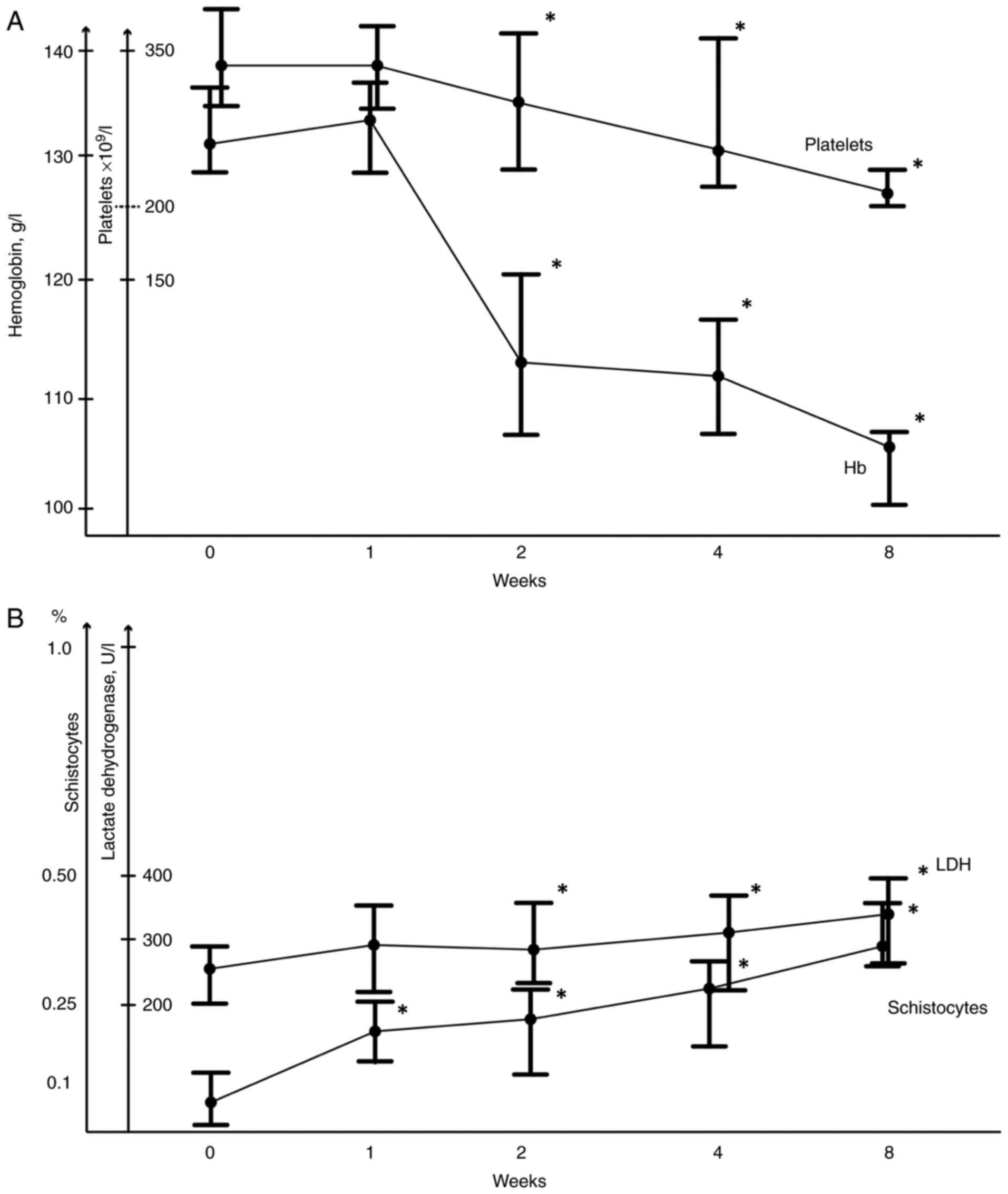
Fig. 1).
Anemia was detected in all 50 patients after 8 weeks
of treatment with antiangiogenic drugs, while a decrease in
hemoglobin <105 g/l was observed in 24 (48%) patients. The
median hemoglobin level in the entire sample of patients was 132
(129-136) g/l before treatment and 104 (99-107) g/l after 8 weeks
of treatment; a significant decrease in hemoglobin level was
observed after 2 weeks of therapy (Fig.
1A). In parallel with the decrease in hemoglobin, an increase
in the number of schistocytes was observed by the 8th week of
treatment, although the increase was ≤1%. There was also a slight
increase in LDH; 260 (202-296) U/l before the start of treatment
and 319 (259-390) U/l 8 weeks after the first injection of the
antiangiogenic drug. The changes became significant 2 weeks after
the start of therapy. The median platelet count in patients was 347
(292-429) x103/µl before the start of treatment and 256
(202-257) x103/µl 8 weeks after the first dose of the
antiangiogenic drug; that is, it decreased by 26% from the initial
levels (Fig. 1B).
Along with a decrease in GFR, an increase in urinary
biomarkers of renal damage was also observed. Associations were
revealed between the following factors: Hemoglobin and LDH levels
after 4 weeks of treatment, and hemoglobin and LDH with the number
of schistocytes after 8 weeks of treatment (Table II).
| Table IIAssociation between routine
laboratory parameters and urinary biomarkers after 8 weeks of
treatment. |
Table II
Association between routine
laboratory parameters and urinary biomarkers after 8 weeks of
treatment.
| Urinary
markers | Hemoglobin | Schistocytes | Platelets | Lactate
dehydrogenase | Urine Albumin | D-dimer |
|---|
| NGAL |
P=0.009b |
P=0.003b |
P<0.001c |
P=0.030a |
P=0.002c |
P=0.026a |
| | Rs=-0.367 | Rs=0.416 | Rs=-0.453 | Rs=0.306 | Rs=0.433 | Rs=0.423 |
| KIM-1 |
P=0.010b |
P=0.003b |
P<0.001c |
P=0.030a |
P=0.002c |
P=0.028a |
| | Rs=-0.360 | Rs=0.418 | Rs=-0.452 | Rs=0.304 | Rs=0.432 | Rs=0.414 |
| HIF-1A |
P=0.050a |
P=0.002b |
P<0.001c |
P=0.038a |
P=0.002c |
P=0.026a |
| | Rs=-0.367 | Rs=0.435 | Rs=-0.460 | Rs=0.294 | Rs=0.432 | Rs=0.421 |
| Nephrin |
P=0.008b |
P=0.003b |
P=0.001c |
P=0.030a |
P=0.002c |
P=0.024a |
| | Rs=-0.372 | Rs=0.418 | Rs=-0.448 | Rs=0.307 | Rs=0.435 | Rs=0.426 |
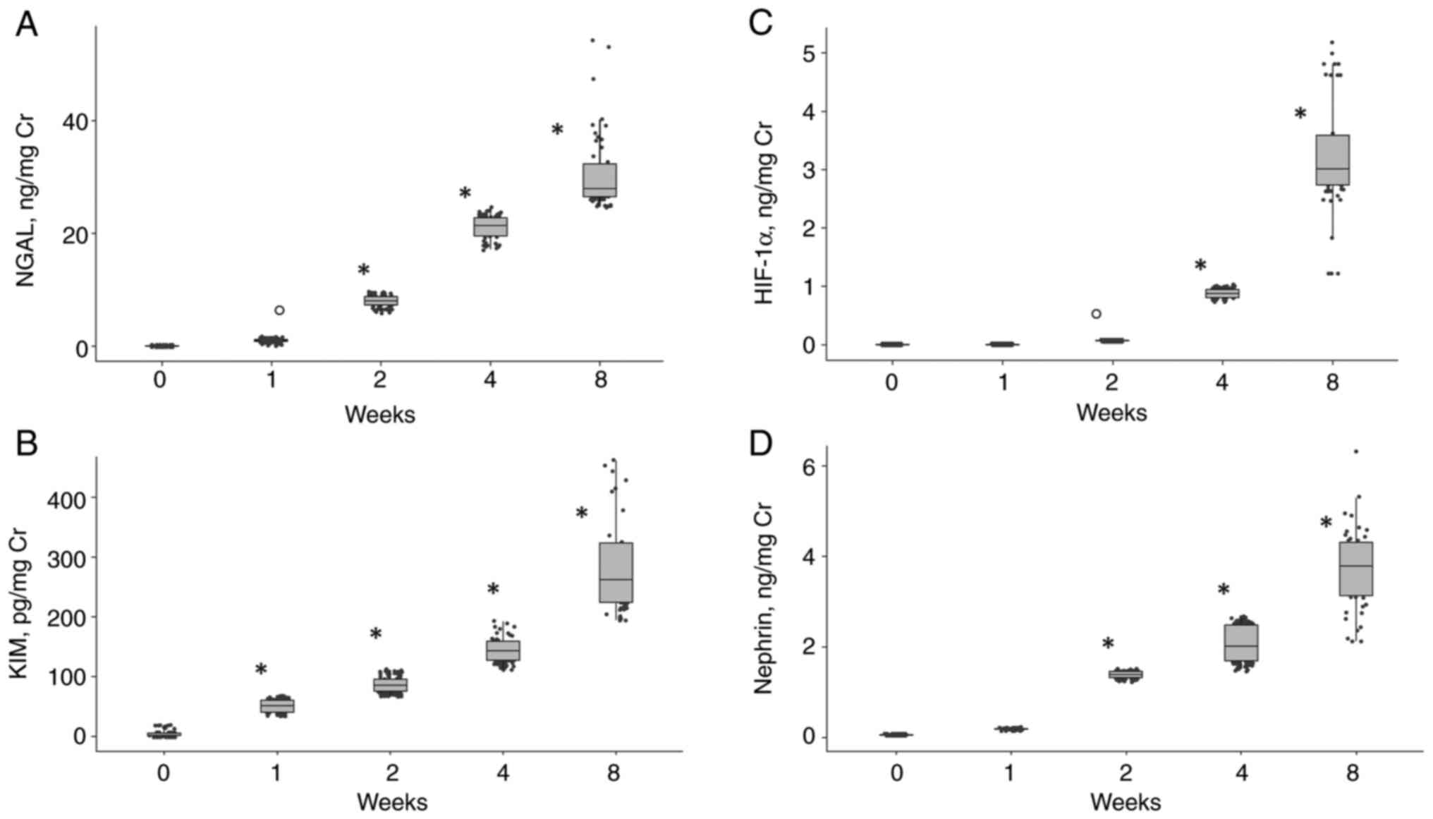
NGAL and KIM-1, HIF-1α and nephrin in
urine before and after 1, 2, 4 and 8 weeks of treatment
A significant increase in NGAL and KIM-1 levels in
the urine was noted after 1 week of therapy, and HIF-1α and nephrin
after 2 weeks (Fig. 2). A
significant change in the levels of LDH, the number of schistocytes
and the platelet counts were detected later (2-8 weeks) (Fig. 1).
An increase in biomarkers after 1 week of treatment
preceded a subsequent decrease in GFR. A high sensitivity and
specificity of the studied biomarkers compared to creatinine, urea
levels and GFR, assessed after a week of treatment, for predicting
the nephrotoxicity of antiangiogenic drugs was established by
receiver operating characteristic curve analysis (Table III).
| Table IIIReceiver operating characteristic
curve analyses of urinary biomarkers after 1 and 2 weeks. |
Table III
Receiver operating characteristic
curve analyses of urinary biomarkers after 1 and 2 weeks.
| A, After 1
week |
|---|
| Urinary
markers | Cut-off | Sensitivity
(%) | Specificity
(%) | PPV (%) | NPV (%) | AUC | Juden index |
|---|
| NGAL | 1.045 ng/mg Cr | 68.75 | 67.65 | 50.00 | 82.14 | 0.70 | 0.364 |
| KIM-1 | 54.07 pg/mg Cr | 68.75 | 61.76 | 45.83 | 80.77 | 0.69 | 0.305 |
| HIF-1α |
6.02x10⁻⁵ ng/mg Cr | 68.75 | 67.65 | 50.00 | 82.14 | 0.70 | 0.364 |
| Nephrin | 0.184 ng/mg Cr | 68.75 | 67.65 | 50.00 | 82.14 | 0.70 | 0.364 |
| B, After 2
weeks |
| Urinary
markers | Cut-off | Sensitivity
(%) | Specificity
(%) | PPV (%) | NPV (%) | AUC | Juden index |
| NGAL | 8.11 ng/mg Cr | 68.75 | 64.71 | 47.83 | 81.48 | 0.70 | 0.340 |
| KIM-1 | 80.0 pg/mg Cr | 69.57 | 48.15 | 53.33 | 85.50 | 0.60 | 0.180 |
| HIF-1α | 0.85 ng/mg Cr | 73.91 | 48.15 | 58.84 | 68.42 | 0.59 | 0.220 |
| Nephrin | 1.37 ng/mg Cr | 69.57 | 48.15 | 53.33 | 65.00 | 0.59 | 0.180 |
Risk factors of nephrotoxicity
associated with anti-VEGF drugs
Multivariate logistic regression analysis with the
inclusion of the following factors (age, sex, body mass index, the
presence of cardiovascular diseases (coronary artery disease,
myocardial infarction or stroke), presence of arterial
hypertension, presence of diabetes mellitus, smoking status, the
type of antiangiogenic drug, GFR rates before treatment, and
concentrations of NGAL, KIM-1, nephrin and HIF-1α in the urine
after 1 and 2 weeks from the start of therapy showed an initial
decrease in the eGFR to <80 ml/min/1.73 m2 [OR, 3.250
(1.060-9.967), P=0.039], which was associated with a history of
arterial hypertension [OR, 1.503 (1.135-1.990), P=0.013].
Additionally, the risk (OR) of nephrotoxicity of antiangiogenic
drugs was higher in patients with an increase in KIM-1 in urine
(odds ratio 1.1, 95% CI 1.02-1.183) after 1 week and with an
increase in HIF-1α in urine (odds ratio 5.7, 95% CI 3.601-8.949)
after 2 weeks of treatment (P<0.05; Table IV).
| Table IVLogistic regression model of
nephrotoxicity associated with anti-VEGF treatment. |
Table IV
Logistic regression model of
nephrotoxicity associated with anti-VEGF treatment.
| | CI 95% | | CI 95% | |
|---|
| Variable | OR unadjusted | Lower | Upper | P-value | OR adjusted | Lower | Upper | P-value |
|---|
| Age | 1.061 | 1.007 | 1.119 | 0.026a | 1.018 | 0.965 | 1.075 | 0.51 |
| Female sex | 4.875 | 0.875 | 24.15 | 0.071 | 2.631 | 0.157 | 44.123 | 0.501 |
| Arterial
hypertension | 3.5 | 1.405 | 31.314 | 0.042a | 1.503 | 1.135 | 1.99 | 0.013a |
| Diabetes
mellitus | 0.316 | 0.047 | 2.118 | 0.235 | 0.472 | 0.055 | 4.081 | 0.495 |
| Body mass
index | 1.009 | 0.892 | 1.14 | 0.892 | 1.016 | 0.846 | 1.22 | 0.864 |
| Cardiovascular
disease | 1.5 | 0.267 | 8.411 | 0.645 | 0.374 | 0.01 | 13.68 | 0.592 |
| Smoking status | 6.782 | 1.08 | 42.57 | 0.041a | 0.654 | 0.049 | 8.808 | 0.749 |
| Decrease in
baseline GFR | 7.474 | 1.033 | 59.856 | 0.048a | 3.25 | 1.06 | 9.967 | 0.039a |
| Type of anti-VEGF
drug | 0.974 | 0.385 | 2.464 | 0.955 | 0.35 | 0.082 | 1.493 | 0.156 |
| Type of cancer | 1.269 | 0.795 | 2.062 | 0.318 | 0.519 | 0.114 | 2.366 | 0.397 |
| KIM-1 (week 1) | 1.492 | 1.121 | 1.986 | 0.006b | 1.1 | 1.02 | 1.183 | 0.023a |
| NGAL (week 1) | 7.18 | 0.245 | 21.023 | 0.253 | 9.48 | 0.007 | 12.088 | 0.253 |
| HIF-1α (week
2) | 6.944 | 5.374 | 8.974 | 0.027a | 5.677 | 3.601 | 8.949 | 0.017a |
Outcomes after 9-months of
follow-up
After a 9-month follow-up, the anti-angiogenic
therapy was ended in all patients. A total of 22 (40%) patients
showed signs of nephrotoxicity; of these, 17 patients demonstrated
a decrease in GFR to <60 ml/min 1.73 m2 by the 8th
week of treatment (primary endpoint). Anti-VEGF therapy was
terminated in 14 (28%) patients due to its nephrotoxicity
(proteinuria or decreased eGFR), in 7 (14%) patients due to the
uncontrolled arterial hypertension, in 5 (10%) patients due to the
thrombosis, and in 1 (2%) patient due to the bleeding. In 16 (32%)
patients anti-VEGF therapy was terminated due to the progression of
underlying diseases, in 7 (14%) patients the reason for canceling
the drug remains unknown.
Discussion
Our study included patients treated with one of
three antiangiogenic drugs: Bevacizumab, aflibercept or
ramucirumab. An increase in blood pressure >130/80 mmHg during
the treatment was observed in half of the patients, higher than the
rates previously reported in the literature. For example, a
frequent (42.4%) cause of arterial hypertension is aflibercept;
other drugs show a lower frequency of arterial hypertension,
including ramucirumab (21%) and bevacizumab (23.6%), while severe
hypertension of 3-4 degrees was observed in 7.9% of patients. A
target level of <130/80 mmHg was more stringent than what was
used in other studies (29-31).
By the 8th week of treatment, a decrease in GFR to <60 ml/min in
42% of the examined patients was observed. According to the
logistic regression analysis, the degree of GFR reduction did not
significantly depend on the specific antiangiogenic drug. Despite
the fact that anti-VEGF-antiangiogenic drugs were prescribed to
patients with initially normal renal function, even a small
decrease in eGFR to <80 ml/min, turned out to be a risk factor
for nephrotoxicity. Another risk factor for nephrotoxicity of
anti-VEGF drugs was arterial hypertension before the initiation of
therapy and lack of sufficient correction during the treatment.
However, the achievement of a target pressure ≤130/80 mmHg was a
favorable factor for a stable GFR during treatment with anti-VEGF.
Normal D-dimer levels during the treatment were also beneficial in
maintaining GFR.
In the mechanisms of GFR reduction, endothelial
dysfunction due to blockade of the effects of VEGF with an increase
in vascular tone, a decrease in natriuresis, a regression of
fenestrated capillaries, and ultimately a decrease in intrarenal
blood flow have been reported (32-34).
Thrombotic microangiopathy (TMA) is a serious complication of
therapy. According to several studies evaluating morphological
changes with anti-VEGF drug treatment, TMA of the microvasculature
of the kidneys is most often observed when there is oedema of
endothelial cells with detachment from the basement membrane and
microthrombi in the vessels of the kidneys (35-37).
With prolonged use of anti-VEGF drugs, chronic irreversible
processes are revealed, including thickening of the vascular wall,
fibrous hyperplasia of the intima, arterio- and arteriolosclerosis
and organized blood clots with recanalization, which ultimately
ends with ischemic atrophy of the renal cortex (38). The pathogenesis of anemia is complex
in those patients who receive anti-VEGF drug treatment. Decreased
hemoglobin levels may be due to the concomitant use of anti-VEGF
drugs and other chemotherapeutic drugs with myelosuppressive
effects, such as irinotecan, fluoropyrimidines (5-fluorouracil,
capecitabine), paclitaxel and eribulin. However, we also noted
decreased hemoglobin levels in patients who received antiangiogenic
drugs as a monotherapy. We suggest the contribution of
microangiopathic hemolysis in the development of anemia, due to
excessive schistocytes, as well as increased LDH levels and a low
platelet count. Acute TMA with kidney damage manifests as a result
of microangiopathic hemolytic anemia, thrombocytopenia,
hypertension, moderate urinary syndrome and impaired renal function
(39).
In the present study, cases of acute TMA were not
observed; however, a gradual decrease in GFR was accompanied by a
decrease in hemoglobin and platelet count, as well as increases in
LDH levels, the number of schistocytes and the D-dimer levels,
which indicated the occurrence of endothelial dysfunction and
microangiopathic processes, and the development of microthrombosis
in the renal vessels. There was a significant increase in the
parameters of microangiopathic hemolysis by the 8th week of
treatment; however, in the early stages, a significant increase in
urine markers of hypoxia, tubular damage and podocytic damage were
recorded. It has been hypothesized that the TMA process appears to
be renally localized rather than part of a systemic process in
which thrombocytopenia, schistocytes on peripheral smear and
hemolytic anemia occur (5).
Currently, biomarkers of tubular injury have been
studied in acute toxic injury, for example, in treatment with
cisplatin (9-12).
Markers that predict damage when treated with anti-VEGF drugs have
not been studied. We assessed the levels of NGAL, KIM-1, HIF-1α and
nephrin in urine as factors that may reveal acute or chronic
hypoxic renal injury. In our study, the urinary NGAL levels were
significantly increased 1 week after the start of anti-VEGF therapy
and gradually increased over time, while the eGFR rate remained
normal. An early increase in urinary NGAL reflected tubular damage
and predicted further deterioration in renal function. KIM-1 levels
in urine were also evaluated, and this analysis revealed an early
increase in its levels after drug treatment, and this early
increase was predictive of a gradual deterioration in renal
function.
Considering the role of endothelial dysfunction and
thrombotic microangiopathy in the progression of renal damage
during treatment with antiangiogenic drugs, we assessed the levels
of HIF-1α in the urine. HIF-1α is a protein produced in the cell in
response to a decrease in oxygen concentration (40). Elevated serum HIF-1α levels are
detected in chronic kidney disease, reflecting a loss of
peritubular capillaries and renal tissue hypoxia (41,42).
In the present study, logistic regression analysis revealed that
HIF-1α was a factor involved in nephrotoxicity. The relationship
between HIF-1α indicators and the levels of markers of
microangiopathic hemolysis and GFR suggests the importance of
chronic ischemia/hypoxia in the development of renal dysfunction
during treatment with anti-VEGF drugs. Several experimental studies
on patients with chronic kidney disease have shown that in the
kidney, HIF-1α is the dominant form expressed in tubular epithelial
cells and it acts as the major regulator of hypoxic adaptation
(43-45).
A reno-protective role of HIF against ischemic injury in
ischemia/reperfusion models and toxic nephropathies have been
described. In these cases HIF induction protects tubular cells from
ischemic injury (46-48).
Hypoxia-inducible gene expression in primary renal proximal tubular
epithelial cells is almost completely blocked by inactivation of
HIF-1α, suggesting that their response to hypoxia is largely
dependent on HIF-1α (49). HIF-1α
has been shown to play a role in the pathogenesis of renal
interstitial fibrosis in patients with chronic kidney disease
(45).
Kimura et al (50) showed that hypoxia and the resultant
stabilization of HIF-1α play pivotal roles in the development of
tubulointerstitial fibrosis. HIF-1 induces expression of
profibrogenic genes, including tissue inhibitor of
metalloproteinase 1, connective tissue growth factor and
plasminogen activator inhibitor 1 (49,51,52).
Kidneys are one of the most vascularized organs, and
considerably susceptible to ischemia in patients who take anti-VEGF
drugs. Ischemia clinically manifests as arterial hypertension and
renal vascular disease with a decrease in GFR. Histologically,
there are signs of thrombotic microangiopathy (35-38).
Based on previous studies, it is hypothesized that the primary
source of HIF-1 α in the urine of patients who take anti-VEGF drugs
are renal tissue cells undergoing hypoxia due to the loss of
peritubular capillaries (53,54).
The increased expression of HIF-1α has been suggested to promote
the nephrotoxicity and the decrease in GFR through profibrotic
effects and inflammatory processes (55).
The occurrence of proteinuria during treatment with
anti-VEGF drugs is associated with impaired expression of nephrin,
a transmembrane podocytic protein that forms the basis of the gap
diaphragm and is regulated by VEGF. When anti-VEGF drugs are used,
nephrin molecules are cleaved from the podocyte, and the slit
diaphragm and the glomerular filter is destroyed (56,57).
Proteinuria ranks second amongst the most common side effects of
anti-VEGF drugs, but its values rarely exceed 2 g/day. Proteinuria
>3.5 g per day and nephrotic syndrome are detected on average in
6.5% of patients (58,59). In the present study, an increase in
nephrinuria was observed. However, by the 8th week of treatment,
there were no cases of high proteinuria or nephrotic syndrome.
According to logistic regression models, it was noted that arterial
hypertension, an early decline in GFR, and increased KIM-1 and
HIF-1α levels had a significant impact on the decrease in GFR
within the first 8 weeks of treatment. Age, sex, presence/absence
of CVD, smoking status, type of chemotherapy and type of tumor had
no significant impact on nephrotoxicity development.
The limitations of our study were the relatively
small cohort and the short follow-up period (8 weeks); the
limitations did not allow us to establish the frequency of a
pronounced decrease in GFR to <30 ml/min/1.73 m2,
cases of nephrotic syndrome or high proteinuria, which would
require discontinuation of the drug treatment. Additionally, the
levels of biomarkers in the early stage of kidney damage in
patients who took anti-VEGF drugs were defined by us. Further
studies are required to ascertain more informative values of urine
biomarkers and their concentration changes.
In conclusion, increased levels of NGAL, KIM-1,
HIF-1α and nephrin in the urine reflect the processes occurring
during renal tissue damage; these markers have high sensitivity and
specificity for predicting the nephrotoxicity of anti-VEGF drugs.
The independent risk factors for nephrotoxicity are a decrease in
the GFR and arterial hypertension before the start of therapy, as
well as an early increase in the concentrations of KIM-1 and HIF-1α
in the urine.
Acknowledgements
We would like to that Dorofeev A.S. (Junior
Researcher, Laboratory of Scientific and Diagnostic Research,
Loginov Moscow Clinical Scientific Center), who kindly helped to
perform ELISA.
Funding
Funding: The study was funded by the Loginov Moscow Clinical
Scientific Center (Moscow, Russia).
Availability of data and materials
The datasets used and/or analyzed during the present
study are available from the corresponding author on reasonable
request.
Authors' contributions
NC conceived and designed the study, and drafted the
manuscript. KG analyzed and interpretated the data, and was
involved in drafting the manuscript. VM analyzed data and drafted
the manuscript. LZ acquired and analyzed the data. TK participated
in data collection and revised the manuscript critically for
important intellectual content. KG and NC confirm the authenticity
of all the raw data. All authors have read and approved the final
manuscript.
Ethics approval and consent to
participate
The present study was approved by Local Ethics
Committee A.S. Loginov Moscow Clinical Scientific Center of Moscow
Healthcare Department (approval no. 2/2020, 17th Feb 2020). All
subjects provided informed written consent before participation,
and the study conformed with the guidelines described in the
Declaration of Helsinki.
Patient consent for publication
Not applicable.
Competing interests
The authors declare that they have no competing
interests.
References
|
1
|
Ferrara N, Gerber HP and LeCouter J: The
biology of VEGF and its receptors. Nat Med. 9:669–676.
2003.PubMed/NCBI View Article : Google Scholar
|
|
2
|
Zirlik K and Duyster J: Anti-Angiogenics:
Current situation and future perspectives. Oncol Res Treat.
41:166–171. 2018.PubMed/NCBI View Article : Google Scholar
|
|
3
|
Wang Z, Dabrosin C, Yin X, Fuster MM,
Arreola A, Rathmell WK, Generali D, Nagaraju GP, El-Rayes B,
Ribatti D, et al: Broad targeting of angiogenesis for cancer
prevention and therapy. Semin Cancer Biol. 35 (Suppl 1):S224–S243.
2015.PubMed/NCBI View Article : Google Scholar
|
|
4
|
Toriu N, Sekine A, Mizuno H, Hasegawa E,
Yamanouchi M, Hiramatsu R, Hayami N, Hoshino J, Kawada M, Suwabe T,
et al: Renal-Limited thrombotic microangiopathy due to bevacizumab
therapy for metastatic colorectal cancer: A case report. Case Rep
Oncol. 12:391–400. 2019.PubMed/NCBI View Article : Google Scholar
|
|
5
|
Hayman SR, Leung N, Grande JP and Garovic
VD: VEGF inhibition, hypertension, and renal toxicity. Curr Oncol
Rep. 14:285–294. 2012.PubMed/NCBI View Article : Google Scholar
|
|
6
|
Izzedine H, Escudier B, Lhomme C, Pautier
P, Rouvier P, Gueutin V, Baumelou A, Derosa L, Bahleda R,
Hollebecque A, et al: Kidney diseases associated with anti-vascular
endothelial growth factor (VEGF): An 8-year observational study at
a single center. Medicine (Baltimore). 93:333–339. 2014.PubMed/NCBI View Article : Google Scholar
|
|
7
|
Hanna RM, Tran NT, Patel SS, Hou J,
Jhaveri KD, Parikh R, Selamet U, Ghobry L, Wassef O, Barsoum M, et
al: Thrombotic microangiopathy and acute kidney injury induced
after intravitreal injection of vascular endothelial growth factor
inhibitors VEGF blockade-related TMA after intravitreal use. Front
Med (Lausanne). 7(579603)2020.PubMed/NCBI View Article : Google Scholar
|
|
8
|
Piscitani L, Sirolli V, Di Liberato L,
Morroni M and Bonomini M: Nephrotoxicity associated with novel
anticancer agents (Aflibercept, Dasatinib, Nivolumab): Case series
and nephrological considerations. Int J Mol Sci.
21(4878)2020.PubMed/NCBI View Article : Google Scholar
|
|
9
|
Morales E, Moliz C and Gutierrez E: Renal
damage associated to intravitreal administration of ranibizumab.
Nefrologia. 37:653–655. 2017.PubMed/NCBI View Article : Google Scholar : (In English,
Spanish).
|
|
10
|
Touyz RM, Herrmann SMS and Herrmann J:
Vascular toxicities with VEGF inhibitor therapies-focus on
hypertension and arterial thrombotic events. J Am Soc Hypertens.
12:409–425. 2018.PubMed/NCBI View Article : Google Scholar
|
|
11
|
Florova B, Rajdl D, Racek J, Fiala O,
Matejka VM and Trefil L: NGAL, albumin and cystatin C during
cisplatin therapy. Physiol Res. 69:307–317. 2020.PubMed/NCBI View Article : Google Scholar
|
|
12
|
Khawaja S, Jafri L, Siddiqui I, Hashmi M
and Ghani F: The utility of neutrophil gelatinase-associated
Lipocalin (NGAL) as a marker of acute kidney injury (AKI) in
critically ill patients. Biomark Res. 7(4)2019.PubMed/NCBI View Article : Google Scholar
|
|
13
|
Ghadrdan E, Ebrahimpour S, Sadighi S,
Chaibakhsh S and Jahangard-Rafsanjani Z: Evaluation of urinary
neutrophil gelatinase-associated lipocalin and urinary kidney
injury molecule-1 as biomarkers of renal function in cancer
patients treated with cisplatin. J Oncol Pharm Pract. 26:1643–1649.
2020.PubMed/NCBI View Article : Google Scholar
|
|
14
|
Tekce BK, Uyeturk U, Tekce H, Uyeturk U,
Aktas G and Akkaya A: Does the kidney injury molecule-1 predict
cisplatin-induced kidney injury in early stage? Ann Clin Biochem.
52:88–94. 2015.PubMed/NCBI View Article : Google Scholar
|
|
15
|
Cassidy H, Slyne J, Higgins M, Radford R,
Conlon PJ, Watson AJ, Ryan MP, McMorrow T and Slattery C:
Neutrophil gelatinase-associated lipocalin (NGAL) is localised to
the primary cilium in renal tubular epithelial cells-A novel source
of urinary biomarkers of renal injury. Biochim Biophys Acta Mol
Basis Dis. 1865(165532)2019.PubMed/NCBI View Article : Google Scholar
|
|
16
|
Mishra J, Mori K, Ma Q, Kelly C, Barasch J
and Devarajan P: Neutrophil gelatinase-associated lipocalin: A
novel urinary biomarker for cisplatin nephrotoxicity. Am J Nephrol.
24:307–315. 2004.PubMed/NCBI View Article : Google Scholar
|
|
17
|
Wagener G, Gubitosa G, Wang S, Borregaard
N, Kim M and Lee HT: Urinary neutrophil gelatinase-associated
lipocalin and acute kidney injury after cardiac surgery. Am J
Kidney Dis. 52:425–433. 2008.PubMed/NCBI View Article : Google Scholar
|
|
18
|
Han M, Li Y, Wen D, Liu M, Ma Y and Cong
B: NGAL protects against endotoxin-induced renal tubular cell
damage by suppressing apoptosis. BMC Nephrol.
19(168)2018.PubMed/NCBI View Article : Google Scholar
|
|
19
|
Castillo-Rodriguez E, Fernandez-Prado R,
Martin-Cleary C, Pizarro-Sánchez MS, Sanchez-Niño MD, Sanz AB,
Fernandez-Fernandez B and Ortiz A: Kidney injury marker 1 and
neutrophil gelatinase-associated lipocalin in chronic kidney
disease. Nephron. 136:263–267. 2017.PubMed/NCBI View Article : Google Scholar
|
|
20
|
Ichimura T, Brooks CR and Bonventre JV:
Kim-1/Tim-1 and immune cells: Shifting sands. Kidney Int.
81:809–811. 2012.PubMed/NCBI View Article : Google Scholar
|
|
21
|
Вailly V, Zhang Z, Meier W, Cate R,
Sanicola M and Bonventre JV: Shedding of kidney injury molecule-1,
a putative adhesion protein involved in renal regeneration. J Biol
Chem. 277:39739–39748. 2002.PubMed/NCBI View Article : Google Scholar
|
|
22
|
Vaidya VS, Ford GM, Waikar SS, Wang Y,
Clement MB, Ramirez V, Glaab WE, Troth SP, Sistare FD, Prozialeck
WC, et al: A rapid urine test for early detection of kidney injury.
Kidney Int. 76:108–114. 2009.PubMed/NCBI View Article : Google Scholar
|
|
23
|
Kramer AB, van Timmeren MM, Schuurs TA,
Vaidya VS, Bonventre JV, van Goor H and Navis G: Reduction of
proteinuria in adriamycin-induced nephropathy is associated with
reduction of renal kidney injury molecule (KIM-1) over time. Am J
Physiol Renal Physiol. 296:F1136–F1145. 2009.PubMed/NCBI View Article : Google Scholar
|
|
24
|
Van Timmeren MM, van den Heuvel MC, Bailly
V, Bakker SJ, van Goor H and Stegeman CA: Tubular kidney injury
molecule-1 (KIM-1) in human renal disease. J Pathol. 212:209–217.
2007.PubMed/NCBI View Article : Google Scholar
|
|
25
|
Bonventre JV: Kidney injury molecule-1
(KIM-1): A urinary biomarker and much more. Nephrol Dial
Transplant. 24:3265–3268. 2009.PubMed/NCBI View Article : Google Scholar
|
|
26
|
World Medical Association. World Medical
Association Declaration of Helsinki: Ethical principles for medical
research involving human subjects. JAMA. 310(20):2191–2194.
2013.PubMed/NCBI View Article : Google Scholar
|
|
27
|
Andrassy KM: Comment on KDIGO 2012
Clinical Practice Guideline for the Evaluation and Management of
Chronic Kidney Disease. Kidney Int. 84:622–623. 2013.PubMed/NCBI View Article : Google Scholar
|
|
28
|
KDIGO-Clinical Practice Guideline for
Acute Kidney Injury. Kidney International. 2012, Supplement 1.
Available from: https://kdigo.org/wp-content/uploads/2016/10/KDIGO-2012-AKI-Guideline-English.pdf.
|
|
29
|
Arnold D, Fuchs CS, Tabernero J, Ohtsu A,
Zhu AX, Garon EB, Mackey JR, Paz-Ares L, Baron AD, Okusaka T, et
al: Meta-analysis of individual patient safety data from six
randomized, placebo-controlled trials with the antiangiogenic
VEGFR2-binding monoclonal antibody ramucirumab. Ann Oncol.
28:2932–2942. 2017.PubMed/NCBI View Article : Google Scholar
|
|
30
|
Qi WX, Shen Z, Tang LN and Yao Y: Risk of
hypertension in cancer patients treated with aflibercept: A
systematic review and meta-analysis. Clin Drug Investig.
34:231–240. 2014.PubMed/NCBI View Article : Google Scholar
|
|
31
|
Vaidya VS, Ozer JS, Dieterle F, Collings
FB, Ramirez V, Troth S, Muniappa N, Thudium D, Gerhold D, Holder
DJ, et al: Kidney injury molecule-1 outperforms traditional
biomarkers of kidney injury in multi-site preclinical biomarker
qualification studies. Nat Biotechnol. 28:478–485. 2010.PubMed/NCBI View
Article : Google Scholar
|
|
32
|
Mourad JJ, des Guetz G, Debbabi H and Levy
BI: Blood pressure rise following angiogenesis inhibition by
bevacizumab. A crucial role for microcirculation. Ann Oncol.
19:927–934. 2008.PubMed/NCBI View Article : Google Scholar
|
|
33
|
Horowitz JR, Rivard A, van der Zee R,
Hariawala M, Sheriff DD, Esakof DD, Chaudhry GM, Symes JF and Isner
JM: Vascular endothelial growth factor/vascular permeability factor
produces nitric oxide-dependent hypotension. Evidence for a
maintenance role in quiescent adult endothelium. Arterioscler
Thromb Vasc Biol. 17:2793–2800. 1997.PubMed/NCBI View Article : Google Scholar
|
|
34
|
Steeghs N, Hovens MM, Rabelink AJ, Op 't
Roodt J, Matthys A, Christensen O and Gelderblom H: VEGFR2 blockade
in patients with solid tumors: Mechanism of hypertension and
effects on vascular function. J Clin Oncol. 24 (Suppl
18)(S3037)2006.
|
|
35
|
Bollée G, Patey N, Cazajous G, Robert C,
Goujon JM, Fakhouri F, Bruneval P, Noël LH and Knebelmann B:
Thrombotic microangiopathy secondary to VEGF pathway inhibition by
sunitinib. Nephrol Dial Transplant. 24:682–685. 2009.PubMed/NCBI View Article : Google Scholar
|
|
36
|
Izzedine H, Brocheriou I, Deray G and Rixe
O: Thrombotic microangiopathy and anti-VEGF agents. Nephrol Dial
Transplant. 22:1481–1482. 2007.PubMed/NCBI View Article : Google Scholar
|
|
37
|
Estrada CC, Maldonado A and Mallipattu SK:
Therapeutic inhibition of VEGF signaling and associated
nephrotoxicities. J Am Soc Nephrol. 30:187–200. 2019.PubMed/NCBI View Article : Google Scholar
|
|
38
|
Niu G and Chen X: Vascular endothelial
growth factor as an anti-angiogenic target for cancer therapy. Curr
Drug Targets. 11:1000–1017. 2010.PubMed/NCBI View Article : Google Scholar
|
|
39
|
Fujii T, Kawaasoe K, Tonooka A, Ohta A and
Nitta K: Nephrotic syndrome associated with ramucirumab therapy. A
single-center case series and literature review. Medicine
(Baltimore). 98(e16236)2019.PubMed/NCBI View Article : Google Scholar
|
|
40
|
Shu S, Wang Y, Zheng M, Liu Z, Cai J, Tang
C and Dong Z: Hypoxia and hypoxia-inducible factors in kidney
injury and repair. Cells. 8(207)2019.PubMed/NCBI View Article : Google Scholar
|
|
41
|
Ma C, Wei J, Zhan F, Wang R, Fu K, Wan X
and Li Z: Urinary hypoxia-inducible factor-1alpha levels are
associated with histologic chronicity changes and renal function in
patients with lupus nephritis. Yonsei Med J. 53:587–592.
2012.PubMed/NCBI View Article : Google Scholar
|
|
42
|
Liu J, Wei Q, Guo C, Dong G, Liu Y, Tang C
and Dong Z: Hypoxia, HIF, and associated signaling networks in
chronic kidney disease. Int J Mol Sci. 18(950)2017.PubMed/NCBI View Article : Google Scholar
|
|
43
|
Haase VH: Hypoxia-inducible factors in the
kidney. Am J Physiol Renal Physiol. 291:F271–F281. 2006.PubMed/NCBI View Article : Google Scholar
|
|
44
|
Rosenberger C, Mandriota S, Jurgensen JS,
Wiesener MS, Hörstrup JH, Frei U, Ratcliffe PJ, Maxwell PH,
Bachmann S and Eckardt KU: Expression of hypoxia-inducible
factor-1alpha and -2alpha in hypoxic and ischemic rat kidneys. J Am
Soc Nephrol. 13:1721–1732. 2002.PubMed/NCBI View Article : Google Scholar
|
|
45
|
Hung TW, Liou JH, Yeh KT, Tsai JP, Wu SW,
Tai HC, Kao WT, Lin SH, Cheng YW and Chang HR: Renal expression of
hypoxia inducible factor-1α in patients with chronic kidney
disease: A clinicopathologic study from nephrectomized kidneys.
Indian J Med Res. 137:102–110. 2013.PubMed/NCBI
|
|
46
|
Conde E, Alegre L, Blanco-Sánchez I,
Sáenz-Morales D, Aguado-Fraile E, Ponte B, Ramos E, Sáiz A, Jiménez
C, Ordoñez A, et al: Hypoxia inducible factor 1-Alpha (HIF-1 Alpha)
is induced during reperfusion after renal ischemia and is critical
for proximal tubule cell survival. PLoS One.
7(e33258)2012.PubMed/NCBI View Article : Google Scholar
|
|
47
|
Weidemann A, Bernhardt WM, Klanke B,
Daniel C, Buchholz B, Câmpean V, Amann K, Warnecke C, Wiesener MS,
Eckardt KU and Willam C: HIF activation protects from acute kidney
injury. J Am Soc Nephrol. 19:486–494. 2008.PubMed/NCBI View Article : Google Scholar
|
|
48
|
Bernhardt WM, Gottmann U, Doyon F,
Buchholz B, Campean V, Schödel J, Reisenbuechler A, Klaus S, Arend
M, Flippin L, et al: Donor treatment with a PHD-inhibitor
activating HIFs prevents graft injury and prolongs survival in an
allogenic kidney transplant model. Proc Natl Acad Sci USA.
106:21276–21281. 2009.PubMed/NCBI View Article : Google Scholar
|
|
49
|
Higgins DF, Biju MP, Akai Y, Wutz A,
Johnson RS and Haase VH: Hypoxic induction of Ctgf is directly
mediated by Hif-1. Am J Physiol Renal Physiol. 287:F1223–F1232.
2004.PubMed/NCBI View Article : Google Scholar
|
|
50
|
Kimura K, Iwano M, Higgins DF, Yamaguchi
Y, Nakatani K, Harada K, Kubo A, Akai Y, Rankin EB, Neilson EG, et
al: Stable expression of HIF-1alfa in tubular epithelial cells
promotesinterstitial fibrosis. Am J Physiol Renal Physiol.
295:F1023–F1029. 2008.PubMed/NCBI View Article : Google Scholar
|
|
51
|
Kietzmann T, Roth U and Jungermann K:
Induction of the plasminogen activator inhibitor-1 gene expression
by mild hypoxia via a hypoxia response element binding the
hypoxia-inducible factor-1 in rat hepatocytes. Blood. 94:4177–4185.
1999.PubMed/NCBI
|
|
52
|
Norman JT, Clark IM and Garcia PL: Hypoxia
promotes fibrogenesis in human renal fibroblasts. Kidney Int.
58:2351–2366. 2000.PubMed/NCBI View Article : Google Scholar
|
|
53
|
Haase VH: The VHL/HIF oxygen-sensing
pathway and its relevance to kidney disease. Kidney Int.
69:1302–1307. 2006.PubMed/NCBI View Article : Google Scholar
|
|
54
|
Kairaitis LK, Wang Y, Gassmann M, Tay YC
and Harris DC: HIF-1alpha expression follows microvascular loss in
advanced murine adriamycin nephrosis. Am J Physiol Renal Physiol.
288:F198–F206. 2005.PubMed/NCBI View Article : Google Scholar
|
|
55
|
Tanaka T, Matsumoto M, Inagi R, Miyata T,
Kojima I, Ohse T, Fujita T and Nangaku M: Induction of protective
genes by cobalt ameliorates tubulointerstitial injury in the
progressive Thy1 nephritis. Kidney Int. 68:2714–2725.
2005.PubMed/NCBI View Article : Google Scholar
|
|
56
|
Hauser PV, Collino F, Bussolati B and
Camussi G: Nephrin and endothelial injury. Curr Opin Nephrol
Hypertens. 18:3–8. 2009.PubMed/NCBI View Article : Google Scholar
|
|
57
|
Patrakka J and Tryggvason K: Nephrin-a
unique structural and signaling protein of the kidney filter.
Trends Mol Med. 13:396–403. 2007.PubMed/NCBI View Article : Google Scholar
|
|
58
|
Azad NS, Posadas EM, Kwitkowski VE,
Steinberg SM, Jain L, Annunziata CM, Minasian L, Sarosy G, Kotz HL,
Premkumar A, et al: Combination targeted therapy with sorafenib and
bevacizumab results in enhanced toxicity and antitumor activity. J
Clin Oncol. 26:3709–3714. 2008.PubMed/NCBI View Article : Google Scholar
|
|
59
|
Feldman DR, Baum MS, Ginsberg MS, Hassoun
H, Flombaum CD, Velasco S, Fischer P, Ronnen E, Ishill N, Patil S
and Motzer RJ: Phase I trial of bevacizumab plus escalated doses of
sunitinib in patients with metastatic renal cell carcinoma. J Clin
Oncol. 27:1432–1439. 2009.PubMed/NCBI View Article : Google Scholar
|