Introduction
Malignant mesothelioma (MM) is a highly aggressive
tumor originating from serous mesothelioma cells (1), which is characterized by cryptic
onset, difficult diagnosis, advanced stage upon discovery and short
survival period. In recent years, the number of individuals exposed
to asbestos chemical materials has increased, and the prevalence of
malignant mesothelioma is increasing year by year. According to
previous studies, the median survival of patients with malignant
mesothelioma after diagnosis is only 12-15 months (2,3).
Although the survival of patients can be prolonged by existing
chemotherapy drugs, the 5-year survival rate of patients with
intermediate and advanced mesothelioma remains <15% (4). Early screening and accurate diagnosis
are important means to prolong the survival of patients with
mesothelioma. Poorly differentiated epithelial mesothelioma is
often difficult to distinguish from cancer. Currently, using biopsy
specimens (obtained by puncture and endoscopic methods) to diagnose
mesothelioma is relatively common, but it is extremely difficult to
diagnose mesothelioma in specimens of serous effusion, especially
mesothelial cells in the lungs. It is difficult to differentiate
between adenocarcinoma cells and ovarian serous cancer cells, and
invasive testing is occasionally required. Therefore, in the
present study, the use of small traumatic serous fluid samples from
patients with mesothelioma was considered to improve the
cytological diagnosis accuracy of malignant mesothelioma through
FISH probe detection combined with immunohistochemical
staining.
Early studies have shown that numerous biochemical
molecular markers are involved in the occurrence and development of
tumors and can be used for early screening of tumors (5). Therefore, it is necessary to further
explore new and highly specific diagnostic markers in the
occurrence and development of malignant mesothelioma. According to
the latest diagnosis and treatment and clinical practice guidelines
for malignant mesothelioma in 2022(6), the deletion of BRCA1-associated
protein 1 (BAP1) and multiple tumor suppressor l (MTS1), more often
called CDKN2A gene, is regarded as the gold standard for molecular
pathological diagnosis of malignant mesothelioma, but only biopsy
specimens are mentioned for detection. At present, there are few
cytological methods for the detection of specimens using serous
cavity effusion.
The fragment of chromosome 9P21 is the 21.3 region
of the short arm of chromosome 9, and tumor suppressor genes in
this region mainly include CDKN2A and MTAP genotypes (7). CDKN2A is a cycle-dependent protease
inhibitor, which is closely associated to negative regulation of
the cell cycle (8). Previous
studies have verified the effectiveness of CDKN2A gene deletion,
and found that CDKN2A gene expression is variable and silenced in
the tumor epigenetic mechanism of basal cell carcinoma, breast,
non-small cell lung and colorectal cancer as well as other tumors
(9,10). In the present study, CDKN2A gene
FISH probe was used to detect serous cavity effusion in biopsy
specimens. A previous study revealed that homozygous deletion (HD)
occurs in 60% of malignant mesothelioma (11). Methylthioadenosine phosphorylase
(MTAP), belonging to the PNP/MTAP phosphorylase family, is a gene
with P16 telomeres of ~100 kb (12,13),
and its antibody localization is mainly in the cytoplasm. This
encoding enzyme plays a major role in saving adenine and methylene
during polyamine metabolism. Previous research has shown that the
expression of MTAP is closely related to the malignant
transformation of cells, and there are varying degrees of deletion
in various types of tumors (14). A
previous study has shown that MTAP and CDKN2A are both deleted in
malignant mesothelioma, and ddPCR detection of both genes can
distinguish mesothelioma from benign mesothelioma (15). Cigognetti et al (16) determined that in pancreatic cancer,
the combined deletion of MTAP and CDKN2A protein expression is
considered as a substitute marker for CDKN2A homologous deletion.
BAP1 is a histone deubiquitination enzyme encoded by genes. BAP1 is
an important tumor suppressor gene encoded in the 21.1 region of
the short arm of chromosome 3(17)
and can significantly increase the stability of the KLF5 protein.
Righi et al (18)
established that the most commonly mutated genes in malignant
mesothelioma genome research results are BAP1, NF2 and CDKN2A/B.
BAP1 deletion promotes cell proliferation by upregulating enhancer
of zeste 2 polycomb repressive complex 2 subunit (EZH2). EZH2, a
histone lysine-methyltransferase, is overexpressed in numerous
cancers. A previous study has shown that BAP1 accelerates the
G(1)-S checkpoint process by
influencing the cell cycle and induces cell death through processes
characterized by apoptosis and necrosis (19).
In the present study, FISH was used to detect serous
fluid samples with evident advantages for the diagnosis of
mesothelioma. In addition, MTAP and BAP1 protein expression
combined with FISH 9P21 gene expression were used to distinguish
malignant mesothelioma from reactive mesothelioma, laying the
foundation for a diagnostic method with low trauma and high
sensitivity for malignant mesothelioma.
Materials and methods
Clinicopathological data
The pathological specimens of all enrolled patients
were collected, and questionnaires were issued to obtain
information concerning the general situation, nature of work,
educational level, and personal hygiene habits of the patients. The
present study was approved (ethical approval document no.
AF/SC-08/02.0.) by the Ethics Committee of Cangzhou People's
Hospital (Cangzhou, China). Following screening, 70 patients with
thoracic and peritoneal diseases treated at Cangzhou People's
Hospital from June 2017 to June 2020 were selected. The
pathological diagnosis met the diagnostic criteria of clinical
guidelines for diagnosis and treatment of malignant mesothelioma.
The inclusion criteria were as follows: i) Age range, 18-80 years
old; ii) pathological type, patients with a clear pathological
diagnosis of thoracic and peritoneal mesothelioma (epithelial);
iii) informed consent obtained from patients and their families as
well as follow-up with patients; and iv) following detection of
D2-40, WT1 as well as other markers, the specimen was confirmed as
mesothelioma in the official pathological report. The exclusion
criteria were as follows: i) Patients with incomplete pathological
data and lost to follow-up; ii) patients without hereditary
diseases in their family history medical records; and iii) patients
with contaminated specimens caused by improper specimen handling
(insufficient specimen fixation, antibody cross-binding, etc.) in
the experiment.
Reagents and experimental
equipment
BAP1, a mouse monoclonal antibody (cat. no.
sc-28383) directed against amino acids was obtained from Santa Cruz
Biotechnology, Inc. The dilution performed for immunohistochemistry
(paraffin-embedded sections) was 1:200. MTAP, a rabbit monoclonal
antibody (product code ab126770; dilution, 1:200) was obtained from
Abcam. CDKN2A(P16) gene deletion FISH probe reagent (in situ
hybridization) (cat. no. FP-032) was obtained from Wuhan HealthCare
Biotechnology Co., Ltd. Red fluorescein was used to label the P16
probe, and green fluorescein was used to label the CEP9 probe. P16
and CEP9 probes can be combined to the target detection site
through in situ hybridization.
Research methods
The expression of BAP1 and MTAP proteins in serous
effusion were detected by FISH probe of 9P21 gene and
immunohistochemistry. The enrolled patients were retrospectively
analyzed by diagnostic test evaluation method. The patients meeting
the inclusion and exclusion criteria were assessed by diagnostic
testing, and the enrolled patients were assessed by FISH in
situ hybridization. The test results were recorded. The cases
that did not meet inclusion criteria were removed from the study,
and the cases were re-numbered randomly by double-blind method. A
new round of diagnostic tests were conducted, in which four
pathologists with senior professional titles diagnosed the
pathophysiology of each case, and then all the cases were assessed
by immunohistochemical staining of BAP1 and MTAP, and the
sensitivity and specificity of the new combined diagnosis method
were recorded to evaluate the efficacy of the diagnostic tests. The
improved paraffin sectioning method of pleural effusion was as
follows: No less than 60 ml of serous cavity fluid with natural
settlement over 0.5 h was used. The serous cavity fluid samples
were placed in 6 centrifuge tubes, and centrifuged 3 times
(depending on the size of the centrifuged precipitated cells) at
room temperature (15-25˚C). The first round of centrifugation was
performed at 694 x g for 5 min, followed by smearing and the
supernatant was discarded. At the second centrifugation, several
drops of protein, glycerin and 90% ethanol were added to
resuscitate the precipitate. The precipitate was centrifuged again
at 694 x g for 5 min. The third centrifugation was the same as the
second centrifugation. Following centrifugation, the supernatant
was discarded, cell masses were removed, fixed, dehydrated,
embedded and sectioned. A FISH probe was used to detect 9P21 gene
expression, and the processed paraffin sections were placed in the
hybridization apparatus for denaturation, washing and drying.
4'6-Diamidine-2-phenylindole (DAPI) was added, observed under a
fluorescence microscope, and the results were recorded. FISH
fluorescent sections were stored at -20˚C, and protected from
light. Paraffin sections of mesothelioma tissues and benign
mesothelioma tissues were dewaxed, hydrated with alcohol of
different concentrations, then incubated with 3%
H2O2 at room temperature (15-25˚C) for 10
min, immersed and rinsed with PBS solution, and then incubated with
antibody reagent drops at 37˚C for 60 min. Reagents on tissues were
rinsed 3 times with PBS solution. DAB chromogenic solution was
added for incubation, and hematoxylin was finally applied. For
immunohistochemistry, tissue sections with a thickness of 4 µm were
baked in a 70˚C toaster for 2 h, and then removed and cooled to
room temperature. The paraffin sections were then placed into two
xylene sample bottles successively and each bottle was soaked for
30 min. The samples were then placed into two absolute ethanol
sample bottles successively, and each bottle was soaked for 10 min.
The sections were then placed in sample bottles containing 95, 85
and 75% ethanol successively, and each bottle was soaked for 5 min.
The sections were then rinsed with tap water for 2 min. Citric acid
buffer was added to the pressure cooker for antigen repair, the
slices were added and soaked, heated for 90-180 sec, removed from
the pressure cooker, and washed 3 times for 5 min each time with
PBS buffer. Subsequently, 50 µl endogenous peroxidase blocker was
added to the sections, followed by incubation at room temperature
for 30 min, and washing 3 times for 5 min each time with PBS. A
total of drops of goat serum blocking solution were added to each
section and then incubated in an incubator at 37˚C for 30 min.
After removal of the blocking solution at room temperature, BAP1
and MTAP antibody reagents (cat. no. sc-28383 and product code
ab126770, respectively; as aforementioned) were added (50 drops of
antibody reagents per section). The slices were then placed in a
refrigerator at 4˚C overnight (12-18 h), removed from the wet box,
returned to room temperature, and washed three times with PBS
buffer for 5 min each time. After removal of the PBS buffer, 50
drops of enzyme-labeled goat anti-mouse/rabbit HRP IgG polymer
(product codes ab97040 and ab7090, respectively; Abcam) were added
to each section and incubated for 30 min in a 37˚C incubator.
Subsequently, the sections were washed 3 times with PBS buffer for
5 min each time. A total of 50 drops of newly prepared DAB color
developer was added to each microliter slice. and color was
developed at room temperature for 3-7 min. Following color
development and rinsing with tap water, hematoxylin was used for
staining at room temperature for 5-8 min, and then the sections
were washed again with tap water for 2 min. After differentiation
with hydrochloric acid and alcohol, the sections were placed in
flowing tap water for 30 min. Subsequently, the sections were
dehydrated in 75, 85 and 95% gradient alcohol sample bottles for 3
min, and dehydrated in anhydrous alcohol sample bottles, 2 times
for 5 min each time. The sections were then rendered transparent
using newly configured xylene for 30 min, and finally sealed with
neutral gum.
Interpretation criteria
9P21 gene FISH probe detection results showed that
the CDKN2A locus signal was red, and the CEP9 locus signal was
green; therefore, 2 red:2 green were considered negative cells, 1
red:2 green or 2 green were considered positive cells. When the
positive cells were >15% it indicated that CDKN2A expression was
missing, and malignant mesothelioma in the experimental results
could be diagnosed. BAP1 antibody is positive in the nucleus, and
the positive expression is brown under the microscope. The
expression of BAP1 in malignant mesothelioma was mostly negative.
The positive control for the MTAP antibody is lung adenocarcinoma,
localized in the cytoplasm. Positive staining is brown, and
malignant mesothelioma is mostly positive. The results were
evaluated according to the depth of staining. Any discrepancies
were verified by both observers until a consensus was reached. The
expression positivity was graded and counted as follows: 0,
negative; 1, 1-50%; 2, 51-74%; 3, ≥75%. The staining intensity
score was graded as follows: 1, weak; 2, intermediate; 3, strong.
The scores for BAP1 and MTAP expression positivity and staining
intensity were multiplied to obtain a final score categorized as:
(-), 0; (+), 1-2; (++), 3-5; and (+++), 6-9. A score of 0-3 was
considered negative and 4-9 as positive.
Statistical analysis
Statistical software SPSS 21.0 (IBM Corp.) was used
for statistical analysis of data. Association analysis was used to
detect the association between BAP1 and MTAP protein expression
levels and clinicopathological features of mesothelioma. The
χ2 test was used to assess the association between BAP1
and MTAP protein expression and clinicopathological characteristics
of mesothelioma. P<0.05 was considered to indicate a
statistically significant difference. Kaplan-Meier plotter with
log-rank testing was used to analyze the association between the
expression levels of MTAP and BAP1 in mesothelioma tissues and the
overall survival (OS) of patients.
Results
Protein expression and diagnostic
results
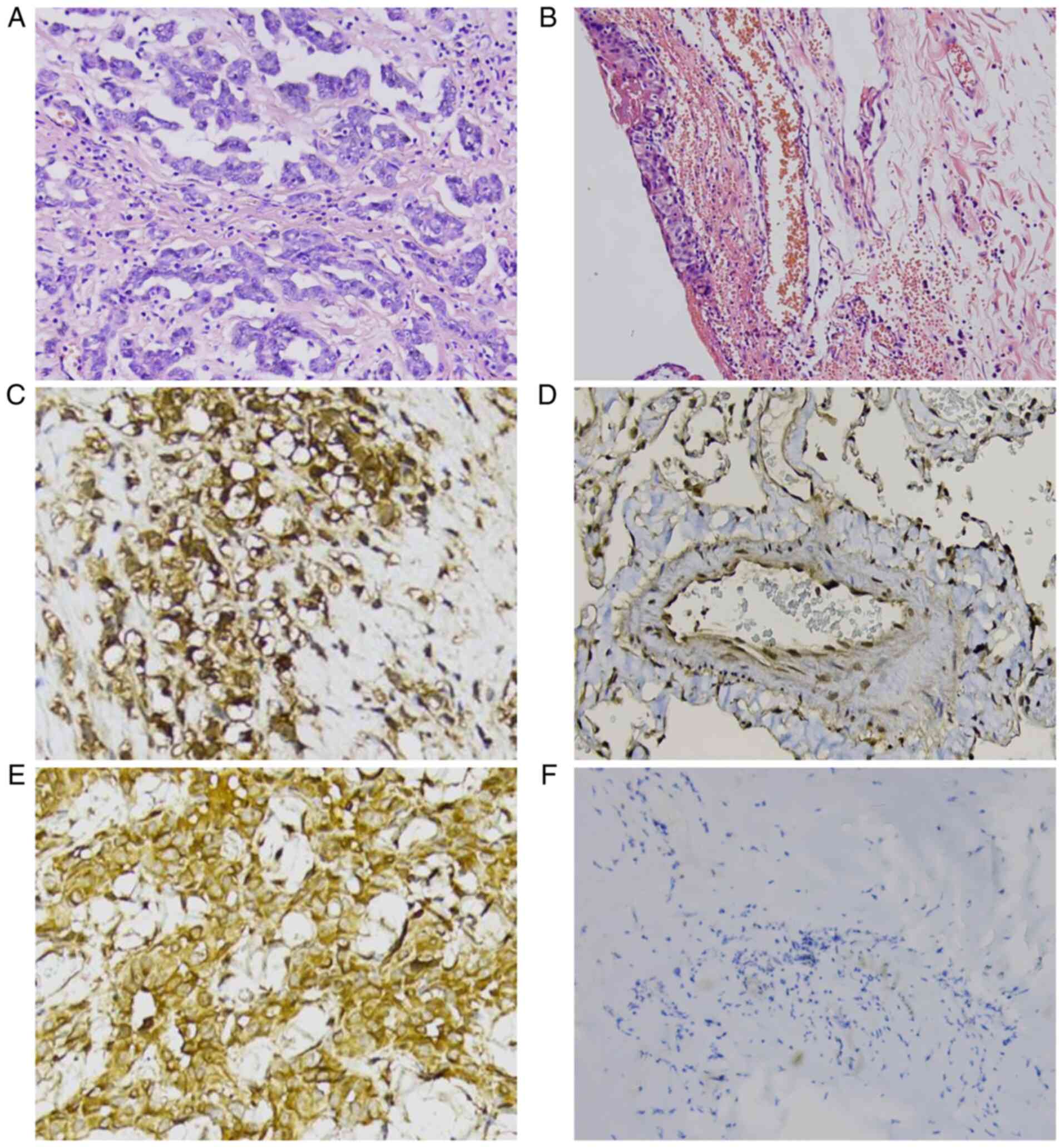
The experimental results demonstrated that the
CDKN2A gene on the 9P21 gene fragment was lost in malignant
mesothelioma, BAP1 protein expression level in malignant
mesothelioma was lower than that in normal serous tissue, and MTAP
gene protein expression level in malignant mesothelioma was higher
than that in normal serous tissue. In biopsy specimens, the
positive rates of CDKN2A FISH, BAP1 and MTAP were 98.00, 94.00 and
90.00%, respectively (Fig. 1A-F and
Table I), with specificity of
96.00, 85.71 and 77.27%, and sensitivity of 90.00, 95.92 and
93.75%. The positive rate of the combined test of biopsy specimens
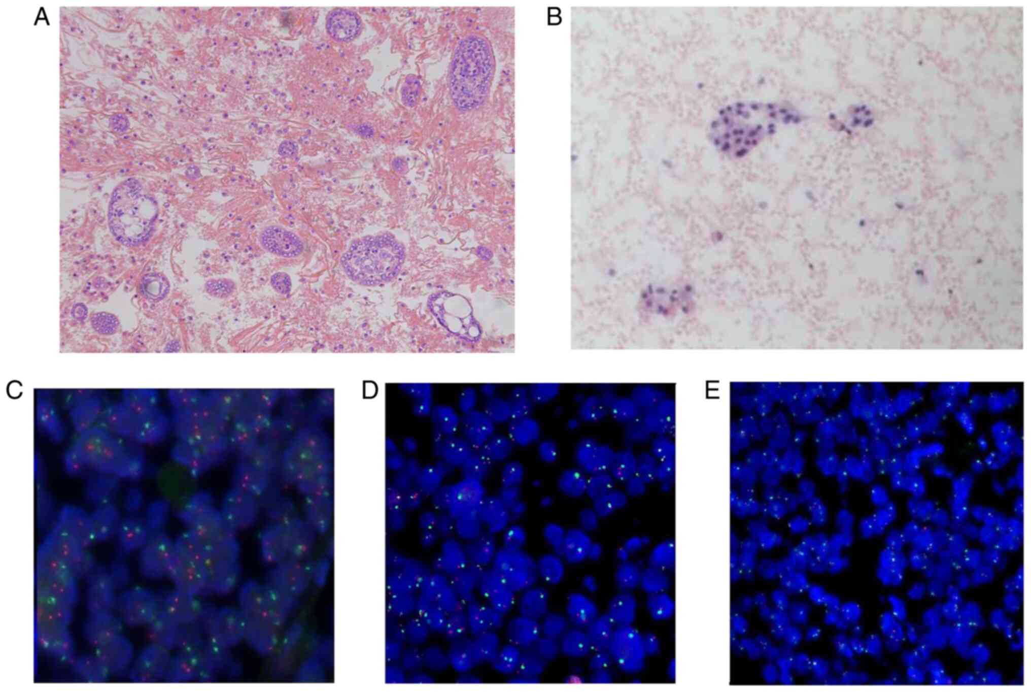
was 93.33% (Table I). The positive
rates of CDKN2A FISH, BAP1 and MTAP in serous fluid samples were
96.00, 90.00 and 88.00% respectively (Fig. 2A and B and Table
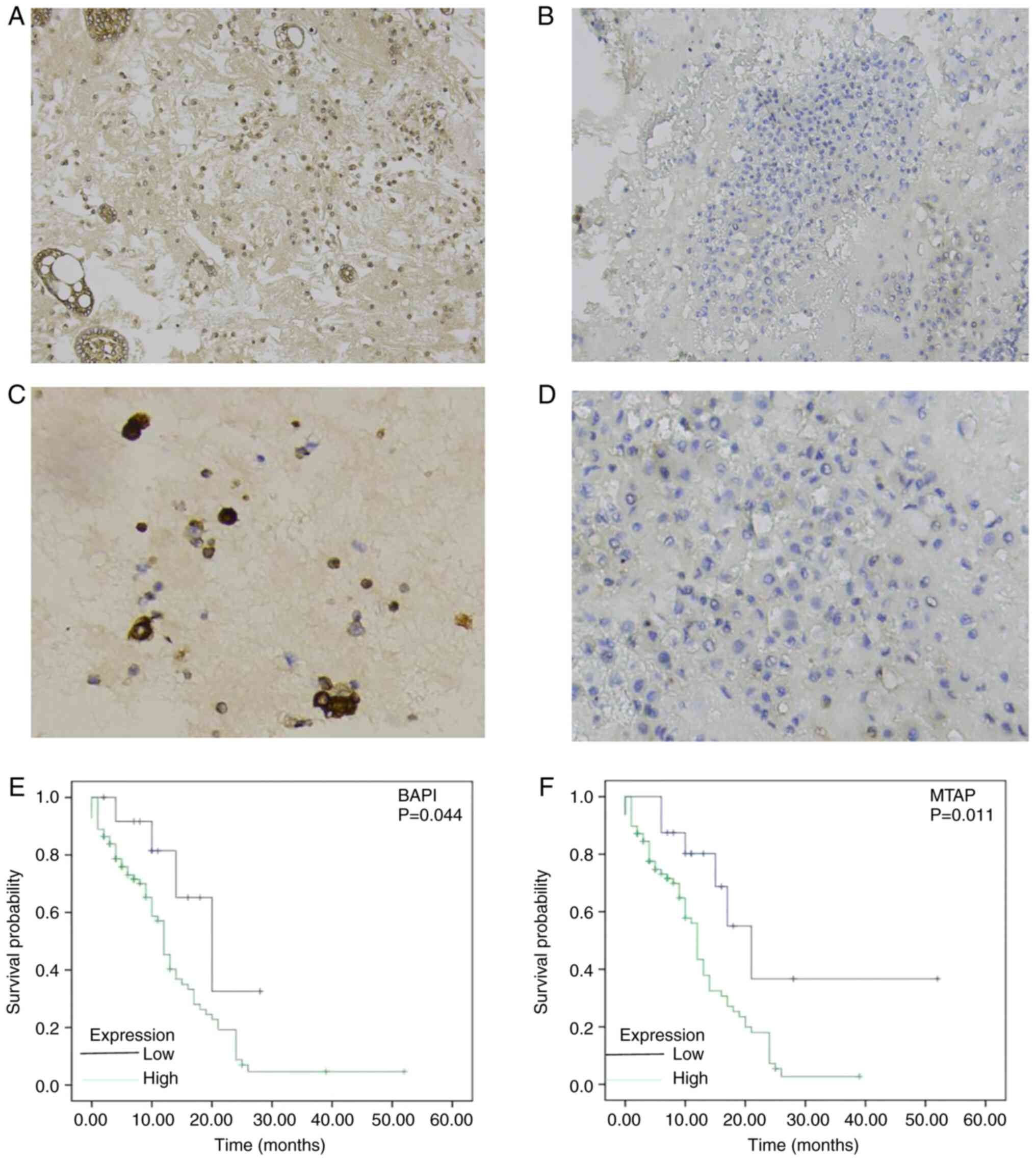
II), with specificity of 90.00, 77.27 and 71.43% (Fig. 2C-E and Table II). The sensitivity was 96.00,
93.75 and 89.80%, respectively (Fig.
3A-D and Table II). The
positive rate of serous cavity effusion specimens diagnosed using
the combination of the three methods (CDKN2A by FISH probe, and
BAP1 and MTAP by immunohistochemistry) was 91.33% (Table II). The number of gene copies
[CDKN2A (FISH) fluorescence signals] is presented in Table III. By comparing the diagnosis of
malignant mesothelioma with the combination of serous fluid
specimens and biopsy specimens, there was a high consistency. The
positive rate of the combination of serous fluid specimens and
biopsy specimens was 91.33 and 93.33%, respectively. The positive
rate of FISH detection of serous fluid in specimens was also highly
consistent with that of biopsy specimens. Comprehensive analysis
revealed that this new combined diagnosis has high clinical
application value, as well as high sensitivity and specificity.
 | Table IBiopsy specimen pathological
morphology combined with immunohistochemical staining of BAP1 and
MTAP, and FISH diagnosis results. |
Table I
Biopsy specimen pathological
morphology combined with immunohistochemical staining of BAP1 and
MTAP, and FISH diagnosis results.
| | Malignant
mesothelioma | Benign mesenchymal
tissue | |
|---|
| Detection method | Negative | Positive | Negative | Positive | Positive rate
(%) | Specificity (%) | Sensitivity (%) |
| CDKN2A | 2 | 48 | 18 | 2 | 98.00 | 96.00 | 90.00 |
| BAP1 | 47 | 3 | 2 | 18 | 94.00 | 85.71 | 95.92 |
| MTAP | 5 | 45 | 17 | 3 | 90.00 | 77.27 | 93.75 |
| Table IIResults from the diagnosis of serous
effusion samples detected by FISH and immunohistochemical staining
of BAP1 and MTAP. |
Table II
Results from the diagnosis of serous
effusion samples detected by FISH and immunohistochemical staining
of BAP1 and MTAP.
| | Malignant
mesothelioma | Benign mesenchymal
tissue | |
|---|
| Detection
method | Negative | Positive | Negative | Positive | Positive rate
(%) | Specificity
(%) | Sensitivity
(%) |
|---|
| CDKN2A | 2 | 48 | 18 | 2 | 96.00 | 90.00 | 96.00 |
| BAP1 | 45 | 5 | 3 | 17 | 90.00 | 77.27 | 93.75 |
| MTAP | 6 | 44 | 15 | 5 | 88.00 | 71.43 | 89.80 |
| Table IIIResults of CDKN2A (FISH) fluorescence
signals. |
Table III
Results of CDKN2A (FISH) fluorescence
signals.
| Specimen type | Signal position
point | Malignant
mesothelioma | Benign mesenchymal
tissue | Total |
|---|
| Biopsy
specimens | 1 red:2 green
(+) | 31 | 2 | 33 |
| | 2 green (+) | 17 | 0 | 17 |
| | 2 red:2 green
(-) | 2 | 18 | 20 |
| Samples of serous
cavity effusion | 1 red:2 green
(+) | 26 | 1 | 27 |
| | 2 green (+) | 22 | 1 | 23 |
| | 2 red:2 green
(-) | 2 | 18 | 20 |
Analysis of clinicopathological
features and survival prognosis
According to the statistical analysis of BAP1 and
MTAP protein expression and clinicopathological data, the
development and prognosis of malignant mesothelioma were positively
associated with asbestos chemical exposure history (P<0.05), and
had no association with sex, age, lesion site and Ki67 expression
(Tables IV and V). The results of these three detection
methods (CDKN2A by FISH probe, and BAP1 and MTAP by
immunohistochemistry) in biopsy specimens and effusion specimens
were all associated (P<0.01; Table
VI), which confirmed that the detection results of the biopsy
specimens and effusion specimens had high consistency, and provided
guidance and basis for clinical diagnosis. The survival of patients
with malignant mesothelioma was observed. Kaplan-Meier plotter was
used to analyze the association between the expression levels of
MTAP and BAP1 in mesothelioma tissues and the OS of patients, and
it was determined that BAP1 and MTAP genes were closely associated
to the prognosis of patients with mesothelioma. The higher the
expression level of MTAP gene, the worse the survival prognosis of
mesothelioma patients (P<0.05). Similarly, when BAP1 gene was
highly expressed, the survival prognosis of mesothelioma patients
was poor (P<0.05; Fig. 3E and
F).
| Table IVBAP1 expression and
clinicopathological features of patients with malignant
mesothelioma. |
Table IV
BAP1 expression and
clinicopathological features of patients with malignant
mesothelioma.
| | Expression of
BAP1 |
|---|
| Clinicopathological
features | No. of
patients | - | + | PR (%) | P-value |
|---|
| Sex | | | | | |
|
Male | 22 | 2 | 20 | 90.91 | 0.639 |
|
Female | 28 | 3 | 25 | 89.29 | |
| Age (years) | | | | | |
|
<60 | 12 | 4 | 8 | 66.67 | 0.816 |
|
≥60 | 38 | 1 | 37 | 97.37 | |
| Asbestos exposure
history | | | | | |
|
Yes | 26 | 1 | 25 | 96.15 |
<0.01 |
|
No | 24 | 4 | 20 | 83.33 | |
| Pathological
changes | | | | | |
|
Pleural | 17 | 2 | 15 | 88.24 | 0.980 |
|
Peritoneal | 25 | 2 | 23 | 92.00 | |
|
Other
(greater omentum, ovary, appendix, etc.) | 8 | 1 | 7 | 87.50 | |
| Ki67 | | | | | |
|
Negative | 11 | 3 | 8 | 72.73 | 0.356 |
|
Positive | 39 | 2 | 37 | 94.87 | |
| Table VMTAP expression and
clinicopathological features of patients with malignant
mesothelioma. |
Table V
MTAP expression and
clinicopathological features of patients with malignant
mesothelioma.
| | Expression of
BAP1 |
|---|
| Clinicopathological
features | No. of
patients | - | + | PR (%) | P-value |
|---|
| Sex | | | | | |
|
Male | 22 | 2 | 20 | 90.91 | 0.10 |
|
Female | 28 | 4 | 24 | 85.71 | |
| Age (years) | | | | | |
|
<60 | 12 | 4 | 8 | 66.67 | 0.927 |
|
≥60 | 38 | 2 | 36 | 94.74 | |
| Asbestos exposure
history | | | | | |
|
Yes | 26 | 1 | 25 | 96.15 |
<0.01 |
|
No | 24 | 5 | 19 | 79.17 | |
| Pathological
changes | | | | | |
|
Pleural | 17 | 2 | 15 | 88.24 | 0.831 |
|
Peritoneal | 25 | 3 | 22 | 88.00 | |
|
Other
(greater omentum, ovary, appendix, etc.) | 8 | 1 | 7 | 87.50 | |
| Ki67 | | | | | |
|
Negative | 11 | 4 | 7 | 63.64 | 0.461 |
|
Positive | 39 | 2 | 37 | 94.87 | |
| Table VIAssociation of the expression of
CDKN2A, BAP1 and MTAP genes in pathological biopsies and effusion
specimens of patients with malignant mesothelioma. |
Table VI
Association of the expression of
CDKN2A, BAP1 and MTAP genes in pathological biopsies and effusion
specimens of patients with malignant mesothelioma.
| | Sample of serous
cavity effusion | |
|---|
| Measurements | | Positive | Negative | No. of cases |
χ2/Fisher | P-value |
|---|
| Biopsy
specimen | Positive | 46 | 2 | 48 | 35.280 | 0.001 |
| (FISH) | Negative | 2 | 0 | 2 | | |
| Biopsy
specimen | Positive | 44 | 3 | 47 | 106.000 | 0.001 |
| (BAP1) | Negative | 1 | 2 | 3 | | |
| Biopsy
specimen | Positive | 43 | 4 | 47 | 92.462 | 0.001 |
| (MTAP) | Negative | 3 | 2 | 5 | | |
Discussion
Malignant mesothelioma is one of the most common
fatal primary pleural tumors, and the degree of disease is closely
related to asbestos exposure (20,21).
Mesothelioma is difficult to distinguish from reactive mesothelioma
(RMH), especially in cytology, with pleural mesothelioma having the
highest incidence (81%) and the worst prognosis. The mesothelioma
mediators can also occur in other membranous structures, including
the peritoneum (9%), pericardium and testicular sheath (22). WHO divides malignant mesothelioma
into epithelioid (the most common), sarcomatoid and biphasic. The
sensitivity of the cytological diagnosis commonly used in clinic is
only 30-75% (23). Since
mesothelioma cells are difficult to distinguish from the
degenerative or proliferative mesothelioma mesenchymal cells, most
patients with serous effusion of malignant mesothelioma are already
in an advanced stage at the time of diagnosis. FISH probe combined
with an immunohistochemical technique can easily identify these
cells. Moreover, cell slices also have disadvantages such as
overlapping cell blocks, a high false positive rate, and easy
removal of slices. The present improved approach addresses some of
these deficiencies. The serous cavity effusion specimens were
centrifuged and then paraffin-embedded into sections. The obtained
cells were smooth and clearly stained, which provided more accuracy
and the diagnostic results were easily identified. This combined
method provides an important basis for the accurate cytological
diagnosis of malignant mesothelioma. At present, the clear
diagnosis of mesothelioma is mostly obtained by endoscopic surgery
or puncture specimens, which are difficult to obtain and harmful to
patients. The sample of serous cavity effusion is easy to obtain
through FISH, it does little harm to the patient, and it can reduce
the clinical symptoms of the patient after extracting the effusion,
which has evident advantages. Therefore, detection of serous fluid
by FISH is a novel method, which has higher sensitivity,
specificity and accuracy than clinical methods.
The CDKN2A gene is 8.5 kb in length and encodes 148
amino acids. It is a cell cycle-dependent protease inhibitor, which
is closely related to negative regulation of the cell cycle. If the
CDKN2A gene is mutated or lost, the inhibition of the cyclin D-CDK4
complex will be relieved, CDKN2A will function as a tumor
suppressor gene, and an abnormal cell cycle will occur. Cells will
acquire infinite proliferation (24). CDKN2A has been confirmed to be
negatively correlated with BAP1 gene expression in colorectal
cancer and non-small cell lung cancer (25). FISH probes detected homozygous
deletion of CDKN2A gene in up to 80% of mesotheliomas, but not in
reactive mesotheliomas (26). The
results of the present study revealed that 9P21 gene was lost in
malignant mesothelioma, with a positive rate and specificity
>80% in biopsy specimens and serous effusion specimens. Chapel
et al (27) determined that
MTAP expression is deficient in non-small cell lung cancer and has
a role in transcriptional expression change. MTAP is highly
expressed in breast cancer, liver cancer, multiple myeloma as well
as other cancer cells. A previous study revealed that the
expression of MTAP gene in hepatocellular carcinoma cells was
decreased after treatment with pathway inhibitors (28). In contrast, in a primary
hepatocellular carcinoma model, the expression levels of VEGF and
microvessel density in rats decreased after treatment with growth
inhibitors, while tumor growth was also inhibited (29). However, there is no clear study on
malignant mesothelioma. The present study also found that the
expression level of MTAP in malignant mesothelioma was higher than
that in normal serous tissue, and the high expression of MTAP was
closely associated with the poor prognosis of mesothelioma
(P<0.05).
BAP1 also mediates deubiquitination and nuclear
localization. BAP1 mutations are localized on chromosome 17q, a
protein that contains a ring domain and has been shown to encode
inhibitors in breast and ovarian tumors. A previous study revealed
that BAP1 lost nuclear expression in malignant mesothelioma. The
occurrence of mesothelioma is related to the failure of the
inhibition of oncogene activity when the expression of the BAP1
gene cluster is insufficient or missing (30). Seastedt et al (31) proposed that the increased risk of
skin melanoma, meningioma and renal cell carcinoma is associated
with germline mutations of BAP1, which is a marker of tumor
susceptibility syndrome. Henderson et al (32) reported that BAP1 mutation is
associated with HPV-associated squamous cell disease, which is of
great significance for the diagnosis of pathological grade, depth
of invasion and lymph node metastasis, and is an important
molecular marker and potential therapeutic target. Previous studies
have shown that BAP1 has a high specificity (81-99%) in detecting
mesothelioma, but a general sensitivity (30-67%), and thus is
generally not used for diagnosis alone (33,34).
The present study revealed that the expression of BAP1 gene was
different in mesothelioma and normal serous tissue, and the
expression of BAP1 was closely related to the survival time of
patients with mesothelioma (P<0.05). In recent years, tumor
suppressor genes have been considered as important biomarkers for
the progression and prognosis of various malignant tumors and
precancerous lesions (35). In the
present study, on the basis of traditional paraffin sections, the
expression of 9P21 gene was detected immunohistochemically by MTAP
and BAP1 protein expression combined with FISH in serous cavity
fluid samples, and quantitative analysis was performed at different
levels, providing a theoretical basis for accurate diagnosis of
mesothelioma pathological sections.
Due to the characteristic industrial and economic
development of the Cangzhou region (China), which has a large
number of factories with asbestos chemical raw materials, the
number of cases of malignant mesothelioma is larger in this
population base than that of other regions. Therefore the present
study is of great clinical significance. In the present study, it
was determined that FISH test combined with BAP1 and MTAP
immunohistochemical detection had high specificity for needle
biopsy in the diagnosis of mesothelioma, with a positive rate,
specificity and sensitivity, all >91%, which are standard
detection values. The detection rate, sensitivity and specificity
of serous cavity effusion specimens in FISH specimens were highly
consistent with the results of the biopsy specimens. However, due
to the small number of cells contained in the plasma cavity exudate
specimen, the atypical nature of the exudate cells in general, the
fact that the cells in the exudate were not extracted
simultaneously, and the different positions (sitting, supine,
lateral, standing) in which the exudate specimen was extracted from
the patient. Both will affect the diagnostic accuracy of serous
cavity effusion specimens. Therefore, the diagnostic accuracy is
slightly lower, thus the combination of BAP1 and MTAP protein
expression method can render the detection results more reliable,
and the serous fluid sample is superior to the puncture biopsy. It
is non-invasive, easy to perform and obtain clinical samples, and
the patient experiences less pain. The present findings revealed
that FISH combined with BAP1 and MTAP has a positive rate of
>90% in the diagnosis of mesothelioma in the thorax, ascites or
biopsies. Differences in immunohistochemical conditions, such as
antibody cloning or fixation and/or staining procedures used, may
contribute to the observed differences. Another possibility is that
the CDKN2A protein may not be continuously expressed. It plays an
important role in cell proliferation and its expression is strictly
regulated. Hypermethylation of the promoter region is also the
mechanism of reduced CDKN2A gene expression in some tumors
(36). It was observed from
comparison Tables I and II, that the new combined diagnosis method
has high sensitivity and specificity and a high diagnostic value as
well, which is expected to be applied in future clinicopathological
diagnosis.
The diagnosis and differential diagnosis of
mesothelioma is a difficult issue in pathology, thus biomarkers
which have high specificity, good sensitivity and short half-life,
can provide the basis for diagnosis and prognosis. Mesothelioma is
a malignant tumor with strong heterogeneity, and it is difficult to
achieve good diagnostic sensitivity with single biomarkers. In
conclusion, in serous cavity effusion specimens, FISH probe (CDKN2A
gene) detection combined with immunohistochemistry (BAP1 and MTAP
genes) can be used for the diagnosis of malignant mesothelioma.
Compared with needle biopsy, serous effusion specimen is easier to
obtain and to perform, with less trauma to patients. It is expected
to be applied in pathological diagnosis in future clinical work and
provide an effective screening method for early detection and
diagnosis of malignant mesothelioma. It also lays a foundation for
future research on the prognosis of patients with malignant
mesothelioma with BAP1 and CDKN2A genes.
Acknowledgements
Not applicable.
Funding
Funding: The present study was supported by the Innovation
Funding Program of The Education Department of Hebei Province,
China (grant no. CXZZSS2022140).
Availability of data and materials
The data sets generated and/or analyzed in the
present study can be obtained from the Data storage repository of
Cangzhou People's Hospital (Cangzhou, China) upon reasonable
request.
Author's contributions
GYM and ZGZ conceived and designed the study and
evaluated the results. Fluorescence in situ hybridization
and immunohistochemical staining were performed by GYM and XGW. SS
and PW collected tissue samples and evaluated the data of patients.
XGW assisted in the evaluation of the results. GYM and SS
participated in the theoretical organization, study design,
manuscript modification and editing of the study. ZGZ and PW
confirm the authenticity of all original raw data. All authors read
and approved the final manuscript.
Ethics approval and consent to
participate
The present study was approved (approval no.
AF/SC-08/02.0) by the Institutional Review Committee of Cangzhou
People's Hospital (Cangzhou, China). All tissue samples were
provided from the biobank maintained by the Department of Pathology
of Cangzhou People's Hospital, which is in accordance with the
guiding principles of the Declaration of Helsinki. The study
obtained written informed consent for the comprehensive study of
clinical samples from all patients.
Patient consent for publication
Not applicable.
Competing interests
The authors declare that they have no competing
interests.
References
|
1
|
Visci G, Rizzello E, Zunarelli C, Violante
FS and Boffetta P: Relationship between exposure to ionizing
radiation and mesothelioma risk: A systematic review of the
scientific literature and meta-analysis. Cancer Med. 11:778–789.
2022.PubMed/NCBI View Article : Google Scholar
|
|
2
|
Tandon RT, Jimenez-Cortez Y, Taub R and
Borczuk AC: Immunohistochemistry in peritoneal mesothelioma: A
single-center experience of 244 cases. Arch Pathol Lab Med.
142:236–242. 2018.PubMed/NCBI View Article : Google Scholar
|
|
3
|
Calabrò L, Rossi G, Morra A, Rosati C,
Cutaia O, Daffinà MG, Altomonte M, Di Giacomo AM, Casula M, Fazio
C, et al: Tremelimumab plus durvalumab retreatment and 4-year
outcomes in patients with mesothelioma: A follow-up of the open
label, non-randomised, phase 2 NIBIT-MESO-1 study. Lancet Respir
Med. 9:969–976. 2021.PubMed/NCBI View Article : Google Scholar
|
|
4
|
Venur VA, Santagata S, Galanis E and
Brastianos PK: New molecular targets in meningiomas: The present
and the future. Curr Opin Neurol. 31:740–746. 2018.PubMed/NCBI View Article : Google Scholar
|
|
5
|
Blee TK, Gray NK and Brook M: Modulation
of the cytoplasmic functions of mammalian post-transcriptional
regulatory proteins by methylation and acetylation: A key layer of
regulation waiting to be uncovered? Biochem Soc Trans.
43:1285–1295. 2015.PubMed/NCBI View Article : Google Scholar
|
|
6
|
Popat S, Baas P, Faivre-Finn C, Girard N,
Nicholson AG, Nowak AK, Opitz I, Scherpereel A and Reck M:
Malignant pleural mesothelioma: ESMO Clinical Practice Guidelines
for diagnosis, treatment and follow-up. Ann Oncol. 33:129–142.
2022.PubMed/NCBI View Article : Google Scholar
|
|
7
|
Galateau-Salle F, Churg A, Roggli V and
Travis WD: World Health Organization Committee for Tumors of the
Pleura. The 2015 world health organization classification of tumors
of the pleura: Advances since the 2004 Classification. J Thorac
Oncol. 11:142–154. 2016.PubMed/NCBI View Article : Google Scholar
|
|
8
|
Chapel DB, Husain AN, Krausz T and
McGregor SM: PAX8 expression in a subset of malignant peritoneal
mesotheliomas and benign mesothelium has diagnostic implications in
the differential diagnosis of ovarian serous carcinoma. Am J Surg
Pathol. 41:1675–1682. 2017.PubMed/NCBI View Article : Google Scholar
|
|
9
|
Lemen RA: Mesothelioma from asbestos
exposures: Epidemiologic patterns and impact in the United States.
J Toxicol Environ Health B Crit Rev. 19:250–265. 2016.PubMed/NCBI View Article : Google Scholar
|
|
10
|
Tsao AS, Wistuba I, Roth JA and Kindler
HL: Malignant pleural mesothelioma. J Clin Oncol. 27:2081–2090.
2009.PubMed/NCBI View Article : Google Scholar
|
|
11
|
Singhi AD, Krasinskas AM, Choudry HA,
Bartlett DL, Pingpank JF, Zeh HJ, Luvison A, Fuhrer K, Bahary N,
Seethala RR and Dacic S: The prognostic significance of BAP1, NF2,
and CDKN2A in malignant peritoneal mesothelioma. Mod Pathol.
29:14–24. 2016.PubMed/NCBI View Article : Google Scholar
|
|
12
|
Hwang HC, Sheffield BS, Rodriguez S,
Thompson K, Tse CH, Gown AM and Churg A: Utility of BAP1
immunohistochemistry and p16 (CDKN2A) FISH in the diagnosis of
malignant mesothelioma in effusion cytology specimens. Am J Surg
Pathol. 40:120–126. 2016.PubMed/NCBI View Article : Google Scholar
|
|
13
|
Hiroshima K, Wu D, Hamakawa S, Tsuruoka S,
Ozaki D, Orikasa H, Hasegawa M, Koh E, Sekine Y, Yonemori Y, et al:
HEG1, BAP1, and MTAP are useful in cytologic diagnosis of malignant
mesothelioma with effusion. Diagn Cytopathol. 49:622–632.
2021.PubMed/NCBI View
Article : Google Scholar
|
|
14
|
Ordóñez NG: Value of PAX8, PAX2,
claudin-4, and h-caldesmon immunostaining in distinguishing
peritoneal epithelioid mesotheliomas from serous carcinomas. Mod
Pathol. 26:553–562. 2013.PubMed/NCBI View Article : Google Scholar
|
|
15
|
Lim CB, Prêle CM, Cheah HM, Cheng YY,
Klebe S, Reid G, Watkins DN, Baltic S, Thompson PJ and Mutsaers SE:
Mutational analysis of hedgehog signaling pathway genes in human
malignant mesothelioma. PLoS One. 8(e66685)2013.PubMed/NCBI View Article : Google Scholar
|
|
16
|
Cigognetti M, Lonardi S, Fisogni S,
Balzarini P, Pellegrini V, Tironi A, Bercich L, Bugatti M, Rossi G,
Murer B, et al: BAP1 (BRCA1-associated protein 1) is a highly
specific marker for differentiating mesothelioma from reactive
mesothelial proliferations. Mod Pathol. 28:1043–1057.
2015.PubMed/NCBI View Article : Google Scholar
|
|
17
|
Zhang X, Yang L, Chen W and Kong M:
Identification of potential hub genes and therapeutic drugs in
malignant pleural mesothelioma by integrated bioinformatics
analysis. Oncol Res Treat. 43:656–671. 2020.PubMed/NCBI View Article : Google Scholar
|
|
18
|
Righi L, Duregon E, Vatrano S, Izzo S,
Giorcelli J, Rondón-Lagos M, Ascoli V, Ruffini E, Ventura L,
Volante M, et al: BRCA1-associated protein 1 (BAP1)
immunohistochemical expression as a diagnostic tool in malignant
pleural mesothelioma classification: A large retrospective study. J
Thorac Oncol. 11:2006–2017. 2016.PubMed/NCBI View Article : Google Scholar
|
|
19
|
Zhao X and Wu X: Polycomb-group proteins
in the initiation and progression of cancer. J Genet Genomics.
48:433–443. 2021.PubMed/NCBI View Article : Google Scholar
|
|
20
|
Mohamed IZB, Idle M, Bates T and Yahya S:
Tongue: The unusual site in malignant pleural mesothelioma. BMJ
Case Rep. 14(e241166)2021.PubMed/NCBI View Article : Google Scholar
|
|
21
|
Inoue N, Takumi K, Sone D, Shirakawa A,
Kawamura T and Uno K: Malignant peritoneal mesothelioma with latent
tuberculosis infection. Intern Med. 60:3409–3412. 2021.PubMed/NCBI View Article : Google Scholar
|
|
22
|
Pazyar N, Yaghoobi R, Rafiee E, Mehrabian
A and Feily A: Skin wound healing and phytomedicine: A review. Skin
Pharmacol Physiol. 27:303–310. 2014.PubMed/NCBI View Article : Google Scholar
|
|
23
|
Mazurek JM, Syamlal G, Wood JM, Hendricks
SA and Weston A: Malignant mesothelioma mortality-United States,
1999-2015. MMWR Morb Mortal Wkly Rep. 66:214–218. 2017.PubMed/NCBI View Article : Google Scholar
|
|
24
|
Meyerhoff RR, Yang CF, Speicher PJ, Gulack
BC, Hartwig MG, D'Amico TA, Harpole DH and Berry MF: Impact of
mesothelioma histologic subtype on outcomes in the surveillance,
epidemiology, and end results database. J Surg Res. 196:23–32.
2015.PubMed/NCBI View Article : Google Scholar
|
|
25
|
Brich S, Bozzi F, Perone F, Tamborini E,
Cabras AD, Deraco M, Stacchiotti S, Dagrada GP and Pilotti S:
Fluorescence in situ hybridization (FISH) provides estimates
of minute and interstitial BAP1, CDKN2A and
NF2 gene deletions in peritoneal mesothelioma. Mod Pathol.
33:217–227. 2020.PubMed/NCBI View Article : Google Scholar
|
|
26
|
Henderson DW, Reid G, Kao SC, van Zandwijk
N and Klebe S: Challenges and controversies in the diagnosis of
mesothelioma: Part 1. Cytology-only diagnosis, biopsies,
immunohistochemistry, discrimination between mesothelioma and
reactive mesothelial hyperplasia, and biomarkers. J Clin Pathol.
66:847–853. 2013.PubMed/NCBI View Article : Google Scholar
|
|
27
|
Chapel DB, Dubuc AM, Hornick JL and Sholl
LM: Correlation of methylthioadenosine phosphorylase (MTAP) protein
expression with MTAP and CDKN2A copy number in malignant pleural
mesothelioma. Histopathology. 78:1032–1042. 2021.PubMed/NCBI View Article : Google Scholar
|
|
28
|
Frau M, Feo F and Pascale RM: Pleiotropic
effects of methionine adenosyltransferases deregulation as
determinants of liver cancer progression and prognosis. J Hepatol.
59:830–841. 2013.PubMed/NCBI View Article : Google Scholar
|
|
29
|
Husain AN, Colby TV, Ordóñez NG, Allen TC,
Attanoos RL, Beasley MB, Butnor KJ, Chirieac LR, Churg AM, Dacic S,
et al: Guidelines for pathologic diagnosis of malignant
mesothelioma 2017 update of the consensus statement from the
international mesothelioma interest group. Arch Pathol Lab Med.
142:89–108. 2018.PubMed/NCBI View Article : Google Scholar
|
|
30
|
Kukuyan AM, Sementino E, Kadariya Y,
Menges CW, Cheung M, Tan Y, Cai KQ, Slifker MJ, Peri S,
Klein-Szanto AJ, et al: Bap1 inactivation of cooperates with losses
of Nf2 and Cdkn2a to drive the development of pleural
malignant mesothelioma in conditional mouse models. Cancer Res.
79:4113–4123. 2019.PubMed/NCBI View Article : Google Scholar
|
|
31
|
Seastedt KP, Pruett N and Hoang CD: Mouse
models for mesothelioma drug discovery and development. Expert Opin
Drug Discov. 16:697–708. 2021.PubMed/NCBI View Article : Google Scholar
|
|
32
|
Henderson DW, Reid G, Kao SC, van Zandwijk
N and Klebe S: Challenges and controversies in the diagnosis of
malignant mesothelioma: Part 2. Malignant mesothelioma subtypes,
pleural synovial sarcoma, molecular and prognostic aspects of
mesothelioma, BAP1, aquaporin-1 and microRNA. J Clin Pathol.
66:854–861. 2013.PubMed/NCBI View Article : Google Scholar
|
|
33
|
Musk AW, Olsen N, Alfonso H, Reid A, Mina
R, Franklin P, Sleith J, Hammond N, Threlfall T, Shilkin KB and de
Klerk NH: Predicting survival in malignant mesothelioma. Eur Respir
J. 38:1420–1424. 2011.PubMed/NCBI View Article : Google Scholar
|
|
34
|
Andrici J, Jung J, Sheen A, D'Urso L,
Sioson L, Pickett J, Parkhill TR, Verdonk B, Wardell KL, Singh A,
et al: Loss of BAP1 expression is very rare in peritoneal and
gynecologic serous adenocarcinomas and can be useful in the
differential diagnosis with abdominal mesothelioma. Hum Pathol.
51:9–15. 2016.
|
|
35
|
Kapur P, Christie A, Raman JD, Then MT,
Nuhn P, Buchner A, Bastian P, Seitz C, Shariat SF, Bensalah K, et
al: BAP1 immunohistochemistry predicts outcomes in a
multi-institutional cohort with clear cell renal cell carcinoma. J
Urol. 191:603–610. 2014.PubMed/NCBI View Article : Google Scholar
|
|
36
|
Cottone L, Eden N, Usher I, Lombard P, Ye
H, Ligammari L, Lindsay D, Brandner S, Pižem J, Pillay N, et al:
Frequent alterations in p16/CDKN2A identified by
immunohistochemistry and FISH in chordoma. J Pathol Clin Res.
6:113–123. 2020.PubMed/NCBI View
Article : Google Scholar
|