Introduction
Diabetic angiopathy, nephropathy, and dyslipidemia
are important risk factors for multiple organ dysfunctions such as
ischemic heart disease and chronic renal failure. There is growing
evidence that angiotensin (Ang) II plays a key role in the
activation of inflammation and oxidative stress, which accelerate
the progression of such diseases through the Ang II type 1 receptor
(AT1R) (1–3). Based on this viewpoint, the
prevention of Ang II signaling by AT1R blockers (ARBs) has been
extensively used in diabetic patients (1,2).
Among them, irbesartan, a so-called metabosartan, is a unique ARB
with the ability to increase the transcriptional activity of
PPAR-γ, which was found to decrease pro-inflammatory cytokines in
monocytes (4) and prevent
up-regulation of MCP-1 receptor expression in animal studies
(5). Since irbesartan stimulated
PPAR-γ activity in an AT1R-deficient cell model, the stimulatory
effect of irbesartan on PPAR-γ activity is independent of its AT1R
blockade (6). Importantly, two
large prospective, randomized, double-blind clinical trials
demonstrated that irbesartan prevented the onset (7) and progression of chronic kidney
disease (8), independent of its
blood pressure-lowering effect. Additionally, endothelial
dysfunction was significantly improved by short-term atorvastatin
and/or irbesartan in type 2 diabetic patients without affecting
blood pressure (2). These data
suggest that irbesartan could have organ-protective actions
independent of blood pressure lowering, possibly due to PPAR-γ
activation, since PPAR-γ agonists such as pioglitazone demonstrated
prevention of cardiovascular events in several clinical trials
(9). To investigate the potential
beneficial effects of PPAR-γ activation by irbesartan, we examined
whether diabetic complications are improved by irbesartan in ZDF
rats.
Materials and methods
Animals
All procedures were approved by the Institutional
Animal Care and Use Committee of Osaka University. Experiments were
performed in obese ZDF rats (12 weeks old) from Charles River Japan
(Yokohama, Japan). As the control, lean fa/+ Zucker rats were used.
Irbesartan was purchased from Shionogi Co., Ltd. (Osaka, Japan).
The animals were divided into three groups: lean rats, ZDF rats and
ZDF rats administered 30 mg/kg/day of irbesartan by gavage for 12
weeks. Systolic blood pressure was measured at 0, 4, 8 and 12 weeks
by the tail-cuff method (MK-2000; Muromachi Kikai, Ltd., Tokyo,
Japan). Fasting blood samples and 24-h urine samples were collected
at 0, 4, 8 and 12 weeks. Serum total cholesterol, triglyceride,
BUN, creatinine, AST, ALT and urinary protein excretion were
measured using commercially available kits (Wako Pure Chemical,
Osaka, Japan).
Measurement of food intake and water
consumption
For food and water intake measurement, one or two
rats were housed in a cage. A preweighed amount of food was
provided, and the weight consumed (evaluated as the difference
between the original amount and the food left in the cage,
including spillage) was measured carefully every 24 h for 5 days.
Similarly, water intake was determined as the difference in weight
of water in the bottle every 24 h for 5 days.
The blood glucose level (mg/dl) was measured by the
glucose oxidase method using an Antosense (Horiba, Ltd., Kyoto,
Japan). For the glucose tolerance test (GTT), rats were fasted
overnight and injected orally with glucose (2 g/kg). For the
insulin tolerance test (ITT), rats in the fasted state were
injected intravenously with 0.75 U/kg human regular insulin
(Novolin R; Novo Nordisk). Blood samples were collected from the
tail vein before and at different time points after injection as
indicated in the figures.
Preparation of mesenteric artery rings
and evaluation of endothelial function
Rats were sacrificed at 12 weeks, and mesenteric
artery rings were dissected and fixed vertically between hooks in
10-ml organ baths containing Krebs-Henseleit buffer (118 mM NaCl,
4.7 mM KCl, 2.5 mM CaCl2, 1.2 mM
KH2PO4, 1.2 mM MgSO4, 25 mM
NaHCO3 and 11.1 mM glucose) aerated with a mixture of
95% O2 and 5% CO2 and maintained at 37°C. To
prevent the synthesis of vascular prostaglandins, the buffer
contained indomethacin (10−5 M). The hook anchoring the
upper end of the strips was connected to an isometric transducer
(TB-611T; Nihon-Kohden, Tokyo, Japan). The resting tension was
adjusted to 1 g. The preparations were equilibrated for at least 60
min. During this period, the buffer was replaced every 10 min.
Concentration-response curves for acetylcholine (ACh;
10−9 to 10−5 M) were obtained in preparations
partially contracted with L-phenylephrine (3x10⁻⁷ to
3×10⁻⁶ M). The relaxant response to sodium nitroprusside
(SNP; 10−10 to 10−6 M) was also obtained
after 10 min of pretreatment with L-NAME (10−4 M). Ach-
or SNP-induced relaxation was expressed as a percentage of the
papaverine (10−4 M)-induced maximum relaxation.
Statistical analysis
All values are expressed as mean ± SEM. One-way
analysis of variance (ANOVA), followed by Tukey's multiple
comparison test was used to analyze differences. In all cases,
P<0.05 was considered statistically significant.
Results
ZDF rats showed a significant increase in serum
glucose level and food intake during 12 weeks (Table I), while there was no difference
between the control and irbesartan groups. Similarly, water intake
and body weight did not show a significant difference in both the
latter groups (data not shown). In addition, there was no
significant difference in ITT and GTT results at 10 weeks between
the latter groups (Table II).
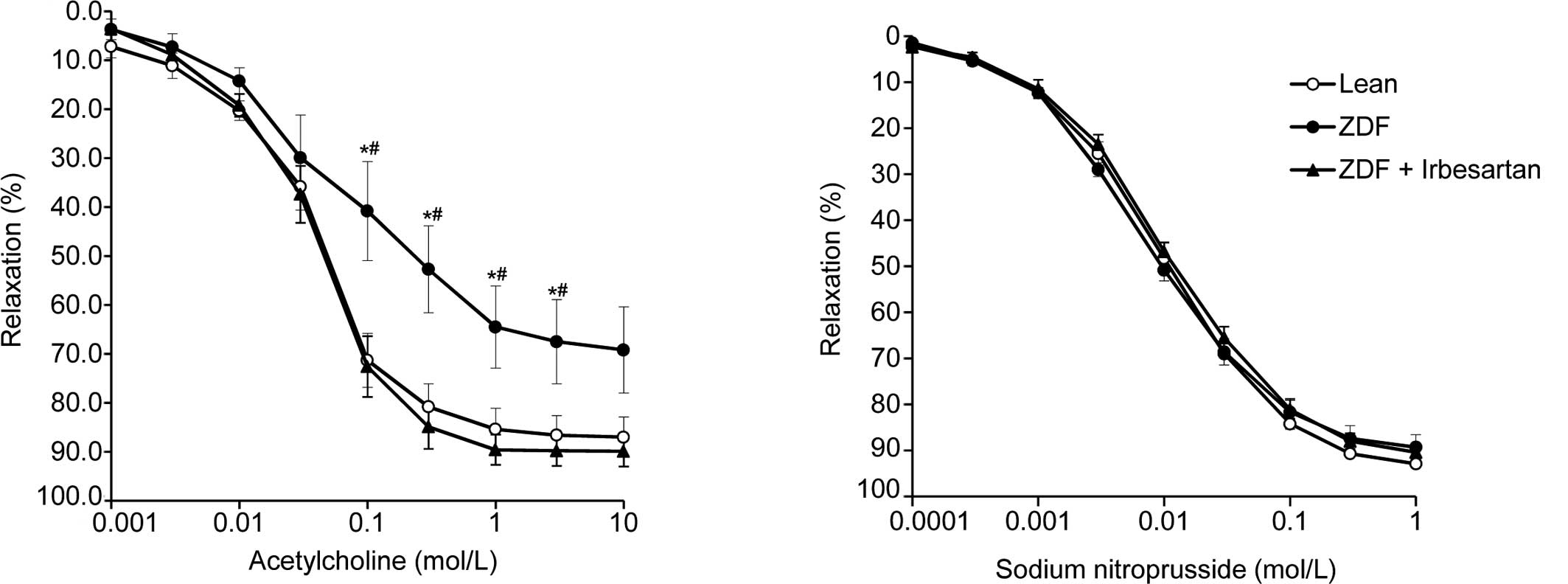
However, treatment with irbesartan significantly improved the
impaired endothelium-dependent relaxation response of isolated
mesenteric artery rings, while the relaxation response to
acetylcholine was significantly impaired in the ZDF rats (Fig. 1A). In contrast, the response to
sodium nitroprusside was the same in all groups (Fig. 1B). Systolic blood pressure was not
different between ZDF rats and lean rats, whereas rats treated with
irbesartan showed lower systolic blood pressure (Table I).
 | Table I.Body weight, blood pressure, food
intake and glucose level. |
Table I.
Body weight, blood pressure, food
intake and glucose level.
| Baseline
| 4 weeks
| 12 weeks
|
|---|
| Lean | ZDF | ZDF+IRB | Lean | ZDF | ZDF+IRB | Lean | ZDF | ZDF+IRB |
|---|
| Body weight | 302.8±1.8 | 382.5±9a | 381±9.3a | 363.5±1.8 | 393.4±10a | 392.3±10.4a | 400.8±3.4 | 403.7±10.4 | 416.7±7.3 |
| SBP (mmHg) | 139±2 | 133±4 | 129±4 | 136±2 | 138±2 | 129±2b | 133±3 | 135±4 | 122±2b |
| Food intake
(g/d) | 16.5±1.1 | 35.6±2.4a | 33.3±1.1a | 16.9±0.6 | 31.4±1.1a | 33.3±1.5a | 17.0±0.6 | 37.6±2.7a | 38.0±2.1a |
| Glucose (mg/dl) | 142±11 | 266±26a | 271±21a | 139±5 | 342±43a | 369±31a | 188±11 | 491±24a | 512±39a |
| Table II.Oral glucose tolerance test (OGTT) and
insulin tolerance test (ITT) after 10 weeks of treatment. |
Table II.
Oral glucose tolerance test (OGTT) and
insulin tolerance test (ITT) after 10 weeks of treatment.
| OGTT glucose (mg/dl)
| ITT glucose % vs. 0
min
|
|---|
| 0 min | 30 min | 60 min | 120 min | 15 min | 30 min | 60 min |
|---|
| Lean | 107±5 | 213±7 | 227±6 | 145±6 | 42±6 | 33±3 | 52±4 |
| ZDF | 368±44a | 656±56a | 653±25a | 535±25a | 80±2a | 44±3a | 37±3a |
| ZDF+IRB | 380±33a | 699±22a | 647±23a | 539±15a | 77±2a | 50±4a | 39±4a |
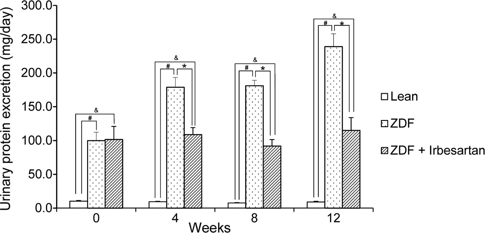
During 12 weeks, urinary protein excretion was also
examined. ZDF rats showed higher excretion than that of the lean
rats (Fig. 2). However, ZDF rats
treated with irbesartan showed no progression of urinary protein
excretion (Fig. 2). Serum BUN and
creatinine were not different between the treated and untreated
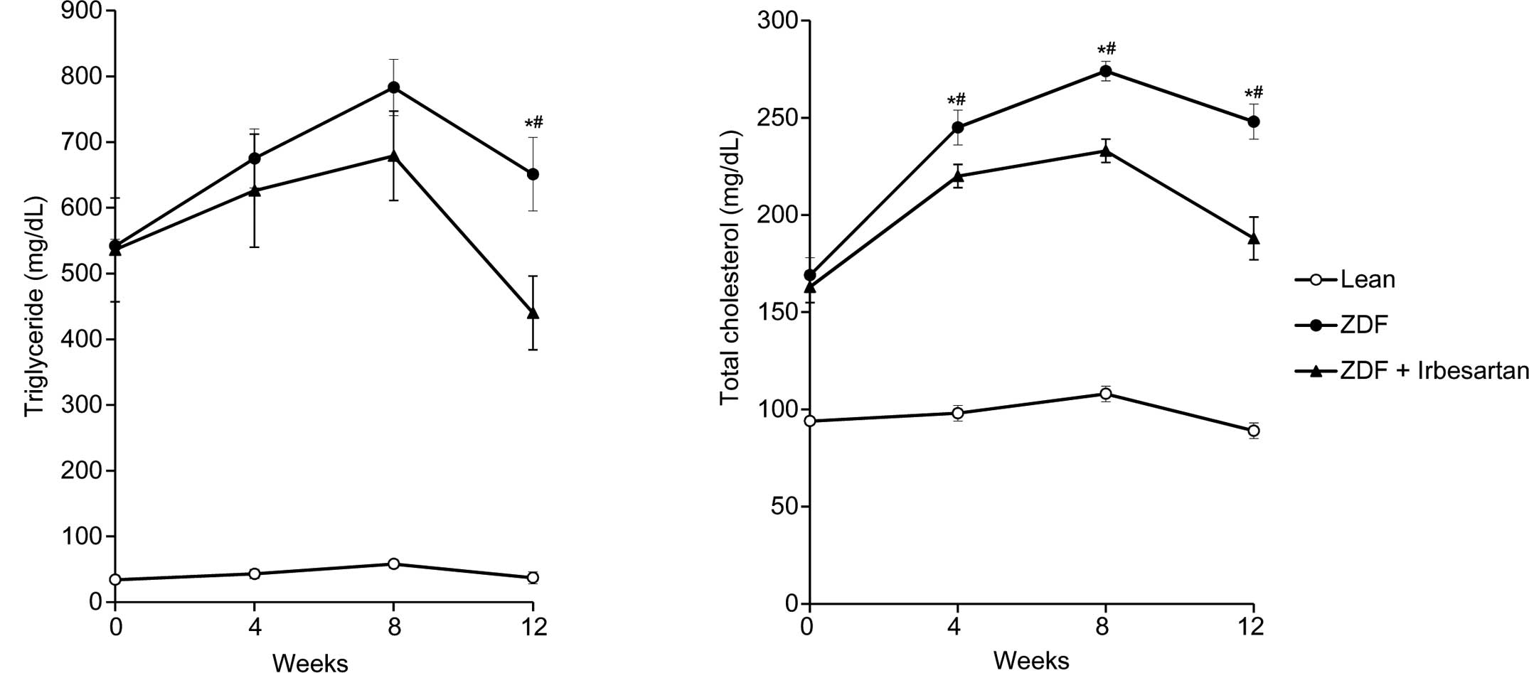
rats (data not shown). Then, we examined whether irbesartan
affected abnormal lipid profile. Serum total cholesterol and
triglyceride gradually worsened until 8 weeks in ZDF rats. A
significantly lower triglyceride level was observed in the
irbesartan-treated rats at 12 weeks (Fig. 3). Similarly, the total cholesterol
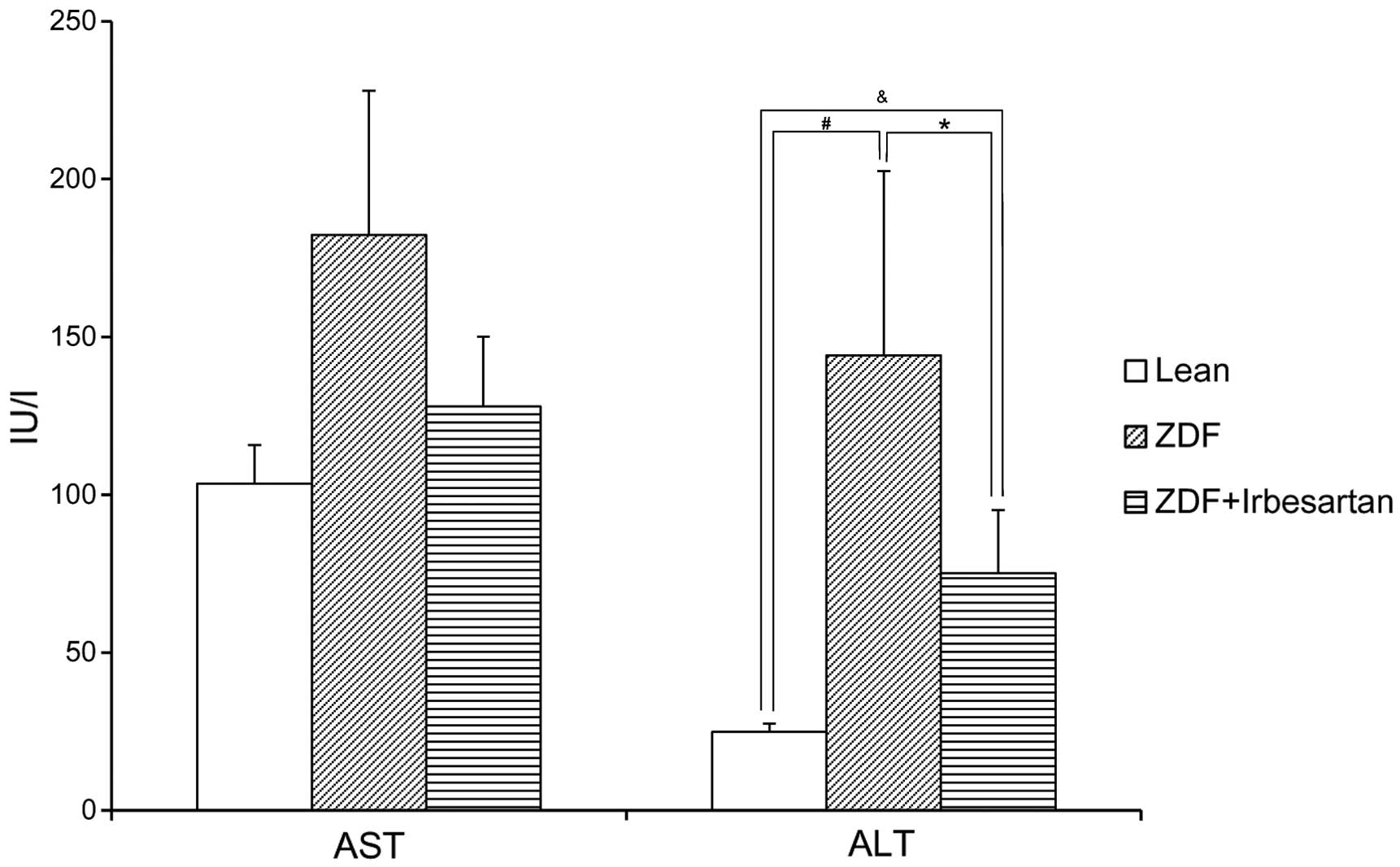
level was also reduced from 4 weeks after treatment (Fig. 3). Of importance, ZDF rats showed a
significant increase in ALT (Fig.
4). However, the level of ALT was significantly decreased by
irbesartan (Fig. 4).
Discussion
The present study demonstrated that endothelial
dysfunction, proteinuria, abnormal lipid profile and liver
dysfunction were significantly improved by irbesartan in ZDF rats,
independent of plasma glucose and insulin levels. Endothelial
dysfunction is caused by diabetes, and also accelerates the
progression of diabetes. This means that the vessel endothelium is
an important target tissue in diabetic patients. Thus, improvement
of endothelial dysfunction is believed to be beneficial for
reducing cardiovascular events in diabetic patients. In this study,
administration of irbesartan for 3 months significantly attenuated
endothelial dysfunction, with no effect on glucose metabolism. This
finding suggests that irbesartan, as a metabosartan that activates
PPARγ, may improve endothelial dysfunction in human hypertensive
patients, even if it does not lower blood glucose or insulin level.
In contrast, previous reports showed that irbesartan improved
plasma glucose level and insulin resistance (10–12)
and decreased body weight (10).
The reason for the lack of effects on glucose metabolism and serum
renal parameters was probably that the dose of irbesartan in the
present study was lower than that in previous reports (50 mg/kg),
and the treatment period was shorter than in previous reports
(12). Our study also showed that
urinary protein was reduced by irbesartan, although serum renal
markers and plasma glucose and insulin were not changed. Similarly,
long-term administration of irbesartan in rats was reported to
diminish the elevation in urinary protein excretion, plasma
creatinine and urea nitrogen levels and reduce the extent of
glomerular and tubule-interstitial lesions (10).
Another important finding of the present study is
that treatment with irbesartan improved the abnormal lipid profile
and liver dysfunction caused by non-alcoholic steatohepatitis
(NASH) in ZDF rats. Since NASH is frequently present in patients
with type 2 diabetes mellitus and leads to liver-related morbidity
and mortality (13), the quick
recovery of steatohepatitis by irbesartan may also be beneficial in
diabetic patients. Longer-term and higher-dose irbesartan (50
mg/kg) was reported to improve fat deposits through recovery of the
insulin signaling pathways in the liver (14). However, the present study
demonstrated that even lower-dose and shorter treatment with
irbesartan may provide beneficial effects on liver dysfunction.
Although irbesartan slightly lowered blood pressure in ZDF rats,
these favorable outcomes may not have been due to blood pressure
lowering, but to improvement of abnormal lipid profile by
irbesartan through activation of PPAR-γ. As a PPAR-γ agonist has
been reported to decrease pro-inflammatory cytokines in monocytes
(4) and prevent up-regulation of
MCP-1 receptor expression in lesional and circulating monocytes
(5), the decrease in
pro-inflammatory gene expression through activation of PPAR-γ by
irbesartan might reduce inflammation in the endothelium, kidney and
liver.
In clinical practice, the effects of irbesartan to
inhibit the onset and progression of diabetic nephropathy were
demonstrated in two prospective, randomized, double-blind clinical
trials, without a reduction in blood pressure (7,8).
Sub-analysis showed that irbesartan treatment exhibited a
significant decrease in hs-CRP, IL-6 and albumin excretion,
indicating that it reduces the risk of microvascular and
macrovascular disease through a reduction of inflammation (1). Also, Ceriello et al (2) reported that the decrease in
endothelial dysfunction, as assessed by flow-mediated vasodilation
(FMD), while the increase in nitrotyrosine, C-reactive protein,
intercellular adhesion molecule-1 and interleukin-6, were
significantly attenuated by short-term atorvastatin and/or
irbesartan treatment in type 2 diabetic patients without affecting
blood pressure. From these viewpoints, the pleiotropic effects of
irbesartan through PPAR-γ activation may contribute to the better
outcome, even in clinical situations.
In summary, the present study demonstrated that
irbesartan significantly improved endothelial dysfunction, abnormal
urinary excretion, abnormal lipid profile, and liver dysfunction in
ZDF rats, without changes in glucose and insulin levels. These
beneficial effects of irbesartan on diabetic nephropathy and NASH,
in addition to its potent blood pressure-lowering effect, would
provide a much better outcome of reduced cardiovascular events in
diabetic and metabolic disease patients.
Acknowledgements
This study was partially supported by
the Ministry of Education, Culture, Sports, Science and Technology,
the Takeda Science Foundation, the Mitsubishi Research Foundation,
and a Japan Heart Foundation/Novartis Grant for Research Award in
Molecular and Cellular Cardiology, 2011.
References
|
1.
|
Persson F, Rossing P, Hovind P, et al:
Irbesartan treatment reduces biomarkers of inflammatory activity in
patients with type 2 diabetes and microalbuminuria: an IRMA 2
substudy. Diabetes. 55:3550–3555. 2006. View Article : Google Scholar : PubMed/NCBI
|
|
2.
|
Ceriello A, Assaloni R, Da Ros R, et al:
Effect of atorvastatin and irbesartan, alone and in combination, on
postprandial endothelial dysfunction, oxidative stress, and
inflammation in type 2 diabetic patients. Circulation.
111:2518–2524. 2005. View Article : Google Scholar
|
|
3.
|
Fliser D, Buchholz K and Haller H:
Antiinflammatory effects of angiotensin II subtype 1 receptor
blockade in hypertensive patients with microinflammation.
Circulation. 110:1103–1107. 2004. View Article : Google Scholar : PubMed/NCBI
|
|
4.
|
Chen FL, Yang ZH, Liu Y, et al: Berberine
inhibits the expression of TNFalpha, MCP-1, and IL-6 in
AcLDL-stimulated macrophages through PPARgamma pathway. Endocrine.
33:331–337. 2008. View Article : Google Scholar : PubMed/NCBI
|
|
5.
|
Ni W, Kitamoto S, Ishibashi M, et al:
Monocyte chemoattractant protein-1 is an essential inflammatory
mediator in angiotensin II-induced progression of established
atherosclerosis in hypercholesterolemic mice. Arterioscler Thromb
Vasc Biol. 24:534–539. 2004. View Article : Google Scholar
|
|
6.
|
Schupp M, Janke J, Clasen R, Unger T and
Kintscher U: Angiotensin type 1 receptor blockers induce peroxisome
proliferator-activated receptor-gamma activity. Circulation.
109:2054–2057. 2004. View Article : Google Scholar : PubMed/NCBI
|
|
7.
|
Lewis EJ, Hunsicker LG, Clarke WR, et al:
Renoprotective effect of the angiotensin-receptor antagonist
irbesartan in patients with nephropathy due to type 2 diabetes. N
Engl J Med. 345:851–860. 2001. View Article : Google Scholar
|
|
8.
|
Parving HH, Lehnert H, Brochner-Mortensen
J, Gomis R, Andersen S and Arner P: The effect of irbesartan on the
development of diabetic nephropathy in patients with type 2
diabetes. N Engl J Med. 345:870–878. 2001. View Article : Google Scholar
|
|
9.
|
Lincoff AM, Wolski K, Nicholls SJ and
Nissen SE: Pioglitazone and risk of cardiovascular events in
patients with type 2 diabetes mellitus: a meta-analysis of
randomized trials. JAMA. 298:1180–1188. 2007. View Article : Google Scholar : PubMed/NCBI
|
|
10.
|
Janiak P, Bidouard JP, Cadrouvele C, et
al: Long-term blockade of angiotensin AT1 receptors increases
survival of obese Zucker rats. Eur J Pharmacol. 534:271–279. 2006.
View Article : Google Scholar : PubMed/NCBI
|
|
11.
|
Clasen R, Schupp M, Foryst-Ludwig A, et
al: PPARgamma-activating angiotensin type-1 receptor blockers
induce adiponectin. Hypertension. 46:137–143. 2005. View Article : Google Scholar : PubMed/NCBI
|
|
12.
|
Toblli JE, Munoz MC, Cao G, Mella J,
Pereyra L and Mastai R: ACE inhibition and AT1 receptor blockade
prevent fatty liver and fibrosis in obese Zucker rats. Obesity
(Silver Spring). 16:770–776. 2008. View Article : Google Scholar : PubMed/NCBI
|
|
13.
|
Adams LA and Feldstein AE: Nonalcoholic
steatohepatitis: risk factors and diagnosis. Expert Rev
Gastroenterol Hepatol. 4:623–635. 2010. View Article : Google Scholar : PubMed/NCBI
|
|
14.
|
Munoz MC, Argentino DP, Dominici FP, Turyn
D and Toblli JE: Irbesartan restores the in-vivo insulin signaling
pathway leading to Akt activation in obese Zucker rats. J
Hypertens. 24:1607–1617. 2006. View Article : Google Scholar : PubMed/NCBI
|