Introduction
Despite its declining incidence, gastric cancer is
still the second most common cause of death from cancer in Asia and
worldwide (1,2). Even after radical surgery, the
majority of gastric cancer patients develop local or distant
recurrence (3). Recently,
combination chemotherapy has become the standard of care for
patients with advanced gastric cancer, as it has been proven
effective for improving the survival and quality of life of these
patients (4). However, the average
overall benefit of chemotherapy is small, and the majority of
patients with advanced gastric cancer still do not appear to draw
any meaningful benefit from chemotherapy. An objective response
(OR) to chemotherapy is the primary study endpoint and one of the
surrogate markers of clinical benefit. Therefore, monitoring OR to
chemotherapy is essential for assessing prognosis and planning
further treatment. Computed tomography (CT) scans are often adopted
by physicians to evaluate the objective tumor response to
chemotherapy. Due to the high cost and adverse effects of
radioactivity, CT scans are used by physicians to evaluate the
objective tumor response to chemotherapy usually after 2 or 3
cycles of chemotherapy. However, approximately 50% of patients with
advanced gastric cancer do not benefit from any one of the
chemotherapy regimens during the first cycle of chemotherapy. The
early identification of no-response patients is very difficult for
most physicians. Thus, the development of more straighforward, less
costly and safer tools with which to monitor the effects of
chemotherapy in patients with gastric cancer would be extremely
valuable. Serum tumor markers may be used for this purpose.
Recently, among the available serum markers, TK1 has been the most
widely tested in solid tumors, including gastric cancer,
particularly for monitoring the effect of tumor therapies,
prognosis and follow-up. Yet, its role as a marker of treatment
efficacy is still contradictory to date.
The aim of the present study was to investigate the
role of serum TK1 during chemotherapy treatment as an adjuvant or
surrogate marker for monitoring the effects of therapy and
prognosis in patients with advanced gastric cancer who received
chemotherapy.
Patients and methods
In total, 84 advanced gastric cancer patients
previously untreated with chemotherapy were enrolled prospectively
into this single-institution study from September 2009 to May 2010.
These patients were pathologically diagnosed with gastric
adenocarcinoma by endoscopic biopsy or surgical tumor specimens.
Among these patients, 56 patients presenting with at least one
measurable lesion received palliative chemotherapy, and 28 patients
received adjuvant chemotherapy after surgery. All patients were
treated with FOLFOX4 or DCF regimens. FOLFOX4 consisted of L-HOP 85
mg/m2 intravenously (i.v.) only on Day 1, with
leucovorin 100 mg/m2 i.v. as a 2-h infusion followed by
bolus 5-fluorouracil (5-FU) 400 mg/m2/day and a 22-h
infusion of 5-FU 600 mg/m2/day, repeated for two
consecutive days every 2 weeks for a minimum of 6 cycles. DCF
regimen was administered according to the following plan using
docetaxel at a dose of 20 mg/m2 i.v. administered over
30 min on Day 1, with cisplatin at a dose of 20 mg/m2
administered over 1 h on Day 2, and 5-FU 350 mg/m2/day,
repeated for 5 consecutive days every 3 weeks for a minimum of 3
cycles. Written informed consent was obtained from the patients for
chemotherapy, and data regarding the patient clinical and
pathological features were collected for this study.
Analysis of remission
For patients with measurable lesions, the tumor
response was assessed according to Response Evaluation Criteria in
Solid Tumors (RECIST) criteria (5). Accordingly, a complete response (CR)
was defined as the disappearance of all target lesions. Any
pathological lymph nodes (whether target or non-target) were
required to show a reduction in the short axis to <10 mm. A
partial response (PR) was defined as at least a 30% decrease in the
sum of the diameters of the target lesions, taking as reference the
baseline sum diameters. Progressive disease (PD) was defined as at
least a 20% increase in the sum of the diameters of target lesions,
taking as reference the smallest sum during study. In addition to
the relative increase of 20%, the sum must also demonstrate an
absolute increase of at least 5 mm. Stable disease (SD) was defined
as neither sufficient shrinkage to qualify for PR nor sufficient
increase to qualify for PD, taking as reference the smallest sum of
diameters during study. In this study, patients who achieved a CR
or PR were classified as having a chemotherapeutic objective
response (OR), and all remaining patients were considered as
non-responders. For postoperative patients with no measurable
lesions, a chemotherapeutic OR was defined as no discovery of all
objective evidence of new disease, and non-response was defined as
discovery of new disease by CT scan or pathological
examination.
Follow-up
Interim history, physical examination, hematologic
studies, serum thymidine kinase 1 (STK1) levels, carcinoembryonic
antigen (CEA) levels were assessed every 1 month during treatment,
and whole-body CT was performed every 2 months during treatment.
The above tests were carried out every 3 months in the first year
and every 6 months thereafter. Patients underwent upper endoscopy
every 2 months during treatment and every 6 months thereafter. The
progression or recurrence of gastric carcinoma was confirmed by
cytology biopsy, surgery or whole-body CT. The 7th edition of the
UICC TNM staging system for gastric cancer was used for the
classification of each case. The study was carried out in a blinded
manner so that patient outcome was unknown to the investigators
performing the molecular analyses. Progression-free survival (PFS)
was the time from study entry until disease progression or death or
the day of the last follow-up visit whichever came first.
Relapse-free survival (RFS) was the time from study entry until
recurrence or death to the day of the last follow-up visit
whichever came first. Overall survival (OS) was the time from the
day of the confirmation of diagnosis until the date of death
regardless of cause or the most recent documented follow-up.
Detection of serum TK1
Serum samples were obtained immediately before the
start of chemotherapy and every 1 month during the chemotherapy
break. STK1 was analyzed by an ECL dot blot assay. The procedure
was performed according to the manufacturer's protocol (commercial
kit; SSTK, Shenzhen, China) as described elsewhere (6).
Statistical methods
Differences in the values among the groups under
study were evaluated by analysis using the independent-samples
t-test, as indicated. The paired-samples t-test procedure was used
to compare STK1 values prior to treatment and after chemotherapy. A
P-value ≤0.05 was indicative of statistical significance.
Results
STK1 levels of the healthy control and
patients prior to chemotherapy
The average STK1 level in all of the patients with
gastric cancer was 5.57±3.07 pM, which was significantly higher
than that in the healthy controls (1.12±0.57 pM) (P<0.001). The
average STK1 value in patients who did not receive surgery
(6.02±3.12) was significantly higher than that in patients who
received surgery (4.68±2.78) (P<0.05), which may have resulted
from the decrease in tumor volume burden.
Associations between STK1 levels and
clinicoppathological features
There was a significant increase in the average STK1
value from stage I + II (2.26±1.09) to stage III + IV (6.28±4.23)
disease (P<0.001). The STK1 value was increased by 1.78 times in
patients with distant metastasis compared with patients without
metastasis. A high STK1 value was also correlated with a poor
Eastern Cooperative Oncology Group performance status (ECOG PS)
(P=0.001) and high serum CEA levels (P=0.004), but not with age and
gender (Table I).
 | Table IAssociations between STK1 levels and
clinicopathological features of the patients. |
Table I
Associations between STK1 levels and
clinicopathological features of the patients.
| Type | n | Mean ± SD | P-value |
|---|
| Agea (years) | | | |
| ≤60 | 43 | 5.03±3.22 | |
| >60 | 41 | 5.92±4.92 | 0.327 |
| Gender | | | |
| Male | 62 | 7.12±6.24 | |
| Female | 22 | 4.88±2.92 | 0.116 |
| CEA | | | |
| ≥5 ng/ml | 28 | 7.41±4.32 | |
| <5 ng/ml | 56 | 4.49±3.71 | 0.004 |
| Stage | | | |
| I+ II | 17 | 2.26±1.09 | |
| III + IV | 67 | 6.28±4.23 | < 0.001 |
| ECOG PS | | | |
| 0,1 | 56 | 4.13±2.60 | |
| 2 | 28 | 8.14±5.26 | 0.001 |
Associations between STK1 levels and
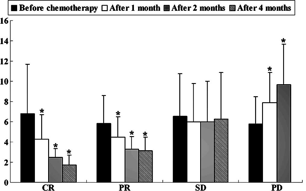
remission in patients treated with palliative chemotherapy
In all of the 56 patients who received palliative
chemotherapy, the mean STK1 value before the start of treatment was
6.02 pM, which was not significantly different from the values
after 2 months of chemotherapy. The mean STK1 value prior to
chemotherapy in the CR patients (n=4) was 6.82 pM, the PR patients
(n=23) 5.84 pM, the SD patients (n=10) 6.55 pM and the PD patients
(n=19) 6.62 pM (no significant difference was noted between any two
groups). The corresponding STK1 values decreased significantly
after 2 and 4 months of treatment in the patients who obtained CR
(P=0.025 and P=0.043, respectively) and PR (P<0.001 and P=0.001,
respectively), while in the patients who obtained PD, the
corresponding STK1 value increased significantly after 2 months of
the treatment (P=0.004) (Fig. 1).
After 1 month of chemotherapy, the corresponding STK1 values
started to decline in the OR groups (P=0.035 in CR group and
P<0.001 in PR group) but increased in the PD group (P=0.011),
respectively. As the patients with PD during the first 2 months of
treatment received other treatment regimens, we did not compare the
difference between the STK1 value prior to chemotherapy and that
after 4 months of treatment. Compared with the value prior to
chemotherapy, the mean STK1 value after 4 months of chemotherapy
did not decrease significantly in patients with SD.
Associations between STK1 levels and
recurrence in patients treated with surgery following by
chemotherapy
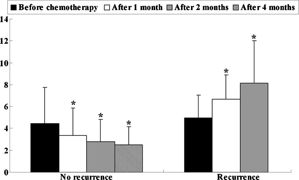
In all 28 patients who received surgery followed by
adjuvant chemotherapy, the mean STK1 value prior to chemotherapy
was 4.68 pM, which was not significantly different from the value
after 2 months of chemotherapy. The mean STK1 values before the
start of chemotherapy were not correlated with clinical response.
After 2 and 4 months of chemotherapy, the STK1 values decreased
significantly in the patients without recurrence (P=0.011 and
P=0.002, respectively), while in patients with recurrence, the STK1
values increased significantly (P=0.003) after 2 months of
chemotherapy, compared with that before chemotherapy (Fig. 2). Particularly, after 1 month of
chemotherapy, the corresponding STK1 values started to decline in
the patients without recurrence (P=0.043) and to increase in the
recurrence group (P=0.003). As patients with recurrence during the
first 2 months of treatment received other treatment regimens, we
did not compare the difference between the STK1 value before
chemotherapy and that after 4 months of treatment. Compared with
the value before chemotherapy, the mean STK1 value after 4 months
of chemotherapy did not decrease significantly in patients with
SD.
Associations between STK1 levels and
survival
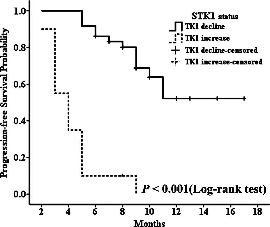
The median progression-free survival of all patients
was 9 months (range, 2–17 months), and due to the short follow-up
period, the median overall survival time could not been defined. In
the patient group receiving palliative chemotherapy, patients with
decreased STK1 levels during the first 2 months of treatment had
significantly longer median PFS than those with increased STK1
levels for the same treatment interval (median PFS, not defined vs.
4 months, P<0.001) (Fig. 3A).
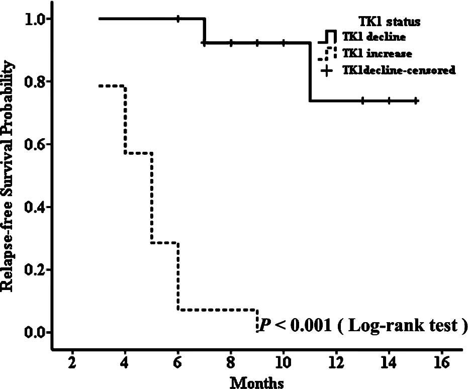
At the same time, we found similar results in the group receiving
adjuvant chemotherapy; patients with decreased STK1 levels during
the first 2 months of treatment had significantly longer median RFS
than those with increased STK1 levels for the same time period
(median RFS, not defined vs. 5 months, P<0.001) (Fig. 4). Although we could not determine
the median overall survival time of the patients with decreased or
increased STK1 levels in both groups, the log-rank test showed that
patients receiving palliative chemotherapy with decreased STK1
levels during the first 2 months of treatment had longer OS
(Fig. 3B). Since only 2 patients
died, a meaningful determination of the association between the
STK1 level and OS was not possible in the adjuvant chemotherapy
patients. Yet, our results indicate that a change in STK1 levels
during the first 2 months of chemotherapy predicts the PFS in
patients receiving palliative chemotherapy or RFS in patients
receiving adjuvant chemotherapy, which may aid clinicians to timely
select new treatment regimens for patients who do not benefit from
first-line chemotherapy regimens.
Discussion
The purpose of cancer chemotherapy is to improve the
overall survival of patients. An objective response (OR) to
chemotherapy often serves as a surrogate marker of clinical
benefit, as it is associated with a better survival outcome
(7). In clinical settings, most
oncologists have adopted the use of repeated imaging studies to
evaluate tumor response and make decisions concerning continued
therapy on the basis of both objective and symptomatic criteria.
Even for patients who undergo sugery yet still require adjuvant
chemotherapy, many clinicians follow up their patients' malignant
disease by means of repeated imaging analysis to evaluate whether
or not the disease has progressed. Although evaluation criteria
based on imaging modalities, including CT, have become standard in
the evaluation of tumor response, serum levels of tumor markers are
sometimes used in clinical settings as adjuvant or surrogate
markers for estimation of tumor response, particularly in patients
with no measurable lesions.
TK1 is a pyrimidine salvage pathway enzyme involved
in DNA synthesis and repair. TK1 activity is low or absent in
resting cells, starts to increase in the late G1 stage, increases
in S-phase, and disappears during mitosis (8). The G2/M specific TK1 activity
disappearance is due to specific degradation of TK1 protein by the
ubiquitin-proteasome pathway (8).
The activity or concentration of TK1 is correlated closely with
cell proliferation of cultured cell lines or tissues (9). It has been reported that TK1 levels
in the serum of patients with malignancies are significantly higher
than those of healthy persons (10), due to the fact that most cancer
cells die in the G2/S phase of the cell cycle while normal cells
often die in Gl. In the present study, the average STK1 level in
all of the patients with gastric cancer was 5.57±3.07 pM, while
that in the healthy controls was 1.12±0.57 pM (P<0.001). Our
results coincided with the above results (11). Base on our results and those of
other studies, we can speculate that STK1 may be an additional
biomarker for cancer diagnosis.
The high levels of STK1 in patients with
malignancies also suggest that TK1 may be a useful proliferation
marker for the assessment of tumor progression or for monitoring
therapy. He et al (11) and
Zou et al (12) reported
that the STK1 activity was elevated in patients before surgery, but
was irregular following surgery and adjuvant treatment. Chen et
al (13) reported that the
mean STK1 values decreased significantly after treatments (surgery
and/or chemotherapy, P<0.01) in 4 types of cancers, apart from
patients with gastric cancer. In our 56 gastric cancer patients who
received only chemotherapy, the mean STK1 value before the start of
treatment was not significantly different from the values after 2
months of chemotherapy. Even in the 28 patients who received
surgery followed by adjuvant chemotherapy, the mean STK1 value
prior to chemotherapy was not significantly different from the
values after chemotherapy. Yet, in patients who obtained CR, PR and
no recurrence, the STK1 values declined significantly after 2
months of treatment (P=0.025, P<0.001 and P=0.011,
respectively), while in patients who obtained PD, the corresponding
STK1 values increased significantly (P=0.004 and p=0.003,
respectively) (Figs. 1 and
2). In both groups, the mean STK1
values before the start of chemotherapy were not correlated to
clinical response. Our results were partially in accordance with
those of Chen et al (13)
and He et al (14). Our
results suggest that the change in STK1 levels during the first 2
months of treatment may be more important than baseline levels of
STK1 in predicting an individual's chemotherapy response. Thus, to
determine changes in STK1, it is necessary for clinicians to
establish a baseline level of the STK1 concentration for every
patient. In the present study, after 1 cycle of chemotherapy, the
levels of STK1 in patients with PD started to increase, which
suggests that clinicians should pay special attention to patients
with elevated levels of STK1 during the first adjuvant/palliative
chemotherapy break. In the event a patient with an increase in STK1
also presents with new emerging clinical symptoms or other elevated
tumor markers, the clinician should adjust the treatment regimen
for this patient early in time.
Many studies have shown that increased serum TK1
levels adversely affect OS (15–18).
In our study, because of the short follow-up period as most of the
patients were followed up for less than 2 years, a meaningful
determination of the association between STK1 level and overall
survival was not possible. Yet, a significant difference in the
median PFS (median PFS, not defined vs. 4 months, P<0.001) or
median RFS (median RFS, not defined vs. 5 months, P<0.001) was
noted between patients with decreased STK1 levels and patients with
increased STK1 levels during the first 2 months of treatment. Our
results suggest that changes in STK1 during the first 2 months of
chemotherapy may be more valuable for evaluating tumor chemotherapy
response, predicting PFS and RFS than baseline values of STK1 in
gastric cancer patients who receive chemotherapy.
We also investigated the association of STK1 with
clinicopathologic characteristics. A significant association was
observed between STK1 values and clinical stage, and there was a
significant increase in the average STK1 value from stage I + II
(2.26±1.09) to stage III + IV (6.28±4.23) (P<0.001). It appeared
that patients with a large tumor burden possess more potential for
cell proliferation in gastric cancer. In our study, a high STK1
value was also correlated with a poor ECOG PS (P=0.001) and high
serum CEA levels (P=0.004), but not with age and gender (Table I). These results partially coincide
with those of Chen et al (13).
The limitations of the present study included the
relatively small size of the sample and the short follow-up period,
which may explain the weak prognostic value of STK1 for overall
survival in our study. To further confirm the prognostic value of
STK1 in gastric cancer patients who receive chemotherapy, a larger
sample and longer period of follow-up is needed in future
studies.
Acknowledgements
The research was supported, in part,
by the Science and Technology Planning Project of Changzhou
(CS20092025) and the Key Medical Innovation Talents Training
Project of Changzhou, Jiangsu Province, China.
References
|
1
|
Leung WK, Wu MS, Kakugawa Y, et al:
Screening for gastric cancer in Asia: current evidence and
practice. Lancet Oncol. 9:279–287. 2008. View Article : Google Scholar : PubMed/NCBI
|
|
2
|
Kamangar F, Dores GM and Anderson WF:
Patterns of cancer incidence, mortality, and prevalence across five
continents: defining priorities to reduce cancer disparities in
different geographic regions of the world. J Clin Oncol.
24:2137–2150. 2006. View Article : Google Scholar
|
|
3
|
Macdonald JS: Treatment of localized
gastric cancer. Semin Oncol. 31:566–573. 2004. View Article : Google Scholar : PubMed/NCBI
|
|
4
|
Wagner AD, Unverzagt S, Grothe W, et al:
Chemotherapy for advanced gastric cancer. Cochrane Database Syst
Rev. CD004064:2010. View Article : Google Scholar
|
|
5
|
Eisenhauer EA, Therasse P, Bogaerts J, et
al: New response evaluation criteria in solid tumours: revised
RECIST guideline (version 1.1). Eur J Cancer. 45:228–247. 2009.
View Article : Google Scholar
|
|
6
|
Xu XH, Zhang YM, Shu XH, et al: Serum
thymidine kinase 1 reflects the progression of pre-malignant and
malignant tumors during therapy. Mol Med Rep. 1:705–711.
2008.PubMed/NCBI
|
|
7
|
Paesmans M, Sculier JP, Libert P, et al:
Response to chemotherapy has predictive value for further survival
of patients with advanced non-small cell lung cancer: 10 years
experience of the European Lung Cancer Working Party. Eur J Cancer.
33:2326–2332. 1997.PubMed/NCBI
|
|
8
|
Ke PY and Chang ZF: Mitotic degradation of
human thymidine kinase 1 is dependent on the anaphase-promoting
complex/cyclosome-CDH1-mediated pathway. Mol Cell Biol. 24:514–526.
2004. View Article : Google Scholar : PubMed/NCBI
|
|
9
|
He Q, Mao Y, Wu J, et al: Cytosolic
thymidine kinase is a specific histopathologic tumour marker for
breast carcinomas. Int J Oncol. 25:945–953. 2004.PubMed/NCBI
|
|
10
|
O'Neill KL, Buckwalter MR and Murray BK:
Thymidine kinase: diagnostic and prognostic potential. Expert Rev
Mol Diagn. 1:428–433. 2001.PubMed/NCBI
|
|
11
|
He Q, Zou L, Zhang PA, Lui JX, Skog S and
Fornander T: The clinical significance of thymidine kinase 1
measurement in serum of breast cancer patients using anti-TK1
antibody. Int J Biol Markers. 15:139–146. 2000.PubMed/NCBI
|
|
12
|
Zou L, Zhang PG, Zou S, Li Y and He Q: The
half-life of thymidine kinase 1 in serum measured by ECL dot blot:
a potential marker for monitoring the response to surgery of
patients with gastric cancer. Int J Biol Markers. 17:135–140.
2002.PubMed/NCBI
|
|
13
|
Chen Y, Ying M, Hu M, et al: Serum
thymidine kinase 1 correlates to clinical stages and clinical
reactions and monitors the outcome of therapy of 1,247 cancer
patients in routine clinical settings. Int J Clin Oncol.
15:359–368. 2010. View Article : Google Scholar : PubMed/NCBI
|
|
14
|
He Q, Fornander T, Johansson H, et al:
Thymidine kinase 1 in serum predicts increased risk of distant or
loco-regional recurrence following surgery in patients with early
breast cancer. Anticancer Res. 26:4753–4759. 2006.PubMed/NCBI
|
|
15
|
Kallander CF, Simonsson B, Hagberg H and
Gronowitz JS: Serum deoxythymidine kinase gives prognostic
information in chronic lymphocytic leukemia. Cancer. 54:2450–2455.
1984. View Article : Google Scholar : PubMed/NCBI
|
|
16
|
Hallek M, Langenmayer I, Nerl C, et al:
Elevated serum thymidine kinase levels identify a subgroup at high
risk of disease progression in early, nonsmoldering chronic
lymphocytic leukemia. Blood. 93:1732–1737. 1999.
|
|
17
|
Hallek M, Wanders L, Ostwald M, et al:
Serum beta(2)-microglobulin and serum thymidine kinase are
independent predictors of progression-free survival in chronic
lymphocytic leukemia and immunocytoma. Leuk Lymphoma. 22:439–447.
1996. View Article : Google Scholar : PubMed/NCBI
|
|
18
|
Magnac C, Porcher R, Davi F, et al:
Predictive value of serum thymidine kinase level for Ig-V
mutational status in B-CLL. Leukemia. 17:133–137. 2003. View Article : Google Scholar : PubMed/NCBI
|