Introduction
Ischemia results from obstruction of the vessels
supplying blood to the tissues due to a number of reasons, and
leads to deficient tissue nutrition. If this occurs blood flow
should be restored (reperfusion) via medicines or mechanical
interventions to prevent ischemia-related cell and tissue damage.
Ischemia-related damage can either be reversible or irreversible. A
series of complex events that occur during reperfusion can
occasionally cause greater damage than the ischemia itself
(1). Renal ischemia/reperfusion
(I/R) injury may occur due to systemic hypotension, hypovolemic
shock, cardiac arrest, renovascular surgery and aortic clamping.
The severity of the damage is increased with the duration of
ischemia. As a result, different clinical entities, ranging from
prerenal azotemia without marked tissue damage, to severe acute
renal failure (ARF) due to tubular or cortical necrosis, may occur
(2). Despite advances in critical
care medicine, ARF remains a clinical problem, as it is a
significant cause of morbidity and mortality. A number of
pharmacological approaches have been investigated for the treatment
of renal I/R injury. Substances including, novel antioxidants and
antioxidant enzyme mimetics, nitric oxide and nitric oxide synthase
inhibitors, erythropoietin (EPO), peroxisome-proliferator activated
receptor agonists, inhibitors of poly (ADP-ribose) polymerase,
carbon monoxide-releasing molecules, statins and adenosine, have
been investigated for their efficacy against renal I/R injury and
ischemic ARF in experimental studies (2). Although a number of promising
pharmacological agents have been developed and demonstrated to be
beneficial in experimental studies, the majority of clinical
studies have yielded unsuccessful results. For these reasons, the
search for new pharmacological agents for the treatment of renal
I/R injury is ongoing, and studies in this field continue to
attract attention (2). A number of
substances have become the subject of studies due to their effects
on the mechanisms involved in the pathophysiology of I/R
injury.
Leptin is a hormone that is synthesized primarily by
adipocytes and is found in the systemic circulation. It mainly
affects body mass index by controlling food intake and energy
expenditure (3). In vitro
studies have demonstrated that leptin has marked mitogenic effects
on endothelial and glomerular cells (4,5).
Moreover, leptin may affect the synthesis of nitric oxide through
the activation of nitric oxide synthase (6). Studies employing a rat model of
intestinal I/R injury have reported that leptin demonstrates a
time-dependent response to acute inflammatory stimuli and acts as
an anti-inflammatory cytokine (7,8).
Apelin is a newly identified adipokine, which is
synthesized in a number of tissues, including the gastrointestinal
system, brain, kidney and liver. Apelin primarily affects the
cardiovascular system. In vivo studies have demonstrated
that apelin also has an endothelium-dependent vasodilator effect,
in addition to its regulatory effects on arterial blood pressure
(9).
The present study aimed to demonstrate the favorable
and unfavorable effects of two adipokines, apelin and leptin, on
renal functions following renal I/R.
Materials and methods
Study design
The present study was conducted in accordance with
the Guidelines for the Care and Use of Experimental Animals
established by the local committee on animal research ethics of the
University. The committee approved the study design. A total of 32
male Sprague-Dawley rats aged between 6 and 8 weeks and weighing
280±20 g were used. Using a computer generated table of random
numbers, rats were assigned to one of the following four groups,
each containing eight animals: i) control group, administered
normal saline solution intraperitoneally and subjected to aorta
mobilization without any clamping of the aorta; ii)
ischemia/reperfusion (I/R) group, administered normal saline
solution intraperitoneally and induced with ischemia by clamping of
the aorta and reperfusion; iii) ischemia/reperfusion and apelin
(I/R+A) group, administered apelin intraperitoneally and induced
with ischemia by clamping of the aorta and reperfusion; iv)
ischemia/reperfusion and leptin (I/R+L) group, administered leptin
intraperitoneally and induced with ischemia by clamping of the
aorta and reperfusion.
All animals were maintained under a controlled
temperature (22±2°C) and relative humidity of 55±15% under 12 h
light/dark cycles. All animals were fed with chow and tap water
ad libitum throughout the acclimatization and study
periods.
Chemicals and reagents
Apelin-13 (200 μg/flacon; Apelin®,
Phoenix Pharmaceuticals Inc., Belmont, CA, USA) and leptin (200
μg/flacon; Leptin®, Phoenix Pharmaceuticals Inc.,
Belmont, CA, USA) were commercially purchased. The I/R+A group was
administered apelin intraperitoneally at a dose of 1.5 μg/kg. The
I/R+L group was administered leptin intraperitoneally at a dose of
100 μg/kg. Apelin and leptin were administered for three
consecutive days prior to the surgical procedure. The control and
I/R groups were administered normal saline solution
intraperitoneally (10,11).
Surgical procedure
Following a night of fasting, each animal was
anesthetized with intraperitoneal xylazine (5 mg/kg) and ketamine
hydrochloride (30 mg/kg). The abdomen was shaved and cleaned with
povidone-iodine solution. Using a sterile technique, all animals
underwent a laparotomy through a 3-cm midline incision. The aorta
and visceral arteries were exposed. The control group underwent
suprarenal aorta mobilization without clamping of the aorta. In the
I/R, I/R+A and I/R+L groups, the suprarenal aorta was clamped with
an atraumatic microvascular bulldog clamp. Ischemia was confirmed
via visual inspection of the kidneys. The clamp remained in place
for 40 min. A total of 5 min prior to removal of the clamp, a right
nephrectomy was performed. The midline incision was closed, and the
rats were immediately returned to their cages for recovery.
Metabolic cage and biochemical
assessment
Following the induction of ischemia, the animals
were placed into individual metabolic cages for 24 h. Urine
samples, which were free of food and feces, were collected in
graduated cylinders. Urine volume, urea, creatinine, osmolality,
and glomerular filtration rate (GFR) were assessed during the 24th
h of reperfusion. Blood samples were collected for measurements of
serum blood urea nitrogen (BUN), creatinine, alanine transaminase
(ALT), aspartate transaminase (AST) and γ-glutamyl transpeptidase
(GGT).
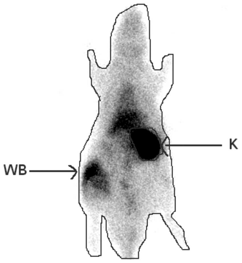
Scintigraphic examination
A 99mTc dimercaptosuccinic acid (DMSA) scintigraphy
was performed during the 24th h of reperfusion. Imaging was
performed, 2 h after the intravenous injection of 1 mCi 99mTc DMSA
via a 24F catheter placed in the tail vein under general
anesthesia. A total of 2 h after 99mTc DMSA was administered,
whole-body images were acquired for 300 sec (matrix, 512 x 512;
zoom factor, 1.55) in the posterior projection using a single-head
γ-camera equipped with a low energy all-purpose collimator. For
semi-quantitative evaluation, regions of interest (ROIs) were drawn
around the whole body to represent the total injected dose, and
around the kidney to represent the renal function (Fig. 1). Background activity was measured
using a ROI, which was drawn over a region outside of the body
contour. The single kidney DMSA uptake (%) was calculated using the
following formula (12): DMSA
uptake (%) = 100 x (kidney/whole body).
Histopathological examination
Harvested kidneys were fixed in 10% formalin for 24
h, then routinely processed and embedded in paraffin. Sections (5
μm) were cut from paraffin blocks and stained with hematoxylin and
eosin (H&E). Slides were then examined under a light microscope
(Nikon E600W, Japan) Each slide was evaluated by an expert
investigator blinded to the experiment and data. Slides were
examined for tubular cell swelling, interstitial edema, medullary
congestion, tubular dilatation and necrosis. Renal damage was
graded as none, mild, moderate or severe (13).
Statistical analysis
Statistical analysis was conducted using the
Statistical Package for the Social Sciences (SPSS) for Windows
(version 15.0; SPSS Inc., Chicago, IL, USA). Descriptive statistics
(mean, standard deviation, minimum, maximum and median) were
presented for numerical variables. For independent groups, a
one-way analysis of variance (ANOVA) was used for multiple
comparisons when the data was normally distributed, while the
Kruskal-Wallis test was used when the data were not normally
distributed. In the case of significant differences between groups,
the Bonferroni correction and Tukey’s tests were used when the data
was normally distributed. Otherwise, a post hoc analysis with the
Bonferroni correction and Mann-Whitney U test was carried out.
Categorical variables were compared using the Monte Carlo method
when the Chi-square assumption was not met. P<0.05 was
considered to indicate a statistically significant difference.
Results
There was no significant difference between the
study groups with respect to albumin and chloride levels, however
there were significant differences in terms of urea, creatinine,
total protein, AST, ALT, GGT, sodium and potassium levels.
Laboratory analysis of the blood samples obtained from the study
groups are summarized in Table I.
One animal in the control group and one animal in the I/R+L group
died after blood and urine samples were obtained and scintigraphic
examinations were performed.
 | Table I.Laboratory analysis of the blood
samples obtained from the study groups. |
Table I.
Laboratory analysis of the blood
samples obtained from the study groups.
| Control |
Ischemia/reperfusion | Ischemia/reperfusion
+ leptin | Ischemia/reperfusion
+ apelin | P-value |
|---|
| Urea (mg/dl) |
27.00±7.33b–d |
120.75±15.56a,c,d |
80.00±11.81a,b |
75.50±10.84a,b | <0.001 |
| Creatinine
(mg/dl) |
0.35±0.06b–d | 1.79±0.49a | 1.36±0.45a | 1.40±0.38a | <0.001 |
| Total protein
(g/dl) |
6.29±0.27c,d | 5.90±0.33 | 5.86±0.30a | 5.86±0.23a | 0.014 |
| Albumin (g/dl) | 2.81±0.59 | 2.93±0.22 | 2.96±0.19 | 3.03±0.14 | 0.640 |
| AST (U/l) | 134.75±53.28b |
274.50±50.66a,c,d | 189.25±18.88b | 190.75±19.39b | <0.001 |
| ALT (U/l) |
60.13±16.55b–d |
143.75±24.51a |
106.50±27.54a |
106.62±24.50a | <0.001 |
| GGT (U/l) |
559.00±46.75b–d |
1,112.25±125.45a,c,d |
838.88±138.46a,b |
842.75±135.10a,b | <0.001 |
| Sodium
(mmol/dl) | 136.38±4.00b | 147.25±6.50a | 142.13±3.04 | 142.38±2.00 | <0.001 |
| Potassium
(mmol/dl) | 4.94±0.50b |
5.99±0.38a,c,d | 5.25±0.39b | 5.13±0.34b | <0.001 |
| Chloride
(mmol/dl) | 104.00±3.16 | 102.38±3.29 | 102.25±2.92 | 101.50±3.96 | 0.511 |
No significant differences were found between the
study groups with respect to 24-hour urine protein, sodium,
potassium, chloride, urea and urine density values. There were
significant differences between the study groups in terms of GFR
and DMSA uptake (Table II).
| Table II.Laboratory analysis of the urine
samples and dimercaptosuccinic acid uptake in the study groups. |
Table II.
Laboratory analysis of the urine
samples and dimercaptosuccinic acid uptake in the study groups.
| Control |
Ischemia/reperfusion + leptin |
Ischemia/reperfusion + apelin |
Ischemia/reperfusion | P-value |
|---|
| 24-hour urine
protein (mg/dl) | 114.00±11.86 | 117.88±9.60 | 117.63±9.86 | 117.63±10.61 | >0.05 |
| 24-hour urine
sodium (mmol/l) | 109.63±18.49 | 110.50±20.78 | 110.75±19.35 | 124.12±44.46 | >0.05 |
| 24-hour urine
potassium (mmol/l) | 140.25±14.12 | 140.13±13.80 | 140.88±15.12 | 141.38±13.84 | >0.05 |
| 24-hour urine
chloride (mmol/l) | 127.38±15.76 | 129.25±20.06 | 129.50±19.15 | 130.50±15.37 | >0.05 |
| 24-hour urine urea
(mg/dl) | 1,013.75±6.94 | 1,012.50±6.55 | 1,010.63±6.23 | 1,011.88±7.04 | >0.05 |
| Urine density |
7,568.75±481.77 |
7,405.00±636.04 |
7,405.00±634.58 |
7,400.00±681.60 | >0.05 |
| Glomerular
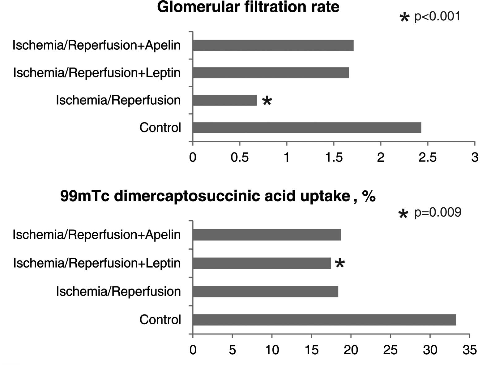
filtration rate (ml/min) | 2.43±1.26b | 0.68±0.35a | 1.66±0.49 | 1.71±0.34 | 0.001 |
| 99mTc
dimercaptosuccinic acid uptake (%) | 33.30±11.06c | 18.37±8.50 | 17.48±5.91a | 18.76±6.98 | 0.03 |
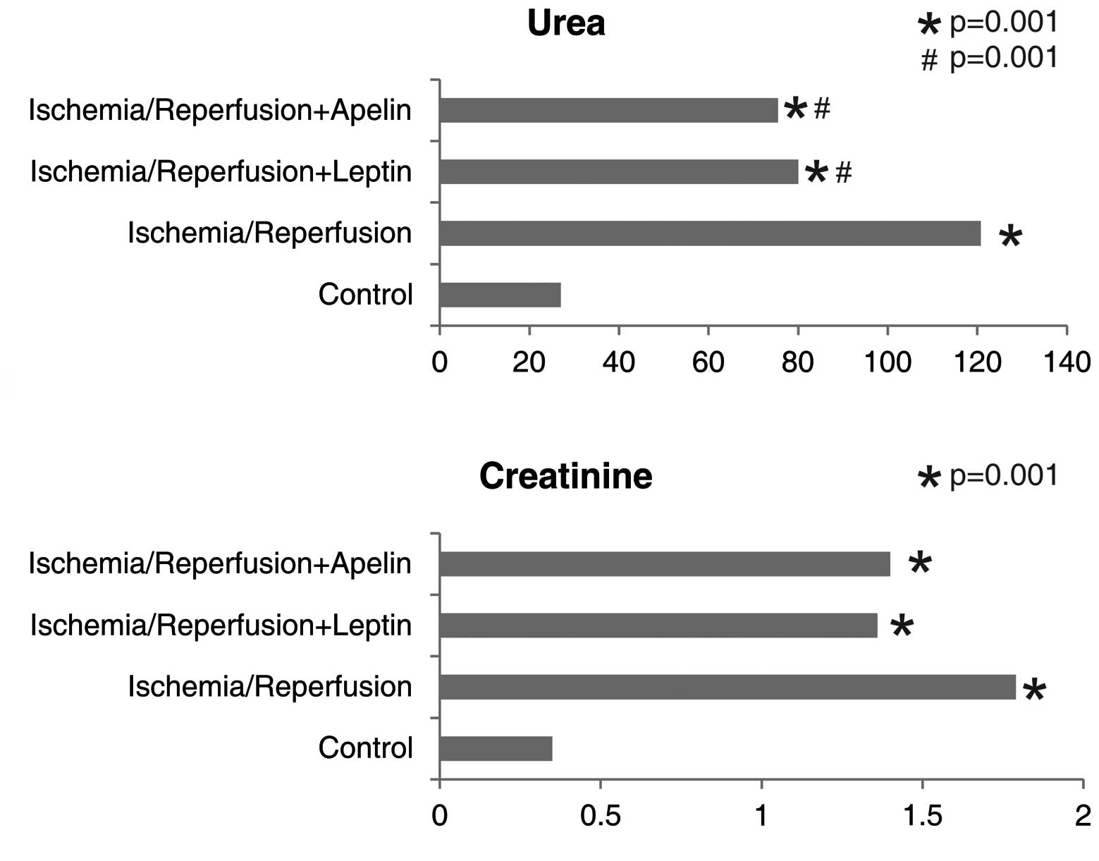
The urea level was significantly higher in the I/R
group compared to the control, I/R+L and I/R+A groups. Urea levels
of the I/R+L and the I/R+A groups were comparable, but were higher
than that of the control and lower than that of the I/R group.
Creatinine levels were higher in all three ischemic groups compared
to the control group (Fig. 2).
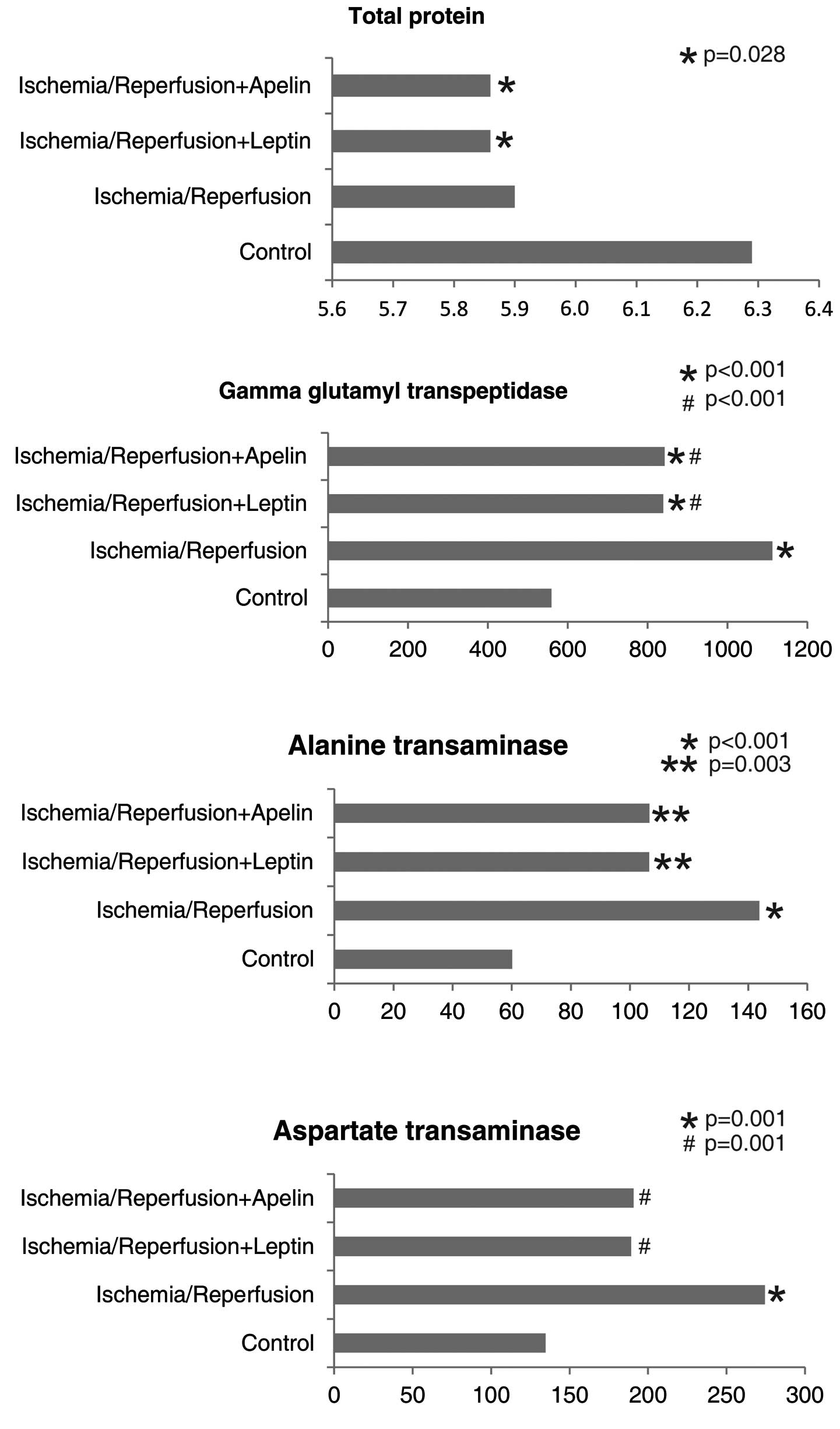
Total protein levels were lower in the I/R+L and
I/R+A groups compared to the control group. GGT levels were higher
in all three ischemic groups compared to the control group. GGT
levels of the I/R+L and I/R+A groups were comparable, but were
significantly lower than that of the I/R group. ALT levels were
higher in all three ischemic groups compared to the control group.
The AST level of the I/R group was higher than that of the control
group, while AST levels of the I/R+L and I/R+A groups were
significantly lower than that of the I/R group (Fig. 3).
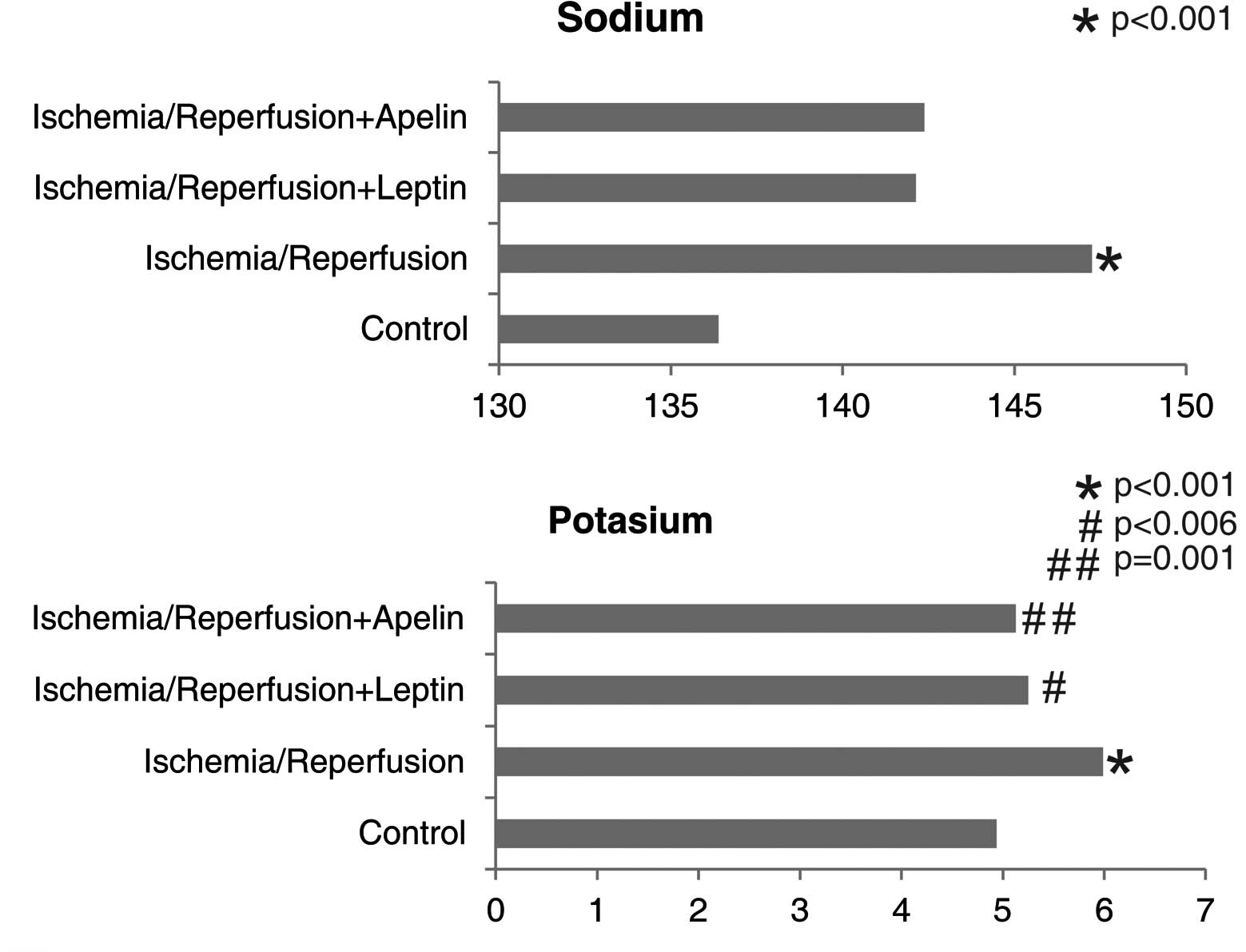
Sodium and potassium levels were significantly
higher in the I/R group compared to the control group. Potassium
levels of the I/R+L and I/R+A groups were significantly lower than
that of the I/R group (Fig.
4).
Glomerular filtration rate was markedly lower in the
I/R group compared to the control group. GFR values of the I/R+A
and I/R+L groups were not significantly, but numerically higher
than that of the I/R group. GFR values of the control, I/R+A and
I/R+L groups were comparable. DMSA uptake was lower in all three
ischemic groups compared to the control group; however, only the
difference between the I/R+L group and the control group reached a
significance level (Fig. 5). There
were significant differences between the study groups with respect
to the degree of renal damage (Table
III).
| Table III.The degree of pathological damage in
the study groups. |
Table III.
The degree of pathological damage in
the study groups.
| Renal damage
(n) | Control (n=7) |
Ischemia/reperfusion (n=8) |
Ischemia/reperfusion + leptin (n=7) |
Ischemia/reperfusion + apelin (n=8) | P-value |
|---|
| None | 7 | 0 | 1 | 3 | <0.001 |
| Mild | 0 | 0 | 3 | 3 | |
| Moderate | 0 | 2 | 3 | 1 | |
| Severe | 0 | 6 | 1 | 0 | |
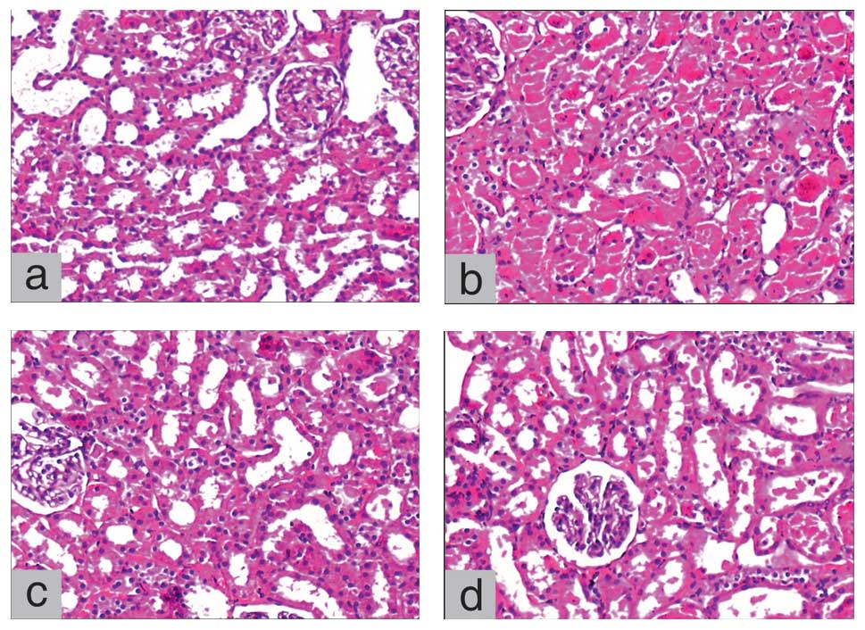
No pathological damage was observed in any of the
animals in the control group. While two animals had moderate and
six animals had severe renal damage in the I/R group, three animals
had moderate and only one animal had severe renal damage in the
I/R+L group. Although moderate damage was observed in one animal in
the I/R+A group, none of the animals in this group had severe renal
damage (Fig. 6).
Discussion
The main finding of the present study was that
pre-operatively administered leptin and apelin had protective,
functional and histopathological, effects against renal I/R injury.
Functional protection against I/R injury was demonstrated through
BUN, creatinine and GFR measurements, while histopathological
protection was demonstrated through examination of the degree of
pathological damage. Moreover, it was found that increments in the
AST, GGT, sodium and potassium levels demonstrated in the I/R group
were decreased in the I/R+A and I/R+L groups.
Tissue damage caused by ischemia is further
increased with reperfusion, and I/R injury occurs. This unfavorable
effect of reperfusion is more pronounced when the ischemic period
is prolonged. Vascular endothelial cells, leukocytes, oxygen
radicals, adhesion molecules and inflammatory mediators play a role
in the complex pathophysiology of I/R injury (1). Reperfusion results in impaired
endothelium-dependent dilation in arterioles, enhanced fluid
filtration and leukocyte plugging in capillaries, and leukocyte
trafficking and extravasation of plasma proteins in postcapillary
venules. Production of oxygen radicals is increased in activated
endothelial cells, and the balance between superoxide and nitric
oxide is impaired, leading to the release of inflammatory mediators
(e.g., platelet-activating factor, tumor necrosis factor).
Moreover, synthesis of adhesion molecules, which mediate
leukocyte-endothelial cell adhesion, is also increased (1).
Protective or therapeutic effects of a number of
substances against renal I/R injury have been investigated using
animal models in experimental studies. Improving effects of
antioxidants, including phosphate ester of vitamin C and vitamin E
(EPC-K1), edaravone (MCI-186), aminoguanidine, ascorbic acid, and
stobadine, against renal I/R injury have been demonstrated in
experimental studies employing rats and dogs (2,14).
Yamamoto et al (14)
investigated the effects of EPC-K1 in a rat model of I/R injury and
histopathologically demonstrated that tissue damage was lower in
rats receiving EPC-K1 prior to I/R compared to those that did not
receive EPC-K1. Moreover, the mean tissue damage score for renal
injury was significantly lower in the EPC-K1-treated group. BUN and
creatinine levels measured to assess the renal function were
significantly higher in the I/R group compared to the control
group, while they were significantly decreased in the
EPC-K1-treated group. Koga et al (15) reported that human atrial
natriuretic peptide (hANP) was effective in the treatment of a
number of types of I/R injuries and that the renal protective
effect of hANP could be associated with its antioxidant properties.
In this particular study on rats, serum BUN and creatinine levels
were demonstrated to be increased in the I/R group, but the
increment was significantly inhibited in the hANP-treated group,
and renal tissue damage was attenuated with hANP administration
(15). In accordance with the two
abovementioned studies, the present study also demonstrated that
urea and creatinine levels were higher in the I/R group compared to
the control group, but lower in the I/R+A and I/R+L groups compared
to the I/R group. These results indicate the protective effects of
leptin and apelin against renal I/R injury. In a study by Medeiros
et al investigating the effect of sildenafil on rat kidneys
subjected to normothermic I/R (16), the protective effect of sildenafil
was scintigraphically and histopathologically demonstrated. In the
present study, the histopathologically protective effects of apelin
and leptin against renal I/R injury were also demonstrated. While
severe renal damage was found in six animals in the I/R group, only
one animal had severe renal damage in the I/R+L group. No severe
damage was observed in any of the rats in the I/R+A group. However,
there was no significant difference between the I/R, I/R+A and
I/R+L groups with respect to scintigraphic findings.
It has been reported that EPO reduces post-ischemic
structural damage and preserves renal functions, particularly in
male rats (17). Prókai et
al (17) demonstrated via BUN
and creatinine measurement and histopathological examination, that
EPO had a protective effect against I/R injury. Lifor®
is a novel artificial organ-preserving solution that comprises
nutrients, growth factors and a non-protein oxygen and nutrient
carrier. It was demonstrated in a rat model, via creatinine
measurement and histopathological examination, that lifor solution
reduced both warm renal I/R and cold storage injury, and that Lifor
may provide protection against apoptosis (18). The protective effect of isoflurane
preconditioning against renal I/R injury was demonstrated via urea
and creatinine measurements and via histopathological examination
in a study conducted on rats (19). In another study on rats, the
beneficial effect of isoflurane plus remifentanil on the kidney was
demonstrated via flow cytometry and serum creatinine levels
(20).
The effects of apelin and leptin against I/R injury
have been investigated in a number of animal models. The effect of
apelin on the myocardium was investigated in vivo using a
mouse model of I/R injury, and in vitro using the
Langendorff-perfused isolated mouse heart model by measuring the
infarct area. Moreover, the direct effects of apelin on rat
cardiomyocytes were evaluated in vitro, and it was
demonstrated that apelin protected the myocardium against I/R
injury (21,22). It has been reported that apelin
plays a role in inflammation (23)
and the pathophysiology of hepatic diseases (24), and that apelin level is predicted
by the presence of coronary artery disease in kidney allograft
recipients (25). It has been
demonstrated that apelin plays a critical role in the development
of a functional vascular network and that adipocyte-derived apelin
is upregulated in case of hypoxia (26). In a study by Hacioglu et al
(10), the protective effect of
leptin against I/R injury in the small intestine of rats was
demonstrated via tissue malondialdehyde and nitric oxide
concentrations and histopathological examination. In another study
employing a rat model of intestinal I/R injury, it was demonstrated
via histopathological examination that pre-treatment with leptin
prevented gut mucosal damage and improved intestinal rehabilitation
(27). Expression of apelin
receptor mRNA in all nephron segments in the rat kidney suggests
that apelin has a modulatory effect on tubular function (28).
Urea and creatinine levels, which were examined in
the majority of the abovementioned studies to assess the renal
function, were also assessed in the present study. In addition, GFR
values were evaluated. Similarly, as performed in the majority of
the studies, histopathological examination was conducted to
demonstrate tissue damage. Moreover, we compared the study groups
via scintigraphic examination. Based on the findings that increased
levels of urea and creatinine in the I/R group demonstrated a
decrease in the I/R+A and I/R+L groups, while low GFR in the I/R
group reached the control levels in the I/R+A and I/R+L groups, and
that severe damage was observed in six animals in the I/R group,
one in the I/R+L group and none in the I/R+A group, it can be
suggested that apelin and leptin have improving effects against
renal I/R injury. It should be considered that the effects of
leptin and apelin on experimental ischemic kidney might be
different from those on the human kidney, and that the findings of
the present study should be supported by clinical studies. Further
experimental and clinical studies are warranted for an improved
understanding of the effects of these substances in the
pre-treatment of ischemic kidney and for the use of these
substances in clinical practice. Currently, surgical procedures and
organ transplantations have increased and become widespread. Thus,
pharmacological approaches with proven clinical efficacy are
required in order to prevent and treat renal I/R injury that might
occur due to peri-operative reasons.
References
|
1.
|
Carden DL and Granger DN: Pathophysiology
of ischaemia-reperfusion injury. J Pathol. 190:255–266. 2000.
View Article : Google Scholar : PubMed/NCBI
|
|
2.
|
Chatterjee PK: Novel pharmacological
approaches to the treatment of renal ischemia-reperfusion injury: a
comprehensive review. Naunyn Schmiedebergs Arch Pharmacol.
376:1–43. 2007. View Article : Google Scholar : PubMed/NCBI
|
|
3.
|
Li MD: Leptin and beyond: an odyssey to
the central control of body weight. Yale J Biol Med. 84:1–7.
2011.PubMed/NCBI
|
|
4.
|
Cao R, Brakenhielm E, Wahlestedt C, et al:
Leptin induces vascular permeability and synergistically stimulates
angiogenesis with FGF-2 and VEGF. Proc Natl Acad Sci USA.
98:6390–6395. 2001. View Article : Google Scholar : PubMed/NCBI
|
|
5.
|
Wolf G, Hamann A, Han DC, et al: Leptin
stimulates proliferation and TGF-beta expression in renal
glomerular endothelial cells: potential role in glomerulosclerosis.
Kidney Int. 56:860–872. 1999. View Article : Google Scholar : PubMed/NCBI
|
|
6.
|
Mastronardi CA, Yu WH and McCann SM:
Resting and circadian release of nitric oxide is controlled by
leptin in male rats. Proc Natl Acad Sci USA. 99:5721–5726. 2002.
View Article : Google Scholar : PubMed/NCBI
|
|
7.
|
Lin J, Yan GT, Wang LH, et al: Leptin
fluctuates in intestinal ischemia-reperfusion injury as
inflammatory cytokine. Peptides. 25:2187–2193. 2004. View Article : Google Scholar : PubMed/NCBI
|
|
8.
|
Shi Y, Yan GT, Lin J, et al: Intestinal
ischemia-reperfusion injury made leptin decreased. Regul Pept.
133:27–31. 2006. View Article : Google Scholar : PubMed/NCBI
|
|
9.
|
Kleinz MJ and Davenport AP: Emerging roles
of apelin in biology and medicine. Pharmacol Ther. 107:198–211.
2005. View Article : Google Scholar : PubMed/NCBI
|
|
10.
|
Hacioglu A, Algin C, Pasaoglu O, et al:
Protective effect of leptin against ischemia-reperfusion injury in
the rat small intestine. BMC Gastroenterol. 5:372005. View Article : Google Scholar : PubMed/NCBI
|
|
11.
|
Pan CS, Teng X, Zhang J, et al: Apelin
antagonizes myocardial impairment in sepsis. J Card Fail.
16:609–617. 2010. View Article : Google Scholar : PubMed/NCBI
|
|
12.
|
Kaya H, Balcı TA, Cobaner A, et al:
Evaluation on the renal tubular damage in rats with acute
haemorrhagic hypovolemic shock using Tc-99m DMSA uptake
scintigraphy. Turk J Nucl Med. 12:85–89. 2003.
|
|
13.
|
Parra C, Salas P and Dominguez J: Effects
of immunosuppressive drugs on rat renal ischemia reperfusion
injury. Transplant Proc. 42:245–247. 2010. View Article : Google Scholar : PubMed/NCBI
|
|
14.
|
Yamamoto S, Hagiwara S, Hidaka S, et al:
The antioxidant EPC-K1 attenuates renal ischemia-reperfusion injury
in a rat model. Am J Nephrol. 33:485–490. 2011. View Article : Google Scholar : PubMed/NCBI
|
|
15.
|
Koga H, Hagiwara S, Kusaka J, et al: Human
atrial natriuretic peptide attenuates renal ischemia-reperfusion
injury. J Surg Res. Nov 4–2010.(Epub ahead of print).
|
|
16.
|
Medeiros PJ, Villarim Neto A, Lima FP, et
al: Effect of sildenafil in renal ischemia/reperfusion injury in
rats. Acta Cir Bras. 25:490–495. 2010. View Article : Google Scholar : PubMed/NCBI
|
|
17.
|
Prókai A, Fekete A, Bánki NF, et al:
Renoprotective effect of erythropoietin in rats subjected to
ischemia/reperfusion injury: gender differences. Surgery.
150:39–47. 2011.PubMed/NCBI
|
|
18.
|
Regner KR, Nilakantan V, Ryan RP, et al:
Protective effect of Lifor solution in experimental renal
ischemia-reperfusion injury. J Surg Res. 164:291–297. 2010.
View Article : Google Scholar : PubMed/NCBI
|
|
19.
|
Zhang L, Huang H, Cheng J, et al:
Pre-treatment with isoflurane ameliorates renal
ischemic-reperfusion injury in mice. Life Sci. 88:1102–1107. 2011.
View Article : Google Scholar : PubMed/NCBI
|
|
20.
|
Vianna PT, Castiglia YM, Braz JR, et al:
Remifentanil, isoflurane, and preconditioning attenuate renal
ischemia/reperfusion injury in rats. Transplant Proc. 41:4080–4082.
2009. View Article : Google Scholar : PubMed/NCBI
|
|
21.
|
Simpkin JC, Yellon DM, Davidson SM, et al:
Apelin-13 and apelin-36 exhibit direct cardioprotective activity
against ischemia-reperfusion injury. Basic Res Cardiol.
102:518–528. 2007. View Article : Google Scholar : PubMed/NCBI
|
|
22.
|
Zeng XJ, Zhang LK, Wang HX, et al: Apelin
protects heart against ischemia/reperfusion injury in rat.
Peptides. 30:1144–1152. 2009. View Article : Google Scholar : PubMed/NCBI
|
|
23.
|
Malyszko J, Malyszko JS, Pawlak K, et al:
Apelin, a novel adipocytokine, in relation to endothelial function
and inflammation in kidney allograft recipients. Transplant Proc.
40:3466–3469. 2008. View Article : Google Scholar : PubMed/NCBI
|
|
24.
|
Kunduzova O, Alet N, Delesque-Touchard N,
et al: Apelin/APJ signaling system: a potential link between
adipose tissue and endothelial angiogenic processes. FASEB J.
22:4146–4153. 2008. View Article : Google Scholar : PubMed/NCBI
|
|
25.
|
Malyszko J, Malyszko JS, Pawlak K, et al:
Visfatin and apelin, new adipocytokines, and their relation to
endothelial function in patients with chronic renal failure. Adv
Med Sci. 53:32–36. 2008. View Article : Google Scholar : PubMed/NCBI
|
|
26.
|
Principe A, Melgar-Lesmes P,
Fernández-Varo G, et al: The hepatic apelin system: a new
therapeutic target for liver disease. Hepatology. 48:1193–1201.
2008. View Article : Google Scholar : PubMed/NCBI
|
|
27.
|
Sukhotnik I, Helou H, Lurie M, et al: The
effect of leptin on intestinal recovery following
ischemia-reperfusion injury in a rat. Pediatr Surg Int. 23:473–478.
2007. View Article : Google Scholar : PubMed/NCBI
|
|
28.
|
Hus-Citharel A, Bouby N, Frugière A, et
al: Effect of apelin on glomerular hemodynamic function in the rat
kidney. Kidney Int. 74:486–494. 2008. View Article : Google Scholar : PubMed/NCBI
|