Introduction
Limb joint and soft tissue injuries are among the
most common conditions in clinical practice and are often followed
by peripheral nerve injury in 5% of cases. The peripheral nerve is
a mixed nerve, and nerve fibers emanating from one side cross over
to the other within nerve bundles. Therefore, the complete recovery
rate is only 10–25% after injury and treatment of peripheral nerve
injuries remains a challenge worldwide.
Several researchers have performed numerous studies
in this field and proposed that the basic condition for peripheral
nerve repair is the construction of the nerve regenerative chamber,
which can create a specific microenvironment for regeneration of
the nerve (1,2). As stem cell research has developed,
study into novel tissue engineering seed cells has received
increasing attention. Muscle-derived stem cells (MDSCs) originate
from muscle tissue. In addition to aspects shared by stem cells,
MDSCs also exhibit such characteristics as harmlessness to the
body, a wide range of sources, ready acceptance by patients, lack
of restrictions of source of donor, lack of immunosuppressive
effects and fewer ethical restrictions. MDSCs are therefore ideal
seed cells for peripheral nerve repair (3–6). As
the study of Chinese traditional medicine has developed, it has
been confirmed that Salvia (Salvia miltiorrhiza) may
be used to induce the differentiation of multi-functional stem
cells into neuron-like cells (7–10).
In the present study, MDSCs whose differentiation
was induced by ciliary neurotrophic factor (CNTF) and Salvia
were used to repair sciatic nerve injury in rats in order to
further confirm their potential as pluripotent stem cells. This
study may contribute to a theoretical concept of more latent
cytokines and novel seed cells for the construction of tissue
engineered peripheral nerve grafts.
Materials and methods
Experimental animals
Adult Sprague-Dawley (SD) rats (n=12) were divided
into 2 groups. The sciatic nerve in the right lower limb was
exposed under the anesthetized condition of 10% chloral hydrate
(0.3 ml/100 g) injection into the abdominal cavity. The tissue,
which was 0.5 cm above the sciatic nerve bifurcation, was broken
using a hemostat. After induction, MDSCs were transferred to sodium
hyaluronate gel and placed into the damaged area. An untreated
control group was also included in this study. The surgical area
was sutured after washing with gentamycin sulfate solution. Animal
experiments were performed in accordance with the Guide for the
Care and Use of Laboratory Animals.
Experimental materials
General observation
The recovery of the wound and the formation of
ulcers in the plantar region were recorded. Under mild anesthesia,
the sensory function recovery was examined following plantar
puncture.
Sciatic nerve function index
(SFI)
Four weeks after surgery, SFI was calculated using
the method described by Reynolds and Weiss (11). Hind legs of the rats were dyed with
ink. When the rats walked on the surface of one piece of white
paper, the footprints of healthy feet (N) and wounded feet (E) were
measured in 3 indices as follows: length of footprint (IPL, from
toe to heel), width of toes (ITS, from the 1st to the 5th toe) and
width of middle toes (IIT, from the 2nd to the 4th toe). The
results should be accurate to 0.1 mm. SFI was calculated according
to the formula described by Bain et al(12): SFI = −38.3 [(EPL - NPL)/NPL] +
109.5 [(ETS - NTS)/NTS] + 13.3 [(EIT - NIT)/NIT] −8.8. A SFI value
between 0 and 11% represented normal nerve function, whereas −100%
represented complete damage of nerve function, and −11 to −100%
incomplete nerve function recovery.
Electrophysiology test
Four weeks after surgery, the sciatic nerve at the
surgical site was exposed under anesthesia. The transplanted
section was linked with an electrode and the gastrocnemius was
linked with a recording electrode to determine the action potential
(AP) and mean conductive velocity (MCV) of the sciatic nerve.
Recovery rate of gastrocnemius wet
weight
Four weeks following surgery, the gastrocnemius was
completely removed and its wet weight was then measured by ER-182
electronic balance (1/2,000 g) to calculate its recovery rate.
Statistical analysis
All statistic data are presented as the means ± SD.
statistical analyses were performed using SPSS 10.0 software. A
P-value <0.05 was considered to indicate a significant
difference and P<0.01 a statistically significant
difference.
Results
General observation
Following surgery it was found that the toes of the
rats were crooked and muscle contraction was not observed when the
toes were punctured. One week after surgery, it was found that the
surgical area had recovered well without infection. However, there
was redness and swelling on the skin of the right foot and
weight-bearing area. All the rats were inactive and the operated
legs were immobile. Two weeks after surgery, ulcers were observed
on the skin behind the ankle of the operated area in some rats,
while other rats exhibited redness and swelling on the soles of
their feet. There were no obvious differences between the MDSC and
control groups.
Three weeks after surgery, the rats’ moods had
recovered and the treated legs became active. However, the toes and
knee joint remained crooked with various degrees of muscle
shrinkage in the treated legs. At the same time, inflammation and
ulcers were found in the plantar region. Four weeks after surgery,
the knee joints of all the rats remained crooked; however, the
inflamed area and ulcers in the plantar region were no longer
observed. All the rats reacted when their toes were punctured. In
the MDSC group, there were 3 rats that generally recovered from
inflammation and ulcers, and partly recovered from muscle
shrinkage. However, all the rats in the control group exhibited
various degrees of muscle shrinkage in the treated legs.
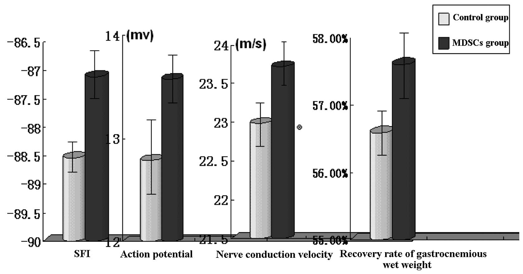
SFI
SFI was statistically higher in the MDSC group in
comparison with the control group (P<0.05; Table I and Fig. 1).
 | Table IExperimental data in the MDSC and
control group. |
Table I
Experimental data in the MDSC and
control group.
| Group | SFI | Action potential | Nerve conductive
velocity | Recovery rate of
gastrocnemius muscle wet weight (%) |
|---|
| MDSC | −87.11±0.88a | 13.58±0.53a | 23.72±0.62a | 57.6±0.0094a |
| Control | −88.54±0.64 | 12.79±0.69 | 22.99±0.61 | 56.59±0.0076 |
Electrophysiology test
Sciatic action potential and nerve conductive
velocity were statistically higher in the MDSC group in comparison
with the control group (P<0.05; Table I and Fig. 1).
Recovery rate of gastrocnemius wet
weight
The recovery rate of gastrocnemius wet weight was
statistically higher in the MDSC group in comparison with the
control group (P<0.05; Table I
and Fig. 1).
Discussion
In Chinese traditional medicine, peripheral nerve
injury is considered to belong to the category of flaccidity.
Flaccidity is a syndrome with such symptoms as weak tendons and
feeble muscle, leading to muscle shrinkage due to lack of bodily
exercise.
Nerve damage can cause blood stasis and meridian
obstruction, leading to poor circulation and malnutrition. Bodily
functions therefore exhibit anomalous changes and pathological
changes occur in such a situation.
Modern experimental research has confirmed that
medicine promoting blood circulation to remove blood stasis can
achieve favorable results in the treatment of disease caused by
peripheral nerve injury. Qian et al(13) demonstrated that Bu Yang Huan Wu
Tang reduces the shrinkage of neurons in injured peripheral nerve
and aids nerve recovery. Fang et al(14) confirmed that such compound Chinese
herbal medicine as ginseng, Astragalus and Salvia can
promote peripheral nerve regeneration.
As a result of technical developments in genetic
engineering, cells with biological activity are used as carriers
for nutritional nerve factor. After modification, the carrier cells
can constantly provide nutritional nerve factor to the gene, which
provides a prospective application for nerve restoration and
regeneration with nutritional nerve factor and which also gives
hope to nerve tissue engineering of cell types.
MDSCs originate from muscle. They are precursor
cells of skeletal muscle cells. Compared with other cells, they
exhibit such characteristics as partial differentiation ability,
favorable histocompatibility, harmlessness to the body, wide range
of sources and ready acceptance by patients. Several investigators
have noted that MDSCs originating from skeletal muscle have the
characteristics of stem cells whose differentiation is induced by
CNTF (15–26). This study used the liquid culture
method allowing the generation of a large number of MDSCs and the
induction of MDSCs to obtain amplification ability. MDSCs after the
3rd generation may be used for cell differentiation and cell
treatment. As seed cells, MDSCs play a substantial role in the
restoration of rat sciatic nerve injury and regeneration of the
peripheral nerve under the effect of Salvia.
Three elements of nerve tissue engineering include
seed cells, nerve carrier and nerve nutritional factor. The source
and large proliferation of seed cells are the most important
problems to be solved. In addition, other research on bionic
scaffold material has received increasing attention. Research on
nerve carriers has also made substantial progress.
Hyaluronic acid is a linear polysaccharide
macromolecular material, which comprises (1→4)-D-glucuronic acid-β
(1→3)-N-acetyl glucosamine disaccharide units and is widely present
in the connective tissue of humans and animals. With a
3-dimensional network structure formed by interaction between
molecules, it has such characteristics as maintaining tissue
formation, important psychological function and good
viscoelasticity. Glucuronic acid is also a main element in
compounding cellular and extracellular matrices, which occupy vital
roles in maintaining normal physiological function, supporting the
growth and differentiation of cells and restoration of injured
tissue. In the early period of wounding, such cells as fibroblast
cells and macrophages exhibit a marked increase in compounding
glucuronic acid in the function of growth factor. It has been shown
in the comparison between the embryo before and after birth that
glucuronic acid maintains high thickness, which prevents
collagenous fibrous tissue from contracting and even aids tissue
recovery without any cicatricial sign. After birth, glucuronic acid
remains at high thickness in the early period; later it is
gradually dissolved by hyaluronidase. At the same time, the
combination of collagenous fiber increases. Finally, cicatricial
restoration is formed. Additionally, glucuronic acid has such
functions as promoting blood vessel growth, partly improving
circulation of injured areas and aiding growth factor expression.
There are high quantities of carboxyl and hydroxyl in glucuronic
acid molecules. These two elements can form an intermolecular and
intramolecular hydrogen bond in water with such potency that it can
hold 1,000 times its weight in water (27,28).
In the present study, sodium hyaluronate gel was
used as a cell carrier. In the recovery process, the extracellular
matrix had an effect on cell performance. Cells also modified the
extracellular matrix in various ways, forming the
micro-environment. The whole recovery process, which was constantly
changing, supported the growth of the cells. In conclusion, the
restoration and regeneration of tissue is a process in which cells
composed of different molecules and macromolecules proliferate,
differentiate and reconstruct the matrix.
References
|
1
|
Mackinnon SE, Dellon AL, Hudson AR and
Hunter DA: Nerve regeneration through a pseudosynovial sheath in a
primate model. Plast Reconstr Surg. 75:833–841. 1985. View Article : Google Scholar : PubMed/NCBI
|
|
2
|
Seckel BR: Enhancement of peripheral nerve
regeneration. Muscle Nerve. 13:785–800. 1990. View Article : Google Scholar : PubMed/NCBI
|
|
3
|
Wallace GQ, Lapidos KA, Kenik JS and
McNally EM: Long-term survival of transplanted stem cells in
immunocompetent mice with muscular dystrophy. Am J Pathol.
173:792–802. 2008. View Article : Google Scholar : PubMed/NCBI
|
|
4
|
Shimazaki T, Shingo T and Weiss S: The
ciliary neurotrophic factor/leukemia inhibitory factor/gp130
receptor complex operates in the maintenance of mammalian forebrain
neural stem cells. J Neurosci. 21:7642–7653. 2001.
|
|
5
|
Helgren ME, Squinto SP, Davis HL, Parry
DJ, Boulton TG, Heck CS, Zhu Y, Yancopoulos GD, Lindsay RM and
DiStefano PS: Trophic effect of ciliary neurotrophic factor on
denervated skeletal muscle. Cell. 11:493–504. 1994. View Article : Google Scholar : PubMed/NCBI
|
|
6
|
Rivera FJ, Kandasamy M, Couillard-Despres
S, Caioni M, Sanchez R, Huber C, Weidner N, Bogdahn U and Aigner L:
Oligodendrogenesis of adult neural progenitors: differential
effects of ciliary neurotrophic factor and mesenchymal stem cell
derived factors. J Neurochem. 107:832–843. 2008. View Article : Google Scholar : PubMed/NCBI
|
|
7
|
Xia W, Xiang P, Zhang L, Chen Z, Yu W,
Zhang X, Li Y and Li S: Human mesenchymal stem cells differentiate
into neuron-like cells with Tanshinone II A. Zongguo Bing Li Sheng
Li Za Zhi. 19:865–869. 2003.(In Chinese).
|
|
8
|
Ma L, Feng X, Yang L, Xie Q, Luo M and
Huang T: Induction of human umbilical cord blood mesenchymal stem
cells into nerve-like cells by Salvia miltiorrhiza. Shiyong
Erke Linchuang Zazhi. 20:1125–1128. 2005.(In Chinese).
|
|
9
|
Sanchez-Ramos J, Song S, Cardozo-Pelaez F,
et al: Adult bone marrow stromal cells differentiate into neural
cells in vitro. Exp Neurol. 164:247–256. 2000. View Article : Google Scholar : PubMed/NCBI
|
|
10
|
Yu Q, Lian J and Guo Y: Experimental study
on differentiation of bone mesenchymal stem cells into neuron-like
cells with Salvia miltiorrhiza injection. Zhongguo Zhong Xi
Yi Jie He Ji Jiu Za Zhi. 13:210–213. 2006.
|
|
11
|
Reynolds BA and Weiss S: Generation of
neurons and astrocytes from isolated cells of the adult mammalian
central nervous system. Science. 255:1707–1710. 1992. View Article : Google Scholar : PubMed/NCBI
|
|
12
|
Bain JR, Mackinnon SE and Hunter DA:
Functional evaluation of complete sciatic, peroneal, and posterior
tibial nerve lesions in the rat. Plast Reconstr Surg. 83:129–138.
1989. View Article : Google Scholar : PubMed/NCBI
|
|
13
|
Qian Y, Tao Y, Huang C and Wu Y:
Experimental study of the survival-promoting effect of Buyang
Huanwu decoction on neurons after peripheral nerve injury. Zhonghua
Shou Waike Zazhi. 9:152–154. 2002.(In Chinese).
|
|
14
|
Fang Y, Chen D and Gu Y: Facilitated
regeneration of rat peripheral nerves with mixture of dangshen,
astragalus root, red sage root. Zhonghua Shou Waike Zazhi.
14:181–183. 1998.(In Chinese).
|
|
15
|
Zeng X, Wang W, Sun L, Zhang L and Zeng L:
Differentiation of muscle-derived stem cells into neuron-like cells
induced by ciliary neurotrophic factor and compound Salvia
miltiorrhiza injection in vitro. Zhonghua Zu Zhi Gong Cheng Yan
Jiu. 13:5336–5340. 2009.(In Chinese).
|
|
16
|
Blanco-Bose WE, Yao CC, Kramer RH and Blau
HM: Purification of mouse primary myoblast based on alpha 7
integrin expression. Exp Cell Res. 265:212–220. 2001. View Article : Google Scholar : PubMed/NCBI
|
|
17
|
Galli R, Borello U, Gritti A, et al:
Skeletal myogenic potential of human and mouse neural stem cells.
Nat Neurosci. 3:986–991. 2000. View
Article : Google Scholar : PubMed/NCBI
|
|
18
|
Kuznetsov SA, Mankani MH, Gronthos S, et
al: Circulating skeletal stem cells. J Cell Biol. 153:1133–1140.
2001. View Article : Google Scholar : PubMed/NCBI
|
|
19
|
Chen S, Zhang Q, Wu X, et al:
Dedifferentiation of lineage-committed cells by a small molecule. J
Am Chem Soc. 126:410–411. 2004. View Article : Google Scholar : PubMed/NCBI
|
|
20
|
Orellana RA, Suryawan A, Kimball SR, Wu G,
Nguyen HV, Jefferson LS and Davis TA: Insulin signaling in skeletal
muscle and liver of neonatal pigs during endotoxemia. Pediatr Res.
64:505–510. 2008. View Article : Google Scholar : PubMed/NCBI
|
|
21
|
Chen X, Mao Z, Liu S, et al:
Dedifferentiation of adult human myoblasts induced by ciliary
neurotrophic factor in vitro. Mol Biol Cell. 16:3140–3151. 2005.
View Article : Google Scholar : PubMed/NCBI
|
|
22
|
Zhu W, Shiojima I, Ito Y, et al: IGFBP-4
is an inhibitor of canonical Wnt signalling required for
cardiogenesis. Nature. 454:345–349. 2008. View Article : Google Scholar : PubMed/NCBI
|
|
23
|
Cebolla B and Vallejo M: Nuclear factor-I
regulates glial fibrillary acidic protein gene expression in
astrocytes differentiated from cortical precursor cells. J
Neurochem. 97:1057–1070. 2006. View Article : Google Scholar : PubMed/NCBI
|
|
24
|
Bhattacharya I and Ullrich A: Endothelin-1
inhibits adipogenesis: role of phosphorylation of Akt and ERK1/2.
FEBS Lett. 580:5765–5771. 2006. View Article : Google Scholar : PubMed/NCBI
|
|
25
|
Xie Y, Yu Z, Liu L, Guo Y and Lou L:
Biphasic regulation of extracellular signal-regulated kinases by
scalaradial, a secretory phospholipase A(2) inhibitor. Cancer Biol
Ther. 5:988–992. 2006. View Article : Google Scholar : PubMed/NCBI
|
|
26
|
Mukherjee A and Rotwein P: Insulin-like
growth factor-binding protein-5 inhibits osteoblast differentiation
and skeletal growth by blocking insulin-like growth factor actions.
Mol Endocrinol. 22:1238–1250. 2008. View Article : Google Scholar : PubMed/NCBI
|
|
27
|
Morrison SJ, Shah NM and Anderson DJ:
Regulatory mechanisms in stem cell biology. Cell. 88:287–298. 1997.
View Article : Google Scholar : PubMed/NCBI
|
|
28
|
Lundberg C, Martínez-Serrano A, Cattaneo
E, et al: Survival, integration, and differentiation of neural stem
cell lines after transplantation to the adult rat striatum. Exp
Neurol. 145:342–360. 1997. View Article : Google Scholar : PubMed/NCBI
|