Introduction
Meningiomas are the most frequent type of
intracranial tumors (1,2). However, treatments for meningiomas
using microsurgical manipulation are highly challenging due to
several factors, such as the location of the meningioma,
complicated surrounding vascular networks including the deep veins
and supplying arteries, and the anatomical obstacles of the
parasagittal and falx (3,4). It is important for the neurosurgeon
to obtain the most precise information concerning the degree of
tumor involvement of critical vascular structures prior to surgery
(5). The advent of spiral computed
tomography has led to the development of 3-dimensional computed
tomographic angiography (3D-CTA) (6). 3D-CTA provides a noninvasive and
rapid diagnosis of intracranial aneurysms (7–9).
3D-CTA is a convenient technique that provides a 3D visual
reconstruction of the tumor and its blood supply. However, few
studies concerning the 3D-CTA imaging of meningioma have been
reported. To investigate the role of 3D-CTA in the preoperative
evaluation of meningioma, we have developed a protocol for
performing 3D-CTA in cranial meningioma. The reliability of 3D-CTA
in the detection and evaluation of meningioma is compared with that
of microsurgical findings. The primary purpose of this study was to
objectively compare the anatomical information provided by 3D-CTA
with the results obtained by surgery.
Materials and methods
Patients
Between October 2001 and May 2012, a total of 331
patients with meningiomas confirmed by CT and MRI were examined by
3D-CTA. The patients comprised 116 men and 215 women, ranging in
age from 34 to 78 years (mean 45.9 years). The locations of the
tumors were observed to be parasagittal and falcine in 125 cases,
sphenoidal in 39 cases, in the olfactory groove in 19 cases,
tentorial in 21 cases, parasellar in 33 cases, petroclival in 29
cases, intraventricular in 7 cases and on the convexity of the
brain in 58 cases. Informed consent was obtained from patients and
the study was approved by the ethics committee of Xuzhou Medical
College.
Methods
3D-CTA was performed on the patients with the use of
a General Electric LightSpeed Plus CT scanner (General Electric,
Milwaukee, WI, USA). An intravenous catheter was inserted in the
antecubital vein. Iobitridol (1.5 ml/kg) was prepared for
administration by a power injector (Medrad, Indianola, PA, USA) at
a rate of 3.0 ml per second. The scans were prescribed starting at
40 sec after the injection. These images were further reconstructed
with the General Electric Advantage Windows 3-D workstation
(General Electric). The reconstructed images were then processed at
the workstation into color-shaded surface display (SSD), maximum
intensity projection (MIP) and shaded volume rendering (SVR)
images. This was performed by the scanner technician, with either a
neurosurgeon or neuroradiologist to provide editing assistance. We
rotated the 3D-CTA images from every point of view in order to
display the meningioma and the relationship of the cranial bone and
vessels surrounding the tumor. 3D-CTA provides imaging features to
suggest the diagnosis of meningioma and to delineate the cortical
and vascular anatomy for preoperative planning.
Results
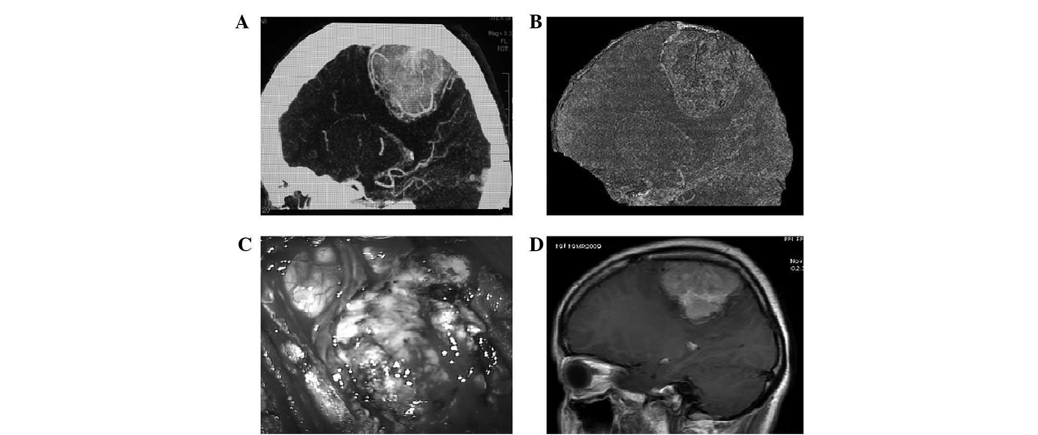
Among the 125 patients with parasagittal and falcine
meningiomas, 3D-CTA demonstrated that the sagittal venous sinuses
were partially occluded in 109 cases (Fig. 1). The anterior third of the
sagittal sinus was completely occluded in 16 cases. 3D-CTA
demonstrated the effect of tumors on major cerebral arteries. Among
the 39 patients with sphenoidal ridge meningioma, displacement of
the internal carotid artery (ICA), anterior cerebral artery (ACA)
and middle cerebral artery (MCA) were shown in 32 cases, and
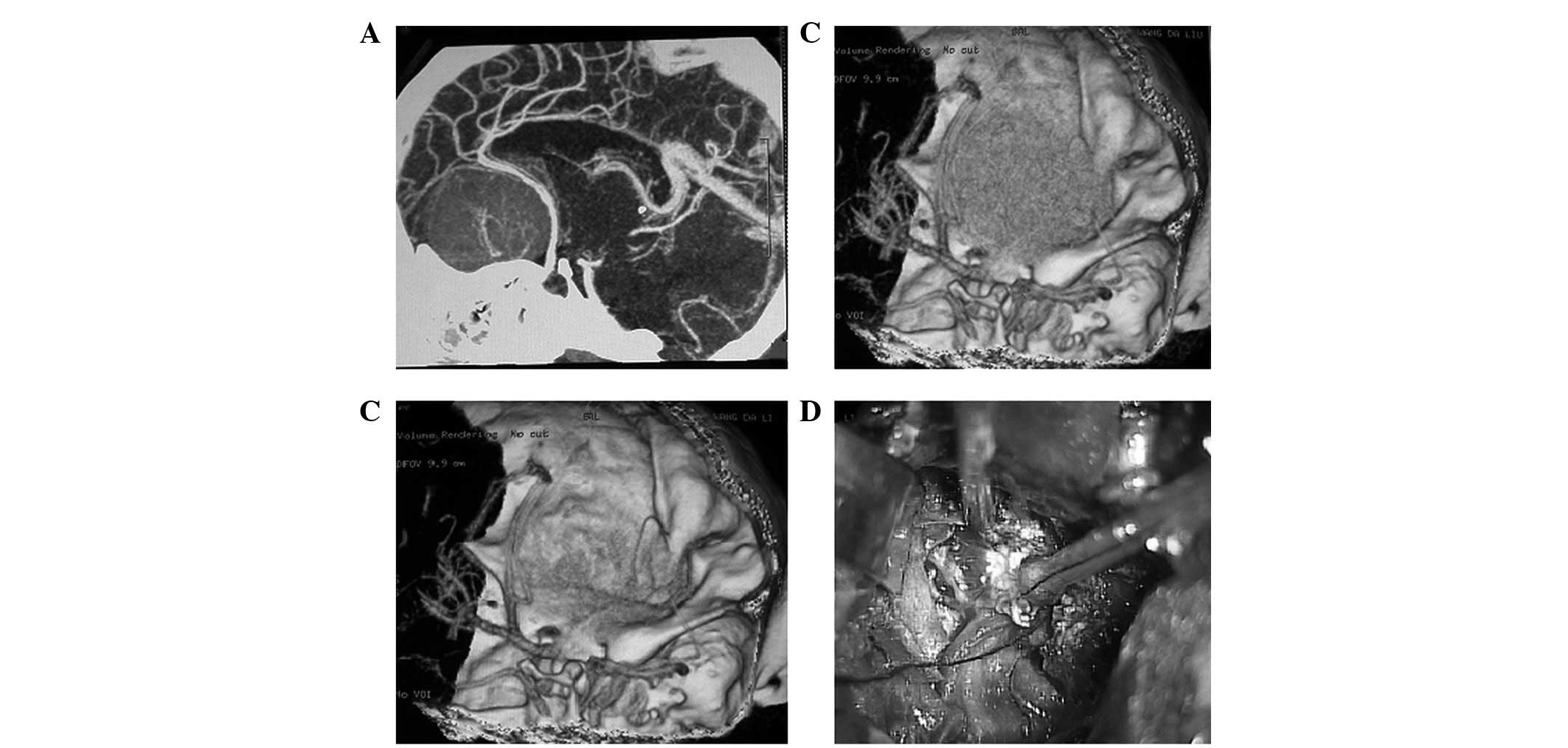
encasement in 7 cases. Among the 19 patients with olfactory groove
meningioma, the ACA, MCA and their proximal branches were displaced
in 16 patients (Fig. 2), and
encased in 3 patients. Among the 33 patients with parasellar
meningioma, displacement of the ICA, ACA and MCA were shown in 29
cases, with encasement in 4 cases. 3D-CTA demonstrated the
relationship of the tumor, bone, clivus and basilar artery clearly
among the 29 patients with petroclival meningiomas. The
relationship of the transverse sinus and tumor were demonstrated
clearly in 21 cases of tentorial meningioma. The relationship of
the cortical vasculature and the tumor were demonstrated clearly in
27 cases of intraventricular and convexity meningioma. The 3D-CTA
images corresponded well to the surgical findings.
Discussion
Meningiomas are the most frequent group of
intracranial tumors, accounting for approximately one-third of all
primary brain tumors (10). The
major problem in management of meningioma is the increased
vascularity of the tumor (1). The
management of intraoperative bleeding during the removal of large
meningiomas is crucial for safe and efficient surgery (11–13).
It is important to the neurosurgeon to obtain the most precise
information concerning the degree of tumor involvement with
critical vascular structures.
3D-CTA performed with a spiral CT scanner has been
used for intracranial aneurysm detection (6–9).
3D-CTA offers a tremendous ability to provide anatomical
information regarding aneurysms. 3D-CTA may provide sufficient
preoperative cranial vascular information about the meningioma. In
the current study, 3D-CTA not only showed the tumors stained by
iobitridol, but also clearly depicted the arteries and veins
surrounding the neoplasms. The division of the sinus into thirds
proves useful in clarifying technical considerations that concern
the operative management of the sagittal sinus itself when it is
involved with the tumor (14). The
middle third of the sagittal sinus lies adjacent to the paracentral
lobule and the motor and sensory cortex for the feet and lower
legs. This area is drained by a cortical vein, or group of veins,
which should be preserved if it is patent at the time of
microsurgery (15). Among the 125
patients with parasagittal and falcine meningioma, 3D-CTA
demonstrated that the sagittal venous sinus was partially occluded
in 109 cases (Fig. 1). The
anterior third of the sagittal sinus was completely occluded in 16
cases. 3D-CTA provides imaging features to suggest the diagnosis of
meningioma and to delineate the cortical and vascular anatomy for
microsurgical planning. 3D-CTA not only demonstrated the
relationship of the tumor and sagittal sinus but also provided the
location of major cortical draining veins. 3D-CTA is useful in
clarifying technical considerations that concern the surgery by
demonstrating the relationship between the tumor and sagittal sinus
(16). According to the images
obtained by 3D-CTA, great care was taken during the dissection of
the posterior portion of the capsule to preserve the cortical
veins, and complete dissection of the tumor was achieved. The
following should also be considered prior to surgery on
parasagittal and falcine meningiomas: i) the patency of the
sagittal venous sinus, including partial or complete occlusion by
tumor; ii) the relationship of major cortical draining veins to the
tumor (15); iii) the relationship
of the branches from the internal carotid artery to the tumor; and
iv) the location of the vessels surrounding the tumor relative to
the planned craniotomy exposure. 3D-CTA provides vascular
information critical for microsurgical planning; it provides data
regarding the feeding vessels to the tumor, tumor staining and
vascular shift. In the current study, these results were observed
to be consistent with the findings during surgery, which
demonstrated that 3D-CTA is a valuable tool for analyzing tumor
blood supply and vascular shift preoperatively. 3D-CTA is a useful
technique for detecting the feeding vessels of the tumor during
tumor removal and for reducing intraoperative blood loss and
operative time. On the basis of 3D-CTA, we conclude that if the
sinus is partially invaded, it may be opened to obtain as complete
a resection as possible and to attempt to preserve the patency of
the sinus. If the sinus is obstructed, the portion of the sinus
involved may be resected completely. In both situations, extreme
care is vital for the preservation of cortical veins, which may
offer important collateral drainage (14–16).
For those patients with parasagittal and falcine meningiomas, the
anatomical information available from 3D-CTA has been of
substantial value. In our experience, the images available from
3D-CTA have been useful for the sophisticated preoperative planning
of the meningioma. 3D-CTA clearly shows the relationship between
vessels and the tumor, and also enables the vessels to be protected
from damage during surgery (17).
In addition to preoperative information, the
application of surgical approach simulation is useful in choosing
an approach for moving the meningioma. Usually, a rim of cerebral
cortex or arachnoid separates the main trunk of the arteries from
the tumor, although occasionally the artery may be engulfed by the
tumor (18). In many cases,
alternative and reasonable surgical approaches are available for
dealing with the meningioma. Olfactory groove meningiomas may be
accessed via a subfrontal or pterional approach (19). 3D-CTA is able to identify the
position of the ICA, MCA and other vessels, and is able to define
the ICA when encased or displaced by the tumor. 3D-CTA depicts the
relationship between skull base meningiomas and neighboring bony
and vascular structures clearly. It is extremely useful to know the
relationship of the ICA to the tumor and the relationship of the
tumor to the cranial bone while planning a patient’s microsurgical
approach (18,19). Among the 19 patients with olfactory
groove meningioma, the anterior, middle cerebral arteries and their
proximal branches were displaced in 16 patients whose tumors were
moved through a subfrontal approach, and the ICA was encased in 3
patients whose tumors were moved through a pterional approach
(Fig. 2). The findings of this
study suggest that preoperative 3D-CTA may greatly aid in the
understanding of the anatomical relationship between the
surrounding venous system, the tumor and its blood supply. 3D-CTA
is able to show clearly the relationship of the petroclival
meningioma attached to the basilar artery and surrounding
structures (20). Thus,
preoperative evaluations using 3D-CTA aided the decisions regarding
the microsurgical approach in the 29 patients with petroclival
meningiomas. The tumor-bone relationships and tumor-vasculature
relationships from 3D-CTA are important to the preoperative
assessment. With 3D-CTA, we were able to obtain clear images
revealing the relationships between the sphenoidal ridge meningioma
and surrounding structures. The microsurgical findings indicated
that 3D-CTA provided useful information concerning the
relationships of parasellar meningioma, bone and blood vessels.
In summary, 3D-CTA is a quick, reliable and
noninvasive diagnostic tool for meningioma, 3D-CTA depicts the
relationship between skull base meningiomas and neighboring bony
and vascular structures clearly. The anatomical information
available from 3D-CTA is useful for surgical planning. Useful
information concerning the cortical venous drainage, sinus patency
and displacement of major arteries in patients with meningioma may
be obtained by 3D-CTA. We suggest that 3D-CTA plays an important
role in the preoperative evaluation of meningiomas.
Acknowledgements
This study was supported by the Health
Department of Jiangsu Province (No. H200818).
References
|
1.
|
Wiemels J, Wrensch M and Claus EB:
Epidemiology and etiology of meningioma. J Neurooncol. 99:307–314.
2010. View Article : Google Scholar
|
|
2.
|
Kotecha RS, Pascoe EM, Rushing EJ, et al:
Meningiomas in children and adolescents: a meta-analysis of
individual patient data. Lancet Oncol. 12:1229–1239. 2011.
View Article : Google Scholar : PubMed/NCBI
|
|
3.
|
Hashemi M, Schick U, Hassler W and Hefti
M: Tentorial meningiomas with special aspect to the tentorial fold:
management, surgical technique, and outcome. Acta Neurochir (Wien).
152:827–834. 2010. View Article : Google Scholar : PubMed/NCBI
|
|
4.
|
Hoover JM, Morris JM and Meyer FB: Use of
preoperative magnetic resonance imaging T1 and T2 sequences to
determine intraoperative meningioma consistency. Surg Neurol Int.
2:1422011. View Article : Google Scholar
|
|
5.
|
Ciurea AV, Iencean SM, Rizea RE and Brehar
FM: Olfactory groove meningiomas: a retrospective study on 59
surgical cases. Neurosurg Rev. 35:195–202. 2012. View Article : Google Scholar : PubMed/NCBI
|
|
6.
|
Matsumoto M, Sato M, Nakano M, et al:
Three-dimensional computerized tomography angiography-guided
surgery of acutely ruptured cerebral aneurysms. J Neurosurg.
94:718–727. 2001. View Article : Google Scholar
|
|
7.
|
Dehdashti AR, Rufenacht DA, Delavelle J,
Reverdin A and de Tribolet N: Therapeutic decision and management
of aneurysmal subarachnoid haemorrhage based on computed
tomographic angiography. Br J Neurosurg. 17:46–53. 2003. View Article : Google Scholar
|
|
8.
|
Otawara Y, Ogasawara K, Ogawa A, Sasaki M
and Takahashi K: Evaluation of vasospasm after subarachnoid
hemorrhage by use of multislice computed tomographic angiography.
Neurosurgery. 51:939–942. 2002.PubMed/NCBI
|
|
9.
|
Abrahams JM, Saha PK, Hurst RW, LeRoux PD
and Udupa JK: Three-dimensional bone-free rendering of the cerebral
circulation by use of computed tomographic angiography and fuzzy
connectedness. Neurosurgery. 51:264–268. 2002. View Article : Google Scholar : PubMed/NCBI
|
|
10.
|
Saloner D, Uzelac A, Hetts S, Martin A and
Dillon W: Modern meningioma imaging techniques. J Neurooncol.
99:333–340. 2010. View Article : Google Scholar : PubMed/NCBI
|
|
11.
|
Hirai T, Korogi Y, Ono K, Uemura S and
Yamashita Y: Preoperative embolization for meningeal tumors:
evaluation of vascular supply with angio-CT. AJNR Am J Neuroradiol.
25:74–76. 2004.PubMed/NCBI
|
|
12.
|
Tsuchiya K, Hachiya J, Mizutani Y and
Yoshino A: Three-dimensional helical CT angiography of skull base
meningioma. AJNR Am J Neuroradiol. 17:933–936. 1996.PubMed/NCBI
|
|
13.
|
Engelhard HH: Progress in the diagnosis
and treatment of patients with meningiomas Part I: diagnostic
imaging, preoperative embolization. Surg Neurol. 55:89–101. 2001.
View Article : Google Scholar : PubMed/NCBI
|
|
14.
|
Nowak A and Marchel A: Surgical treatment
of parasagittal and falx meningiomas. Neurol Neurochir Pol.
41:306–314. 2007.PubMed/NCBI
|
|
15.
|
DiMeco F, Li KW, Casali C, et al:
Meningiomas invading the superior sagittal sinus: surgical
experience in 108 cases. Neurosurgery. 55:1263–1272. 2004.
View Article : Google Scholar : PubMed/NCBI
|
|
16.
|
Pettersson-Segerlind J, Orrego A, Lönn S
and Mathiesen T: Long-term 25-year follow-up of surgically treated
parasagittal meningiomas. World Neurosurg. 76:564–571.
2011.PubMed/NCBI
|
|
17.
|
Li Y, Zhao G, Wang H, et al: Use of
3D-computed tomography angiography for planning the surgical
removal of pineal region meningiomas using Poppen’s approach: a
report of ten cases and a literature review. World J Surg Oncol.
9:642011.PubMed/NCBI
|
|
18.
|
Tsuchiya K, Katase S, Yoshino A and
Hachiya J: MR digital subtraction angiography in the diagnosis of
meningiomas. Eur J Radiol. 46:130–138. 2003. View Article : Google Scholar : PubMed/NCBI
|
|
19.
|
Wu Z, Hao S, Zhang J, et al: Foramen
magnum meningiomas: experiences in 114 patients at a single
institute over 15 years. Surg Neurol. 72:376–382. 2009.PubMed/NCBI
|
|
20.
|
Roberti F, Sekhar LN, Kalavakonda C and
Wright DC: Posterior fossa meningiomas: surgical experience in 161
cases. Surg Neurol. 56:8–20. 2001. View Article : Google Scholar : PubMed/NCBI
|