Introduction
Human aging is a complex series of specific changes
and processes, mainly presenting as organ dysfunction, decreased
repair function and the ready occurrence of pathological changes
(1–3). Cardiac aging involves a number of
senile changes associated with the cardiovascular system and other
organs, which affect the structure and function of the
cardiovascular system in a similar manner to hypertension, with
symptoms including cardiac hypertrophy, reduced ventricular
compliance and increased vascular stiffness (4–6).
Myocardial hypertrophy and the reduction of
ventricular compliance affect each other within the aging process,
and are the pathological basis of diastolic heart failure in the
aged (7,8). One of the key factors for improving
the quality of life of aged individuals is the delaying of cardiac
aging changes, and the subsequent protection of heart function.
Therefore, an understanding of the development of cardiac aging and
the identification of the underlying molecular mechanisms has
become a focus of current medical studies.
Cardiac aging research in recent years has made
great progress, which has developed from the overall and organ
levels to the cellular and molecular levels (9). For example, left ventricular
hypertrophy is one of the most important structural changes in the
aging heart (10). A number of
variables, including inflammatory factors and peroxisome
proliferator-activated receptors (PPARs), have been identified as
being important in the aging process of the heart (11,12).
Statins are currently the most widely used and effective
lipid-lowering drugs in medical practice (13). A number of studies have reported
that in addition to lipid-lowering effects, statins also have a
variety of other functions, including the reduction of
inflammation, anti-oxidation, improvement of endothelial function,
inhibition of cell proliferation and other cholesterol-independent
effects (14–18). Statins are also able to regulate
numerous physiological and pathological processes, including
myocardial hypertrophy, myocardial interstitial collagen remodeling
and oxidative stress (19–21). Therefore, the cardiovascular
protective effects of statins are increasingly attracting the
attention of scholars.
Atorvastatin (AVT), as a statin, has been reported
to dose-dependently reduce the expression of numerous inflammatory
cytokines, attenuate the reduction of PPAR expression levels,
inhibit cardiomyocyte hypertrophy and improve the ventricular
diastolic function in aged hypertensive patients (22–25).
Although there have been several studies on the cardiovascular
protective effects of statins, the roles and mechanisms of statin
intervention in the occurrence and development processes of cardiac
aging have been incompletely investigated. In the current study,
naturally aging Wistar rats were chosen as research subjects and
the regulatory effects of long-term (AVT) intervention on blood
lipids, myocardial cell changes and apoptosis, and oxidative stress
indicators in aged rats were studied to explore the protective
effect of statin intervention on cardiac aging and the mechanism of
action.
Materials and methods
Materials
Wistar rats were purchased from Weitong Lihua
Experimental Animal Technology Co., Ltd. (Beijing, China). AVT was
purchased from Pfizer Pharmaceuticals. Ltd. (Beijing, China). The
terminal deoxynucleotidyl transferase-mediated dUTP-biotin nick end
labeling (TUNEL)assay kit was purchased from Roche (Indianapolis,
IN, USA).
Animal studies
Ninety 20-month-old Wistar rats were randomly
assigned into 3 groups in which the rats were treated with AVT (10
mg/kg/day or 1 mg/kg/day) or saline orally for four months. The
body weights were measured every week following drug
administration. At the end of the experiment, another thirty
3-month-old rats were set as the young control group. The animals
were allowed free access to food and water. Throughout the
experiment, the animals were maintained on a 12 h light/12 h dark
cycle (lights on at 6:00 a.m.) at 22°C. The experiments were
performed in accordance with the Declaration of Helsinki of 1975
and approved by the Ethics Committee of Navy General Hospital of
Chinese PLA, Beijing, China. All the animals used in the study
received humane care.
Measurement of blood lipids
Blood samples were collected and the biochemical
indicators, including triglyceride (TG), total cholesterol (TC),
high-density lipoprotein cholesterol (HDL-C) and low-density
lipoprotein cholesterol (LDL-C) levels, were measured using an
automatic biochemical analyzer (Hitachi 7060, Tokyo, Japan).
Histological analysis
The treatment of the rat heart for histological
analysis was performed as previously described (26). The sections were then stained using
standard hematoxylin and eosin (H&E) protocols to investigate
the changes in cardiac morphology induce by the treatment with
AVT.
Myocardial apoptosis assay
Myocardial apoptosis was determined by the TUNEL
assay. The detailed steps were carried out using the commercial kit
(Roche) following the manufacturer’s instructions.
Biochemical analyses
The isolated rat heart tissues were washed with
saline to remove blood. The tissue was weighed and minced in 9-fold
of the tissue weight pre-cooled saline. The minced tissue was
homogenized in a glass homogenizer and centrifuged at 1,400 × g for
15 min. The supernatant was collected for biochemical analysis. The
content of lipofuscin was estimated by fluorescence analysis using
the commercially available kits obtained from Nanjing Jiancheng
Bioengineering Institute (Nanjing, China) according to the
manufacturer’s instructions.
Statistical analysis
The results are expressed as the means ± SD.
Statistical significance was determined using SPSS 11.0 for Windows
(SPSS, Inc., Chicago, IL, USA). One-way ANOVA was performed for
multiple comparisons followed by Fisher LSD post hoc comparisons.
P<0.05 was considered to indicate a statistically significant
result.
Results
Effect of AVT treatment on the body
weights of rats
The effects of AVT on the weights of all groups are
listed in Table I. There were no
significant differences in average weight among all groups prior to
gavage treatment with AVT (P>0.05). After four months of AVT
treatment, the average weights of the high-dose and low-dose AVT
groups were significantly reduced compared with those of the aging
control group (P<0.01 and P<0.05, respectively), and the
comparison of results between the two AVT groups showed that the
weight loss of the high-dose group was more evident
(P<0.01).
 | Table IEffect of AVT on the body weight of
rats (mean ± SD). |
Table I
Effect of AVT on the body weight of
rats (mean ± SD).
| Group | Weight
(pre-intervention) | Weight
(post-intervention) |
|---|
| Young control
(n=30) | - | 245.83±11.38a |
| Aging control
(n=27) | 586.67±39.40 | 632.50±42.77 |
| Atorvastatin low dose
(n=24) | 581.50±39.46 | 608.00±41.24b |
| Atorvastatin high
dose (n=26) | 588.17±39.23 | 559.17±40.30ac |
Changes of blood lipid levels in aging
rats and the effects of AVT intervention
The blood lipid levels of all groups are listed in
Table II. Compared with the young
control group, the TG levels of the aging control group were
significantly increased (P<0.01). There was no significant
difference in the TG levels between the aging control and low-dose
AVT group (P>0.05). The TG level of the high-dose group was
lower than that of the aging control group, and the difference in
levels was determined to be statistically significant (P<0.05).
Comparison of the two statin groups showed that the mean TG level
of the high-dose group was lower, but the difference was not
statistically significant (P>0.05).
| Table IIEffect of AVT on the blood lipids of
rats (mean ± SD). |
Table II
Effect of AVT on the blood lipids of
rats (mean ± SD).
| Group | TG | TC | HDL-C | LDL-C |
|---|
| Young control
(n=30) | 1.11±0.45a | 1.89±0.56a | 1.22±0.37a | 0.54±0.24a |
| Aging control
(n=27) | 4.32±2.20 | 3.55±0.73 | 1.68±0.42 | 1.61±0.46 |
| AVT low dose
(n=24) | 3.54±1.42 | 3.12±0.35b | 1.77±0.32 | 1.16±0.24a |
| AVT high dose
(n=26) | 3.06±0.85b | 2.81±0.40ac | 1.82±0.27 | 0.96±0.25ac |
The TC levels of the aging control rats were
significantly higher compared with those of the young control rats,
(P<0.01). The TC levels of the high-dose and low-dose AVT groups
were lower than those of the aging control group (P<0.05 and
P<0.01, respectively). Comparison of the two statin groups
showed that the TC levels of the high-dose group were lower than
those of the low-dose group, and the difference was determined to
be statistically significant (P<0.05).
The HDL-C levels of the aging control rats were
significantly higher compared with those of the young control
(P<0.01). The HDL-C levels of high-dose and low-dose groups
presented an elevated trend compared with the aging control group,
but the difference was not statistically significant (P>0.05).
No significant difference was observed between the two statin
groups (P>0.05).
The LDL-C levels of the aging control were
significantly higher than those of the young control (P<0.01).
The LDL-C levels of the two statin groups were significantly lower
than those of the aging group (P<0.01). Comparison of the two
statin groups showed that the LDL-C levels of the high-dose group
were lower with statistical significance (P<0.05).
Changes in cardiomyocyte size in aging
rats and the effects of AVT intervention
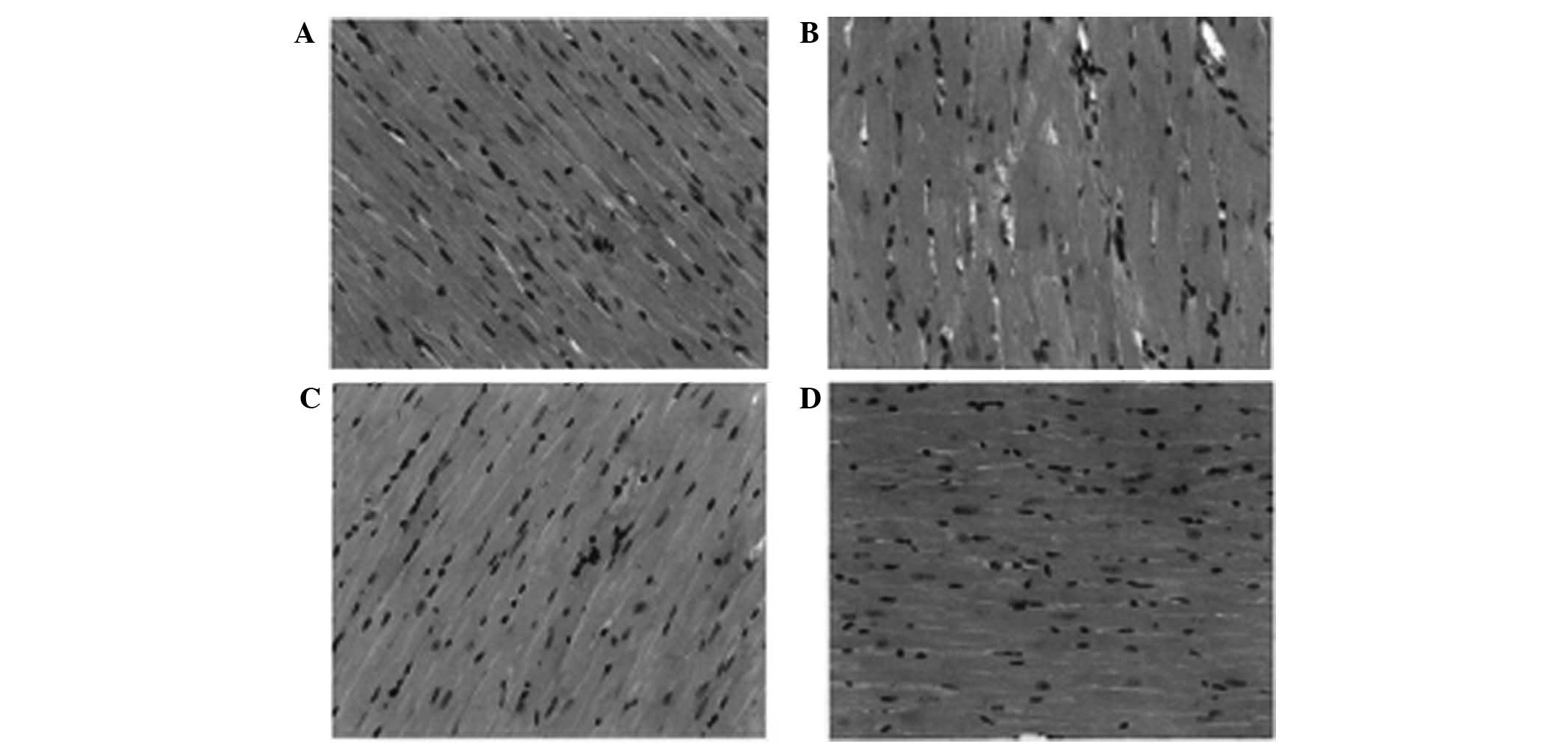
The H&E staining results observed under an
optical microscope are shown in Fig.
1A. The figure shows that compared with the aged control group,
the young rats had myocardial cells with a smaller diameter, a
relatively neat and compact arrangement of the muscle fibers, fewer
deeply stained nuclei and smaller interstitial spaces between the
myocardial cells. The diameter of the myocardial cells was the
largest in the aging control group (Fig. 1B), which showed the larger the
diameter of myocardial cells, the more serious the extent of
apoptosis is, the loose muscle fibers and interstitial spaces also
demonstrated the apoptosis of the cells. By contrast, the apoptosis
of myocardial cells was decreased following treatment with
atorvastatin, and compared with low-dose atorvastatin (Fig. 1C), high-dose atorvastatin (Fig. 1D) was able to inhibit apoptosis
more effectively.
The AVT intervention significantly ameliorated
cardiomyocyte hypertrophy, reduced the number of deeply stained
nuclei and reduced the myocardial interstitial space.
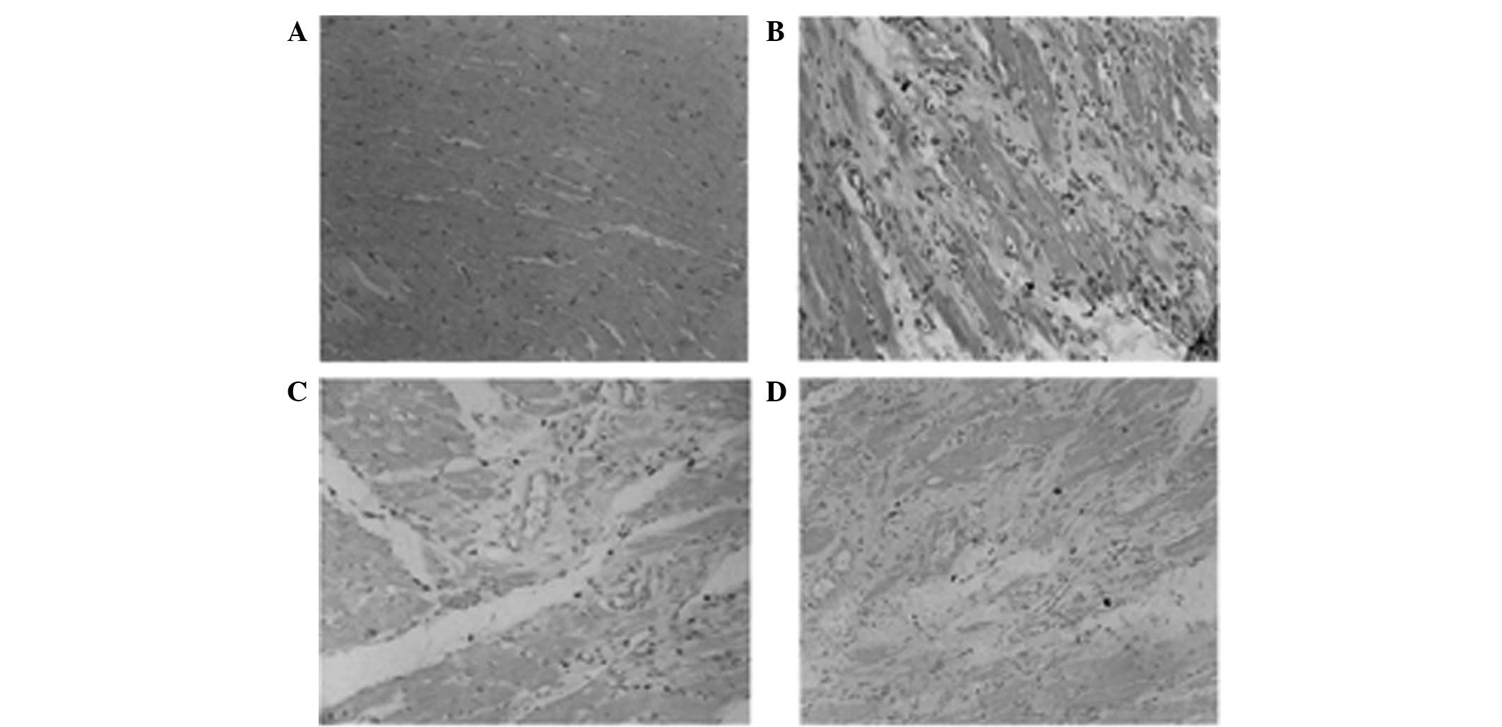
Impact of aging on myocardial apoptosis
and the effects of AVT intervention
The results of the TUNEL staining of cardiomyocyte
apoptosis are shown in Fig. 2.
Under a microscope, the nuclei of apoptotic myocardial cells were
dark and the normal cells were gray, the young rat group(Fig. 2A) has negligible apoptotic cells
while the aging rat group (Fig.
2B) has the largest amount of apoptotic (dark) myocardial
cells. Treatment with atorvastatin reduced the apoptosis in
myocardial cells and the apoptosis in the high-dose group (Fig. 2D) was lower compared with the
low-dose group (Fig. 2C). The
numbers of apoptotic cells are shown in Table III. The apoptotic myocardial
cells of the aging control were significantly increased in number
compared with those of the young control group (P<0.01). The
number of apoptotic cells of the high-dose and low-dose AVT groups
were both fewer than those of the aged control group (P<0.01).
Moreover, compared with the low-dose group, the reduction of
apoptotic cells in the high-dose group was significant
(P<0.05).
| Table IIIEffect of AVT on cardiomyocyte
apoptosis in rats (mean ± SD). |
Table III
Effect of AVT on cardiomyocyte
apoptosis in rats (mean ± SD).
| Group | Number of apoptotic
cardiomyocyte cells |
|---|
| Young control
(n=10) | 8.6±5.1a |
| Aging control
(n=10) | 69.7±25.8 |
| Atorvastatin low dose
(n=10) | 38.4±19.2a |
| Atorvastatin high
dose (n=10) | 21.7±13.6ab |
Lipofuscin levels of aging rats and the
effects of AVT intervention
The lipofuscin levels of the rats are shown in
Table IV. The lipofuscin levels
of the myocardial tissue in the aging control group were
significantly increased compared with those of the young control
group (P<0.01). The lipofuscin levels of the high-dose and
low-dose AVT groups were both lower than those in the aged control
group (P<0.01). Moreover, compared with the low-dose group, the
reduction of lipofuscin contents in the high-dose group was
significant (P<0.01).
| Table IVEffect of AVT on the lipofuscin
levels of rats (mean ± SD, U/mg·prot). |
Table IV
Effect of AVT on the lipofuscin
levels of rats (mean ± SD, U/mg·prot).
| Group | Lipofuscin
level |
|---|
| Young control
(n=30) | 327.7±39.5a |
| Aging control
(n=27) | 504.8±40.3 |
| Atorvastatin low
dose (n=24) | 428.1±33.5a |
| Atorvastatin high
dose (n=26) | 394.1±29.6ab |
Discussion
AVT is a statin, which is the most widely prescribed
type of cholesterol-lowering medication (27). The effects and the benefits of AVT
in regulating blood lipids have been acknowledged by the majority
of scholars. Consistent with numerous previous studies, our results
showed that AVT played an important role in significantly lowering
the lipid levels in aging and young rats, and there were certain
correlations between the regulation and the dosage.
In recent years, studies have shown that in addition
to lipid-lowering effects, AVT has other independent mechanisms of
action, such as the inhibition of vascular smooth muscle cell
proliferation (28). According to
the results of the current study, the cardiomyocyte size in the
aged rat group was markedly different compared with that in the
young rats, and after 4 months of long-term intervention with AVT,
the average cardiomyocyte diameter of the two AVT groups was
significantly lower than that of the aging control group.
The results suggest that the long-term intervention
with AVT was able to significantly reduce the myocardial
hypertrophy level of aging rats. Numerous previous studies, both
in vivo and in vitro, have confirmed that statins
inhibit cardiac hypertrophy by various pathways (29,30).
In the present study, the results indicate that the role of AVT in
inhibiting cardiac hypertrophy may be associated with the
suppression of oxidative stress and inflammatory cytokines.
Apoptosis affects aging in two ways: i) it removes
the injured and dysfunctional cells, such as fibroblasts, and
replaces them with fibrous tissue to maintain the stability of the
body; and ii) it clears the non-renewable cells, such as
cardiomyocytes, to cause pathological changes and dysfunction of
the body (31). The current study
indicates that statins are able to significantly inhibit myocardial
apoptosis, thus protecting the heart function. Statins may play
important roles in the inhibition of apoptosis by various
mechanisms (22).
In the current study, we observed that the
myocardial apoptosis levels of the aged rats were significantly
increased compared with those of the young rats, which indicated
that the increased apoptosis of myocardial cells was an important
manifestation of cardiac aging and it combined with cardiac
hypertrophy and ventricular remodeling to participate in the
pathophysiological process of aging. AVT intervention was able to
significantly inhibit the apoptosis of myocardial cells in the aged
rats, and the effects in the high-dose AVT group were more marked.
The results suggest that AVT may regulate the cardiomyocyte
apoptosis signaling pathway, which may be one of the cardiac
protective mechanisms of statins.
Lipofuscin has been generally accepted by the
majority of scholars to be one of the indicators that reflect
oxidative stress, aging state and the effects of anti-aging
interventions (32). Previous
studies have confirmed that the in vivo level of lipofuscin
gradually increases with increasing age (33). Therefore, in the present study, we
chose lipofuscin as the indicator to reflect the cardiac aging
state of the aged rat group and evaluate the effects of the AVT
intervention.
According to our experimental results, the
lipofuscin levels in the cardiac muscle of the aged rats were
significantly increased compared with those of the young rats.
Following AVT intervention, the myocardial lipofuscin levels of the
aging rats were significantly reduced. The results suggest that AVT
may significantly suppress the myocardial oxidative stress level of
the aging rats and inhibit the aging of the heart, and suppression
of oxidative stress may be one of the cardiac protective
mechanisms.
In summary, long-term AVT intervention may reduce
blood lipid levels, inhibit cardiac hypertrophy, suppress
cardiomyocyte apoptosis and lower the level of oxidative stress to
protect the heart from aging.
The cardiovascular systems of the aged rats showed
significant aging-related changes compared with those of young
rats, mainly presenting as cardiac hypertrophy and markedly
elevated blood lipid levels, myocardial apoptosis and lipofuscin
levels.
AVT intervention is able to significantly reduce the
blood lipid levels of aged rats, relieve myocardial hypertrophy,
inhibit cardiomyocyte apoptosis and improve oxidative stress
indicators to protect the heart.
References
|
1
|
Marchand WR, Lee JN, Suchy Y, et al:
Age-related changes of the functional architecture of the
cortico-basal ganglia circuitry during motor task execution.
Neuroimage. 55:194–203. 2011. View Article : Google Scholar : PubMed/NCBI
|
|
2
|
Goldsmith TC: Mammal aging: Active and
passive mechanisms and their medical implications. Biosci
Hypotheses. 2:59–64. 2009. View Article : Google Scholar
|
|
3
|
Alichniewicz KK, Brunner F, Klünemann HH
and Greenlee MW: Structural and functional neural correlates of
visuospatial information processing in normal aging and amnestic
mild cognitive impairment. Neurobiol Aging. 33:2782–2797. 2012.
View Article : Google Scholar : PubMed/NCBI
|
|
4
|
Fowler MR, Naz JR, Graham MD, Orchard CH
and Harrison SM: Age and hypertrophy alter the contribution of
sarcoplasmic reticulum and Na+/Ca2+ exchange
to Ca2+ removal in rat left ventricular myocytes. J Mol
Cell Cardiol. 42:582–589. 2007. View Article : Google Scholar : PubMed/NCBI
|
|
5
|
Steppan J, Tran H, Benjo AM, et al:
Alagebrium in combination with exercise ameliorates age-associated
ventricular and vascular stiffness. Exp Gerontol. 47:565–572. 2012.
View Article : Google Scholar : PubMed/NCBI
|
|
6
|
Hung CL, Wu YJ, Liu CC, et al: Age-related
ventricular remodeling is an independent risk for heart failure
symptoms in subjects with preserved systolic function. Int J
Gerontol. 5:17–24. 2011. View Article : Google Scholar
|
|
7
|
Fraysse B, Weinberger F, Bardswell SC, et
al: Increased myofilament Ca2+ sensitivity and diastolic
dysfunction as early consequences of Mybpc3 mutation in
heterozygous knock-in mice. J Mol Cell Cardiol. 52:1299–1307.
2012.
|
|
8
|
Chatterjee K: Pathophysiology of systolic
and diastolic heart failure. Med Clin North Am. 96:891–899. 2012.
View Article : Google Scholar : PubMed/NCBI
|
|
9
|
Strait JB and Lakatta EG: Cardiac aging:
From humans to molecules. Muscle 2-Volume Set: Fundamental Biology
and Mechanisms of Disease. Hill JA and Olson EN: 1st edition.
Academic Press; Waltham, MA: pp. 639–659. 2012, View Article : Google Scholar
|
|
10
|
Boyle AJ, Shih H, Hwang J, et al:
Cardiomyopathy of aging in the mammalian heart is characterized by
myocardial hypertrophy, fibrosis and a predisposition towards
cardiomyocyte apoptosis and autophagy. Exp Gerontol. 46:549–559.
2011. View Article : Google Scholar
|
|
11
|
Gonzalez-Aparicio R, Flores JA, Tasset I,
Tunez I and Fernandez-Espejo E: Mice lacking the peroxisome
proliferator-activated receptor alpha gene present reduced number
of dopamine neurons in the substantia nigra without altering motor
behavior or dopamine neuron decline over life. Neuroscience.
186:161–169. 2011. View Article : Google Scholar
|
|
12
|
Wang XJ, Zhang S, Yan ZQ, et al: Impaired
CD200-CD200R-mediated microglia silencing enhances midbrain
dopaminergic neurodegeneration: roles of aging, superoxide, NADPH
oxidase, and p38 MAPK. Free Radic Biol Med. 50:1094–1106. 2011.
View Article : Google Scholar
|
|
13
|
Pisciotta L, Sallo R, Rabacchi C, Wunsch
A, Calandra S and Bertolini S: Leucine 10 allelic variant in signal
peptide of PCSK9 increases the LDL cholesterol-lowering effect of
statins in patients with familial hypercholesterolaemia. Nutr Metab
Cardiovasc Dis. 22:831–835. 2012. View Article : Google Scholar : PubMed/NCBI
|
|
14
|
Owens CD: Statins and other agents for
vascular inflammation. J Vasc Surg. 56:1799–1806. 2012. View Article : Google Scholar : PubMed/NCBI
|
|
15
|
Obregón O, Gestne A, Lares M, et al:
Efectos tempranos de las estatinas: inflamación y oxidación. Clin
Invest Arterioscl. 23:269–274. 2011.(In Spanish).
|
|
16
|
Marrone G, Russo L, Rosado E, et al: The
transcription factor KLF2 mediates hepatic endothelial protection
and paracrine endothelial-stellate cell deactivation induced by
statins. J Hepatol. 58:98–103. 2012. View Article : Google Scholar : PubMed/NCBI
|
|
17
|
Yokomizo A, Shiota M, Kashiwagi E, et al:
Statins reduce the androgen sensitivity and cell proliferation by
decreasing the androgen receptor protein in prostate cancer cells.
Prostate. 71:298–304. 2011. View Article : Google Scholar : PubMed/NCBI
|
|
18
|
Shin SK, Lee YK, Jo YI and Chang TI: The
effects of statins unrelated to cholesterol level on clinical
outcome of continuous ambulatory peritoneal dialysis patients.
Kidney Research and Clinical Practice. 31:A732012. View Article : Google Scholar
|
|
19
|
Choi EY, Chang W, Lim S, et al:
Rosuvastatin inhibits norepinephrine-induced cardiac hypertrophy
via suppression of Gh. Eur J Pharmacol. 627:56–62. 2010. View Article : Google Scholar : PubMed/NCBI
|
|
20
|
Sverdrup FM, Yates MP, Vickery LE, et al:
Protein geranylgeranylation controls collagenase expression in
osteoarthritic cartilage. Osteoarthritis Cartilage. 18:948–955.
2010. View Article : Google Scholar
|
|
21
|
Murrow JR, Sher S, Ali S, et al: The
differential effect of statins on oxidative stress and endothelial
function: atorvastatin versus pravastatin. J Clin Lipidol. 6:42–49.
2012. View Article : Google Scholar : PubMed/NCBI
|
|
22
|
Chuang YH, Chuang WL, Huang SP, Liu CK and
Huang CH: Atorvastatin ameliorates tissue damage of obstructed
ureter in rats. Life Sci. 89:795–805. 2011. View Article : Google Scholar : PubMed/NCBI
|
|
23
|
Yang P, Li Y, Li JJ, Qin L and Li XY:
Up-regulating PPAR-gamma expression and NO concentration, and
down-regulating PAI-1 concentration in a rabbit atherosclerotic
model: the possible antiatherogenic and antithrombotic effects of
atorvastatin. Int J Cardiol. 139:213–217. 2010. View Article : Google Scholar : PubMed/NCBI
|
|
24
|
Garjani A, Andalib S, Biabani S, Soraya H,
Doustar Y, Garjani A and Maleki-Dizaji N: Combined atorvastatin and
coenzyme Q10 improve the left ventricular function in
isoproterenol-induced heart failure in rat. Eur J Pharmacol.
666:135–141. 2011. View Article : Google Scholar : PubMed/NCBI
|
|
25
|
Koh KK, Quon MJ, Han SH, et al: Additive
beneficial effects of atorvastatin combined with amlodipine in
patients with mild-to-moderate hypertension. Int J Cardiol.
146:319–325. 2011. View Article : Google Scholar : PubMed/NCBI
|
|
26
|
Han L, Li M, Liu Y, Han C and Ye P:
Atorvastatin may delay cardiac aging by upregulating peroxisome
proliferator-activated receptors in rats. Pharmacology. 89:74–82.
2012. View Article : Google Scholar : PubMed/NCBI
|
|
27
|
Kobayashi M, Chisaki I, Narumi K, et al:
Association between risk of myopathy and cholesterol-lowering
effect: a comparison of all statins. Life Sci. 82:969–975. 2008.
View Article : Google Scholar : PubMed/NCBI
|
|
28
|
Doyon M, Hale TM, Huot-Marchand JE, Wu R,
de Champlain J and deBlois D: Does atorvastatin induce aortic
smooth muscle cell apoptosis in vivo? Vascular Pharmacol. 54:5–12.
2011. View Article : Google Scholar : PubMed/NCBI
|
|
29
|
Asakawa M, Takano H, Nagai T, Uozumi H,
Hasegawa H, Kubota N, Saito T, Masuda Y, Kadowaki T and Komuro I:
Peroxisome proliferator-activated receptor gamma plays a critical
role in inhibition of cardiac hypertrophy in vitro and in
vivo. Circulation. 105:1240–1246. 2002. View Article : Google Scholar
|
|
30
|
Hsu S, Nagayama T, Koitabashi N, et al:
Phosphodiesterase 5 inhibition blocks-pressure overload-induced
cardiac hypertrophy independent of the calcineurin pathway.
Cardiovasc Res. 81:301–309. 2009. View Article : Google Scholar
|
|
31
|
Bao XM, Wu CF and Lu GP: Atorvastatin
inhibits homocysteine-induced oxidative stress and apoptosis in
endothelial progenitor cells involving Nox4 and p38MAPK.
Atherosclerosis. 210:114–121. 2010. View Article : Google Scholar : PubMed/NCBI
|
|
32
|
Yu X and Li G: Effects of resveratrol on
longevity, cognitive ability and aging-related histological markers
in the annual fish Nothobranchius guentheri. Exp Gerontol.
47:940–949. 2012. View Article : Google Scholar : PubMed/NCBI
|
|
33
|
Höhn A, Sittig A, Jung T, Grimm S and
Grune T: Lipofuscin is formed independently of macroautophagy and
lysosomal activity in stress-induced prematurely senescent human
fibroblasts. Free Radic Biol Med. 53:1760–1769. 2012.PubMed/NCBI
|