Introduction
The Hedgehog (HH) signaling pathway is a crucial
signal transduction pathway involved in the regulation of embryonic
development. The activation of the pathway has shown a correlation
with the medulloblastoma basal cell carcinoma gastrointestinal
tumor and other solid tumors (1,2).
Therefore, the study of the function and regulatory mechanism of
the HH signaling pathway may be beneficial in the elucidation of
the mechanism underlying the development of malignant tumors and in
the diagnosis, prevention and treatment of tumors. The HH family
mainly includes Sonic hedgehog (SHH), hedgehog interacting protein
(HHIP), patched 1 (PTCH1), Smo and Gli. When SHH combines with the
PTCH1 receptor, Smo is released from the combination with PTCH1 to
excite Gli, prior to acting on target genes. The PTCH1 gene is one
of the negative regulatory factors in the HH pathway. The
suppression of PTCH1 expression is able to activate the HH pathway
and is important in carcinogenesis. It has been indicated that the
methylation of the PTCH1 gene may be associated with the
development of certain tumors (3–5).
However, the correlation between the methylation of PTCH1 and
gastric cancer has rarely been explored. In the present study, the
expression of PTCH1 in a human gastric cancer cell line was
investigated prior to and following treatment with a methylation
inhibitor, in order to explore the correlation between PTCH1
expression and the CpG island methylation of the gene promoter
region. The purpose of this investigation was to understand the
role of PTCH1 gene methylation in the development of gastric cancer
and to provide guidance for the clinical diagnosis and treatment of
gastric cancer.
Materials and methods
Cell line and main reagents
The AGS human gastric cancer cell line was purchased
from the Cell Data Center of the Shanghai Institutes for Biological
Sciences, Chinese Academy of Sciences (Shanghai, China). The DNA
methyltransferase inhibitor 5-aza-2′-deoxycytidine (5-Aza-dc), a
demethylation reagent, was purchased from Sigma (St. Louis, MO,
USA), while TRIzol® reagent was purchased from
Invitrogen Life Technologies (Carlsbad, CA, USA) and the RNA
reverse transcription kit was purchased from Jingmei Biological
Engineering Co., Ltd. (Shanghai, China). An EZ DNA
Methylation-Gold™ kit for methylation conversion was purchased from
the Beijing Tianmo Technology Development Co., Ltd. (Beijing,
China) and an ABI 7500 Real-Time polymerase chain reaction (PCR)
instrument was obtained from Applied Biosystems (Life Technologies,
Carlsbad, CA, USA). The mouse anti-human PTCH1 monoclonal antibody
used in the study was purchased from Santa Cruz Biotechnology, Inc.
(Santa Cruz, CA, USA), while the 3,3′-diaminobenzidine (DAB)
chromogenic agent, pure xylene, hydrogen peroxide methanol,
hematoxylin and neutral gum were purchased from Shanghai Ruicong
laboratory equipment Co. Ltd. (Shanghai, China).
AGS cell line culture and 5-Aza-dc
treatment
The AGS cell line was inoculated in a 100 ml culture
flask with a density of 3×10⁵/flask. The cells underwent
conventional culture with Ham's F-12K Medium containing 10% fetal
calf serum in conditions of 37°C, 5% CO2 for ~24 h. Once
the cells had entered the logarithmic growth phase, with an 80%
degree of cell fusion, the cells were treated with 5-Aza-dc
(5×10⁻⁶ mol/l). The treatment medium was changed once
every 24 h and the cells were collected 72 h later. In addition, a
control group without the 5-Aza-dc treatment was cultured and
collected.
Evaluation of PTCH1 mRNA expression using
quantitative PCR (qPCR)
Total RNA from the AGS gastric cancer cell line was
isolated using TRIzol reagent and converted into cDNA. The primer
sequences of PTCH1 were: 5′-TGT GCG CTG TCT TCC TTC TG-3′ and
5′-ACG GCA CTG AGC TTG ATT C-3′, with an amplified fragment length
of 119 bp. The primer sequences of the internal control, β-actin,
were : 5′-GCC ATC CTG CGT CG-3′ and 5′-TGG GCA CCG GAA CCG CT-3′,
with an amplified fragment length of 260 bp. The volume of the
reaction agents was 20 μl, with reaction conditions of 95°C for 5
sec, 55°C for 5 sec and 72°C for 30 sec. The PCR products were
subjected to 1.5% agarose gel electrophoresis analysis.
Evaulation of PTCH1 protein expression
using immunocytochemistry
The S-P method was applied for immunocytochemistry.
A 2×2 cm cover slip was soaked in concentrated sulfuric acid,
autoclaved and then fixed with anticorrosive poly-l-lysine solution
diluted with 1:100 ddH2O. The AGS gastric cancer cell
line was inoculated in the sterile cover slip at a density of
2×10⁴/ml and then placed into a 90 mm petri dish. Cell
specimens were extracted 3 days later and washed three times with
phosphate-buffered saline (PBS). The specimens were subsequently
blocked with 10% normal goat serum blocking fluid at 37°C. Twenty
minutes later, the goat serum solution was disposed of. PTCH1
primary antibody working reagent (working concentration 1:100) was
added into the cell specimens, prior to the specimens being
incubated at 37°C. Following 60 min, the specimens were washed
three times with PBS and the PTCH1 secondary antibody working
solution (Shanghai Yanhui biotechnology Co., Ltd., Shanghai, China)
was added at 37°C for incubation. Thirty minutes subsequently, the
specimens were washed with PBS three times and stained with DAB
chromogenic reagent, counterstained with hematoxylin and mounted
with neutral gum. The criteria for a positive result comprised the
observation of cells with brown particles in the cytoplasm and/or
nucleus under a microscope.
Assessment of CpG island methylation
status of the PTCH1 gene using methylation-specific PCR (MSP) and
sequencing
The DNA of the gastric cancer cell line was
extracted and purified using phenol/chloroform, prior to being
transformed by methylation with a methylation conversion kit. The
PTCH1 MSP primers were designed using ABI Methyl Primer
Express® Software v1.0 (Applied Biosystems). The PTCH1
methylated primer sequences were: 5′-GTT AAT TCG TGA TTT TTC GCA-3′
and 5′-ATA ACA AAC CTA CGA ACC GC-3′, with an amplified fragment
length of 197 bp, while the PTCH1 unmethylated primer sequences
were: 5′-AAT GTT AAT TTG TGA TTT TTT GGA-3′ and 5′-TAA ATA ACA AAC
CTA CAA ACC AC-3′, with an amplified fragment length of 197 bp. The
reaction system included 5 μl 10X PCR buffer, 2 μl deoxynucleotide
triphosphate (dNTP) 1, 1.5 μl methylated or unmethylated upstream
primers and 1.5 μl methylated or unmethylated downstream primers,
1.0 μl TaqDNA polymerase, 32 μl ddH2O and 8 μl DNA
converted using the methylation conversion kit. The reaction
conditions were 94°C for 30 sec, 60°C for 40 sec and 72°C for 50
sec. The amplification procedure was repeated for 40 cycles, prior
to the amplification products being analyzed using electrophoresis
with 1.5% agarose gel. The amplified 197 bp strip was sent to the
Huada Gene Institute (Beijing, China) for sequencing analysis.
Results
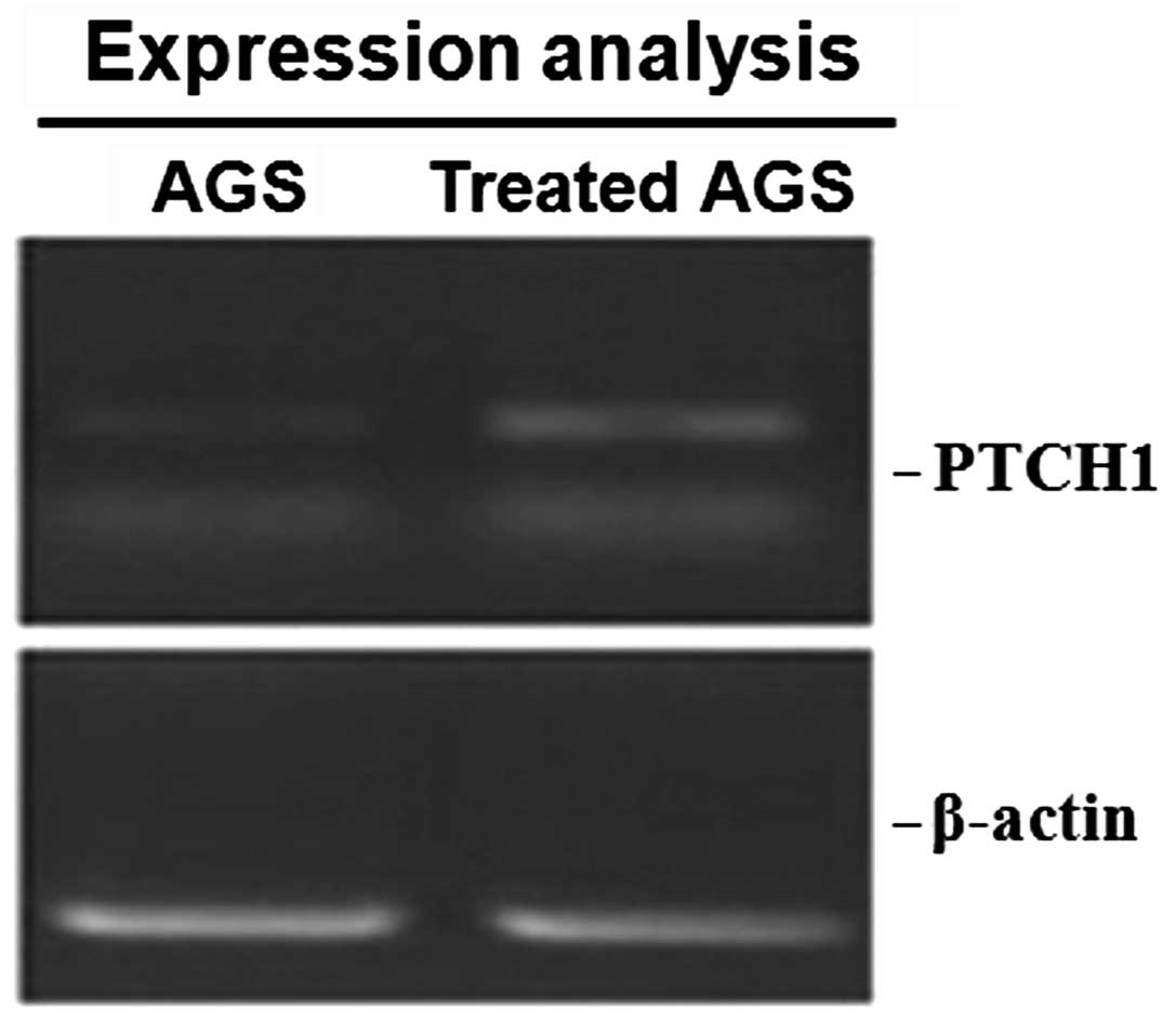
Expression of PTCH1 mRNA
Based on the qPCR, no amplified strip was observed
in the control AGS gastric cancer cell line without the treatment
of 5-Aza-dc, indicating that there was no expression of PTCH1 mRNA
present. However, a 119-bp strip corresponding to PTCH1 mRNA was
observed in the AGS cells following treatment with 5-Aza-dc, which
demonstrated the re-expression of PTCH1 mRNA following the
demethylation process (Fig.
1).
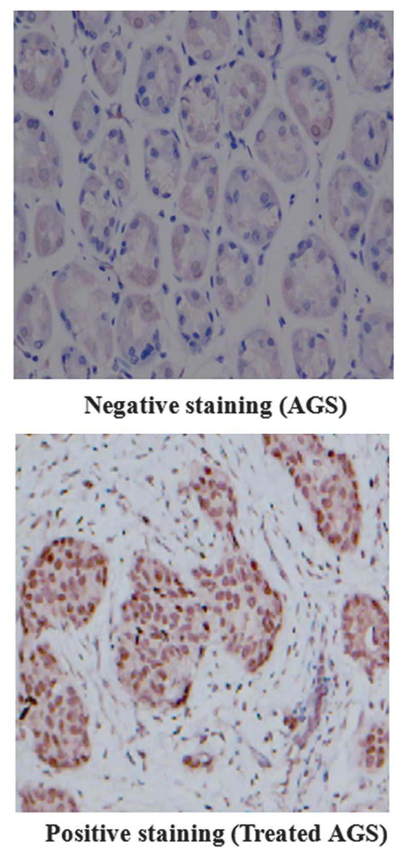
PTCH1 protein expression
According to the results of the immunocytochemistry,
no brown particles were observed in the AGS cells that were not
treated with 5-Aza-dc, indicating that there was no expression of
PTCH1 protein. Following 5-Aza-dc treatment, brown particles were
observed in the AGS cells, which demonstrated the re-expression of
PTCH1 protein subsequent to demethylation treatment (Fig. 2). These results were consistent
with the results of the qPCR.
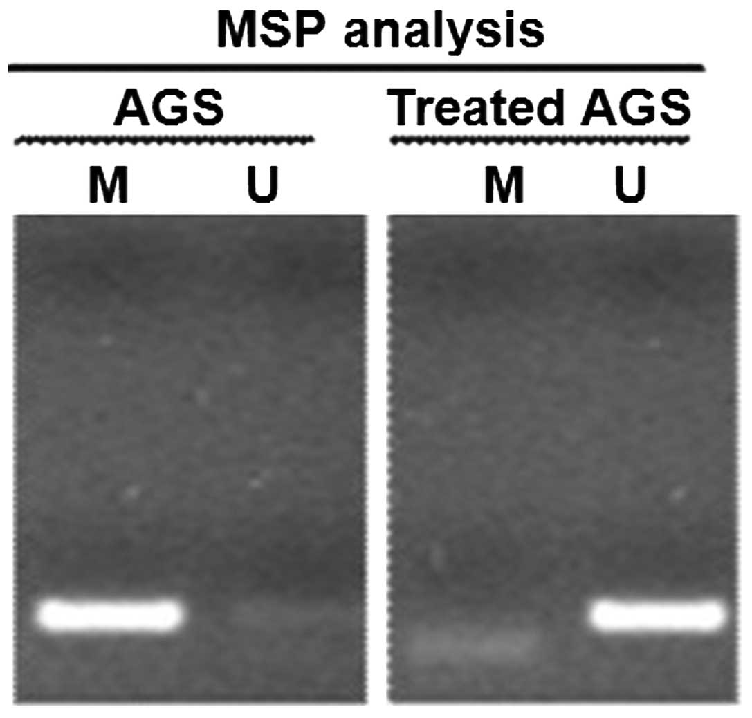
CpG island methylation status of
PTCH1
The amplification procedure using MSP and methylated
primers resulted in the presence of an amplified strip of 197 bp
for the AGS cells; however, no amplified strip was observed for the
AGS cells amplified using unmethylated primers. For the AGS cells
that underwent demethylation treatment with 5-Aza-dc, no amplified
strips were observed when the methylated primers were used;
however, an amplified strip of 197 bp was observed with the use of
unmethylated primers. This indicated that the PTCH1 gene of the AGS
gastric cancer cell line had undergone methylation in the CpG
island (Fig. 3).
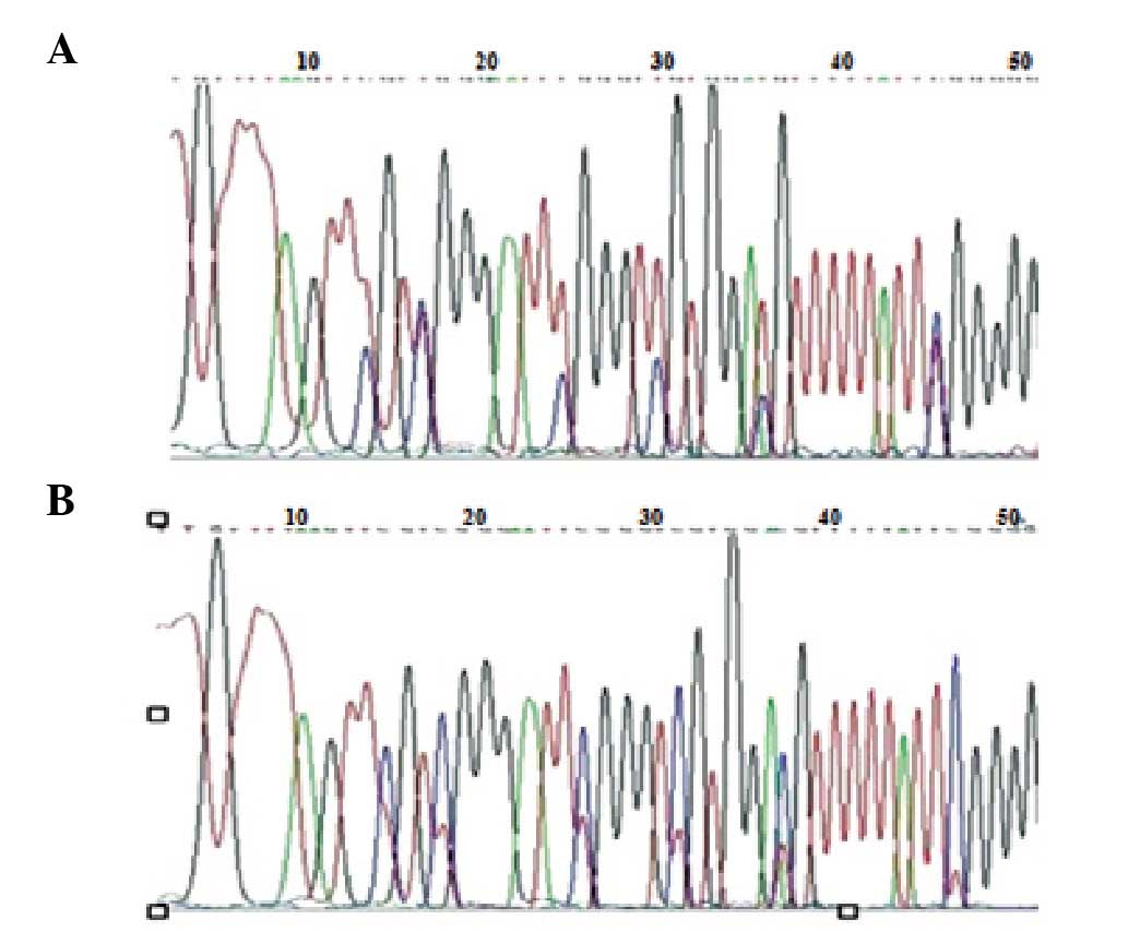
According to the sequencing detection of the
amplification products from the MSP with methylated primers,
methylation was observed in the AGS CpG island and not in the
non-CpG island region (Fig. 4).
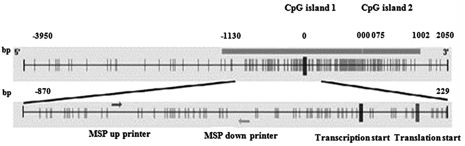
The CpG island and its location sites in the PTCH1 gene promoter
region of the AGS cell line were analyzed. There were multiple
transcriptional initiation sites in the PTCH1 gene. According to
the analysis of the PTCH1 mRNA 1a transcriptional initiation site
(as zero) using Methyl Primer Express® software (Applied
Biosystems), two CpG islands were apparent. One island was located
between −1,139 and 860 bp and the second was located between +875
and +1,692 bp (Fig. 5). The −870
to +229 bp zone of the first CpG island was set as the target
sequence and there were 19 sites in this zone. The quantitative
indicator of the degree of methylation was the number of CpG sites
for methylation in proportion to the total number of CpG sites
detected. Statistics indicated that all methylation in the AGS
gastric cancer cell line occurred in the target sequencing zone.
The results of the sequencing analysis corresponded with the
methylation status of the PTCH1 gene as revealed using MSP.
Discussion
The HH signaling pathway is a critical signal
transduction pathway involved in the regulation of embryonic
development. HH signaling is very active in embryogenesis and then
its activity disappears or reduces in normal mature tissue.
However, HH signaling is extremely active in adult malignancies
(6–8). PTCH1 gene is a tumor suppressor gene,
located at 9q22.3, and is the most widely expressed gene in the HH
family. Studies have shown that the silencing of the functional
PTCH1 allele is the key factor leading to the activation of the HH
pathway and carcinogenesis. The mutation of PTCH1 protein leads to
the loss of its normal inhibitory function on Smo, resulting in the
abnormal activation of the HH pathway and causing carcinogenesis.
The expression of PTCH1 in a number of malignant tumors, such as
breast, liver and esophageal cancer, has been shown to be reduced
compared with that in normal tissues (9–11).
In the present study, qPCR and immunocytochemical staining were
used to detect PTCH1 expression in the AGS gastric cancer cell
line. The results of the two detection methods were consistent.
There was no expression of PTCH1 in the AGS cells that were not
treated with 5-Aza-dc, whereas PTCH1 expression was observed in the
AGS cells that were treated with 5-Aza-dc. This suggested a low
level of PTCH1 expression in gastric cancer.
Studies on cancer epigenetics have demonstrated that
there exists wide hypomethylation and some regional
hypermethylation of CpG islands in the genomic DNA of tumor cells.
The abnormally high methylation of the CpG island is associated
with the transcriptional silencing of the gene expression and may
lead to the partial inactivation of certain tumor suppressor genes.
This is an important mechanism causing the malignant transformation
of cells (12,13). These highly methylated genes are
able to re-express gene product following the use of a
demethylating agent to act on the tumor cell lines, which is
important in tumor suppression (14). As such, the establishment of a DNA
methylation spectral type of multiple tumor-related genes for a
particular tumor, which facilitates the early diagnosis or
differential diagnosis of the tumor, has been proposed (14). In a study of PTCH1 gene promoter
methylation, only the regulatory sequences on mRNA 1b were
assessed. The methylation analysis results for upstream regulation
(−1,593 bp, with 1b transcription initiation site count)
demonstrated that significantly high levels of methylation were
observed in ovarian tumors; however, there were no significantly
high levels of methylation in basal cell carcinoma, which retained
a demethylation status (15). The
methylation analysis results of the regulatory sequences (−776 to
+1,238 bp, with 1b transcription initiation site count) showed that
a high methylation status existed in breast cancer. The methylation
level was negatively correlated with the expression of PTCH1b
(9). In the present study, MSP
amplification was performed to detect the CpG island methylation
status of the PTCH1 gene in the AGS gastric cancer cell line. The
results demonstrated that the gastric cancer cell line methylated
primers amplified the corresponding-sized fragment. No
appropriately-sized fragments were amplified using unmethylated
primer. This indicated that the CpG island of the PTCH1 gene in the
AGS cells existed in a highly methylated state. The sequences of
the methylation products from MSP amplification were analyzed to
investigate whether there was CpG dinucleotide methylation in the
amplified fragments. This study was aimed at methylation analysis
of the PTCH1a mRNA regulatory sequence. The methylation analysis of
the PTCH1a upstream regulatory sequence (−870 to +229 bp, with 1a
transcription initiation site count) indicated that a high
methylation status existed in PTCH1 in the AGS cells, and that this
high methylation status was negatively correlated with gene
expression. The sequencing results were consistent with those from
the MSP test. The high level of methylation of the PTCH1 gene in
the gastric cancer cell line may be one of the pathogenic
mechanisms of certain forms of gastric cancer. The methylation zone
in the gastric cancer cell line was different from those of
previous studies, indicating that the high-methylation mechanism of
the PTCH1 gene promoter may be different for different types of
carcinogenesis.
In conclusion, this study showed that the PTCH1 gene
expression in a gastric cancer cell line was reduced. The gene
expression recovered following treatment with 5-Aza-dc. MSP
amplification detection and DNA sequencing showed that multiple
sites of CpG island hypermethylation existed in the PTCH1 gene of
the gastric cancer cell line. PTCH1 gene expression was negatively
correlated with a high level of methylation. The high level of
methylation in the CpG islands of the PTCH1 gene may be associated
with the occurrence and development of gastric cancer. The
detection of methylation of the PTCH1 gene may become a diagnostic
marker of gastric cancer, which may provide guidance for the
treatment and evaluation of gastric cancer.
References
|
1
|
Saqui-Salces M and Merchant JL: Hedgehog
signaling and gastrointestinal cancer. Biochim Biophys Acta.
1803:786–795. 2010. View Article : Google Scholar : PubMed/NCBI
|
|
2
|
Shahi MH, Lorente A and Castresana JS:
Hedgehog signaling in medulloblastoma, glioblastoma and
neuroblastoma. Oncol Rep. 19:681–688. 2008.PubMed/NCBI
|
|
3
|
Cul'bová M, Lasabová Z, Stanclová A, et
al: Methylation of selected tumor-supressor genes in benign and
malignant ovarian tumors. Ceska Gynekol. 76:274–279. 2011.(In
Slovak).
|
|
4
|
Wolf I, Bose S, Desmond JC, et al:
Unmasking of epigenetically silenced genes reveals DNA promoter
methylation and reduced expression of PTCH in breast cancer. Breast
Cancer Res Treat. 105:139–155. 2007. View Article : Google Scholar : PubMed/NCBI
|
|
5
|
Pritchard JI and Olson JM: Methylation of
PTCH1, the Patched-1 gene, in a panel of primary medulloblastomas.
Cancer Genet Cytogenet. 180:47–50. 2008. View Article : Google Scholar : PubMed/NCBI
|
|
6
|
Merchant JL, Saqui-Salces M and
El-Zaatari: Hedgehog signaling in gastric physiology and cancer.
Prog Mol Biol Transl Sci. 96:133–156. 2010. View Article : Google Scholar : PubMed/NCBI
|
|
7
|
Kawahira H, Ma NH, Tzanakakis E, et al:
Combined activities of hedgehog signaling inhibitors regulate
pancreas development. Development. 130:4871–4879. 2003. View Article : Google Scholar
|
|
8
|
Karhadkar SS, Bova GS, Abdallah N, et al:
Hedgehog signaling in prostate regeneration, neoplasia and
metastasis. Nature. 431:707–712. 2004. View Article : Google Scholar : PubMed/NCBI
|
|
9
|
Wolf I, Bose S, Desmond JC, et al:
Unmasking of epigenetically silenced genes reveals DNA promoter
methylation and reduced expression of PTCH in breast cancer. Breast
Cancer Res Treat. 105:139–155. 2007. View Article : Google Scholar : PubMed/NCBI
|
|
10
|
Fu X, Wang Q, Chen X, et al: Expression
patterns and polymorphisms of PTCH in Chinese hepatocellular
carcinoma patients. Exp Mol Pathol. 84:195–199. 2008. View Article : Google Scholar : PubMed/NCBI
|
|
11
|
Ishiyama A, Hibi K, Koike M, et al: PTCH
gene expression as a potential marker for esophageal squamous cell
carcinoma. Anticancer Res. 26:195–198. 2006.PubMed/NCBI
|
|
12
|
Tada M, Kanai F, Tanaka Y, et al:
Down-regulation of hedgehog-interacting protein through genetic and
epigenetic alterations in human hepatocellular carcinoma. Clin
Cancer Res. 14:3768–3776. 2008. View Article : Google Scholar : PubMed/NCBI
|
|
13
|
Caffarelli E and Filetici P: Epigenetic
regulation in cancer development. Front Biosci (Landmark Ed).
16:2682–2694. 2011. View
Article : Google Scholar
|
|
14
|
To KF, Leung WK, Lee TL, et al: Promoter
hypermethylation of tumor-related genes in gastric intestinal
metaplasia of patients with and without gastric cancer. Int J
Cancer. 102:623–628. 2002. View Article : Google Scholar : PubMed/NCBI
|
|
15
|
Cretnik M, Musani V, Oreskovic S, et al:
The Patched gene is epigenetically regulated in ovarian dermoids
and fibromas, but not in basocellular carcinomas. Int J Mol Med.
19:875–883. 2007.PubMed/NCBI
|