Introduction
The lung is one of the most radiosensitive organs,
yet is occasionally irradiated as part of therapy programs for
tumors of the lung, esophagus, breast and lymphatic system.
Radiation-induced lung injury (radiation pneumonitis and radiation
pulmonary fibrosis) is radioactive pulmonary damage which occurs
when normal lung tissues are irradiated, such as in actinotheraphy
for chest tumors, bone marrow treatment prior to transplantation
and nuclear accidents, and is characterized by hypoxemia,
non-cardiogenic pulmonary edema, low lung compliance and widespread
capillary leakage. Radiation-induced lung injury is a common and
critical problem that limits the doses that may be delivered in
radiotherapy (1–3).
There are a number of possible etiologies for lung
injury, but a clear cause for each patient may not usually be
ascertained (4–5). The pathogenesis of pulmonary fibrosis
remains unclear, although it has been widely accepted that the
abnormal activation of certain inflammatory cells and cytokines in
the lung is critical (6). In the
initiating stage of the disease, numerous types of inflammatory
cells, including macrophages, T lymphocytes, neutrophils and
fibroblasts, are activated to release a variety of cytokines which
contribute to cell aggregation. Subsequently, large amounts of
inflammatory cytokines, such as tumor necrosis factor (TNF)-α,
transforming growth factor (TGF)-β and interleukin-1β (IL-1β) are
expressed by these cells. These cells and cytokines are responsible
for abnormal airway tissue repair, which may lead to lung fibrosis
(7–9). In the development of pulmonary
fibrosis, proinflammatory cytokines and growth factors are
considered to be significant in cell-cell and cell-matrix
interactions (10–12).
Radiotherapy is a basic and important mode of
treatment for nearly half of all tumor patients. Ionizing radiation
kills tumor cells by inducing DNA damage, since non- or incorrectly
repaired DNA results in lethal chromosomal aberrations which cause
a loss of proliferative capacity. The formation of free radicals,
such as the superoxide anion radical (O2•−)
and hydroxyl radical (OH•−), is an unavoidable
consequence of radiotherapy. These free radicals are extremely
unstable and react rapidly with other groups or substances in the
body, leading to cell or tissue injury. This may be explained by
considering one of the numerous mechanisms by which oxidative
stress causes damage by stimulating the free radical chain
reaction. Free radicals activate TGF-β1, one of the most
significant growth factors in the pathogenesis of fibrotic lung
diseases, which promotes epithelial cell apoptosis (13–15).
Heavy ions, ionized atoms which are heavier than
helium, are key components of cosmic rays and have linear energy
transfer (LET) and high relative biological effectiveness (RBE)
values. Heavy ions are also significantly more deleterious at the
cellular and molecular level than low-LET ionizing radiation,
including X-rays and γ-rays (16).
Therefore, heavy ion irradiation is able to induce more
unrepairable breaks in DNA damage than low-LET rays (17). It has also been demonstrated that
heavy ion radiation produces chromosomal breakage and
rearrangements and a greater degree of abnormal differentiation
(18). Based on the excellent
properties of heavy-ion beams, such as an energy deposition peak
(Bragg peak) at the end of its range and an increased RBE within
the peak, heavy ion radiotherapy is attracting growing interest
worldwide (19) and is becoming
one of the most promising therapeutic approaches for malignant
tumors (20). However, heavy ion
radiation not only destroys the tumor but may also potentially
damage the normal tissue around the tumor. The objective of the
present study was to evaluate the time, radiation dose, damage and
repair effects of heavy ion radiation on lung injury and pulmonary
fibrosis and provide experimental evidence for cancer therapy with
heavy ions.
Materials and methods
Chemicals
Hydroxyproline (HP) assay kits were supplied by
Nanjing Jiancheng Bioengineering (Nanjing, China). All other
reagents were of analytical purity.
Animals
SPF-class Kunming mice (male and female; age, 6–7
weeks; weight, 20±2 g) were provided by Lanzhou Medical College
(Lanzhou, Gansu, China). The animals were housed in cages with
ad libitum access to food and water and were kept in an
environmentally controlled room (temperature, 23±1°C; humidity,
40±10%) with a 12 h light/dark cycle. All animal care and
experiments were consistent with the Public Health Guide for the
Care and Use of Laboratory Animals (National Research Council,
1996) and in accordance with protocols approved by the
International Institutional Animal Care and Use Committee.
Pretreatment for irradiation
A total of 144 mice, with an equal number of males
and females, were randomly divided into 4 groups: a normal control
(CK) group and 2, 4 and 6 Gy groups, with 36 animals per group. The
CK group did not receive any treatment. The experimental groups
received whole-body uniform carbon ion beam irradiation with 2, 4
or 6 Gy, at a dose rate of 0.5 Gy/min.
Carbon ion irradiation and experimental
design
The mice were positioned in a chamber fixed to the
irradiation equipment at the Heavy Ion Research Facility in Lanzhou
(HIRFL, Institute of Modern Physics, Chinese Academy of Sciences,
Lanzhou, China). Each animal was placed in a cloth bag and
underwent whole-body irradiation using a
12C6+ ion beam (235 MeV/u primary energy,
∼14.55 keV/μm LET in water), at a dose rate of ∼0.5 Gy/min.
Collimation of the beam to the irradiation location and the
acquisition of data (preset numbers converted by doses of
irradiation), were automatically performed by a microcomputer
during the irradiation. The particle fluence was determined from an
air-ionization chamber signal according to the calibration of the
detector (PTW-UNIDOS; PTW-Freiburg Co., Wiesbaden, Germany).
Sham-treated animals did not undergo irradiation.
Lung index determination and
histopathological examination
At months 1, 2, 3, 4, 5 and 6 after radiation
exposure, the mice were weighed and then sacrificed by cervical
dislocation (n=24, 6 per group). The lungs were excised immediately
on an ice-cold plate, weighed and washed with physiological saline
solution to prepare them for the subsequent experiments. Certain
lung tissues were fixed with 10% formaldehyde in PBS buffer,
dehydrated and embedded in paraffin. Next, 3–4-mm thick tissue
sections were cut and stained with hematoxylin and eosin (H&E)
for a histopathological observational study. Lung index = weight of
lungs (mg) × 10 / body weight (g)
HP measurement
Certain lung tissues were triturated and filtered
through a 200-μm pore mesh to remove debris or cell
clusters, then used to analyze the HP levels. The lung HP content,
considered as a biochemical index for the parenchymal collagen
content, was measured in the lung tissue homogenate using a
diagnostic reagent kit according to the manufacturer’s instructions
(Nanjing Jiancheng Bioengineering) with analysis using a
colorimetric method at 550 nm. The residual lung tissue homogenates
were stored and frozen at −80°C until the biochemical analyses.
Enzyme-linked immunosorbent assay (ELISA)
for cytokines in lung tissues
The concentrations of TNF-α and TGF-β in the whole
mouse lung tissue homogenates stored at −80°C at various time
points were measured using a commercially available ELISA kit
according to the manufacturer’s instructions (Nanjing Jiancheng
Bioengineering Institute, Nanjing, China).
Statistical analysis
Each experiment was repeated twice, with three
samples for each treatment. The data were expressed as the mean ±
standard error of the mean (SEM). Analysis of variance (ANOVA) with
multiple comparison tests was used to determine the significance of
differences between the groups. P<0.05 was considered to
indicate a statistically significant difference. The correlation
statistical analyses were performed using SPSS 11.5 for Windows
(SPSS, Inc., Chicago, IL, USA).
Results
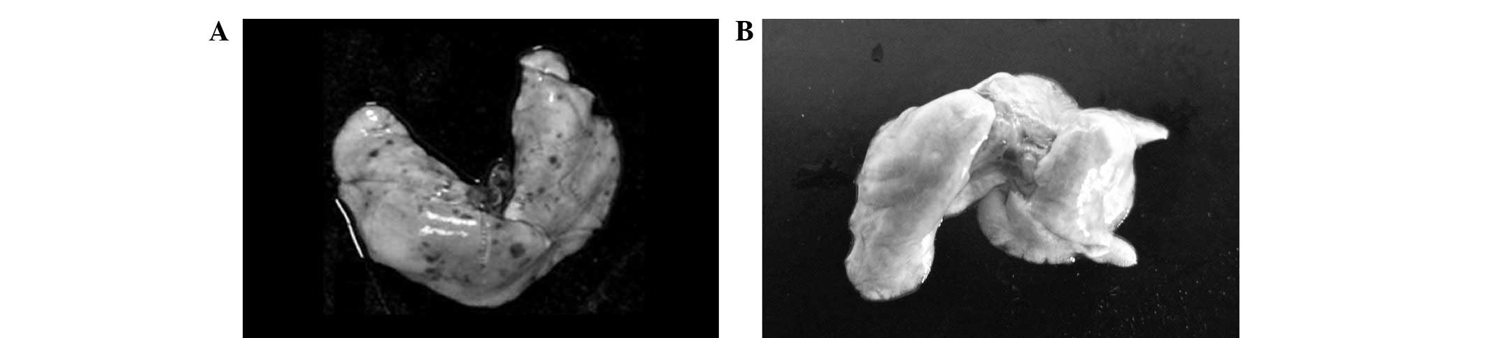
Morphological changes of the lungs
The changes in the appearance of the lungs of the
mice undergoing carbon ion radiation began as acute lung injury
followed by widespread acute pulmonary inflammation, indicated by
increases in pulmonary edema. The lungs of the normal mice had a
pink, soft, smooth and glossy surface, as well as fair elasticity
upon touching. Compared with the CK group, the lungs of the mice
irradiated with heavy ion beams were swollen with collapsed
surfaces and poor elasticity. The irradiated lungs exhibited
limited fibrotic involvement and appeared edematous, with
noticeable hemorrhaging (Fig.
1).
Changes in body weight of mice following
carbon ion beam irradiation
As shown in Table
I, during the 1st month following carbon ion beam irradiation
(2, 4 and 6 Gy), the body weights of the mice decreased
significantly due to the acute injury (P<0.05) compared with the
CK group. Following month 1, the irradiated mice gradually regained
weight, but remained significantly lighter than the CK group
(P<0.05) during the whole 6-month experimental period. This
result clearly indicated that 12C6+
irradiation caused a significant body weight loss compared with the
normal mice, although the body weight increased continuously with
time in all experimental animals.
 | Table I.Changes in the body weight of mice
following carbon ion beam irradiation. |
Table I.
Changes in the body weight of mice
following carbon ion beam irradiation.
| Body weight (g)
|
|---|
| Group | 1 month | 2 months | 3 months | 4 months | 5 months | 6 months |
|---|
| CK | 30.3±2.1 | 35.5±5.0 | 39.1±4.7 | 41.6±1.9 | 44.8±1.4 | 44.5±3.2 |
| 2 Gy | 25.1±3.4a | 32.9±2.3 | 35.3±2.5 | 38.4±6.2 | 39.7±2.4a | 42.3±2.7 |
| 4 Gy | 24.2±1.1a | 29.8±2.2a | 33.7±1.6a | 36.1±4.2a | 37.3±1.6a | 36.9±4.8a |
| 6 Gy | 23.9±1.9a | 29.5±4.4a | 34.1±1.3a | 34.8±2.7a | 37.6±3.3a | 35.8±4.1a |
Changes in pulmonary index following
carbon ion beam irradiation
The pulmonary index showed a marked increase
(P<0.05 and P<0.01) after irradiation with various carbon ion
beam doses with a clear dose-effect association compared with the
CK group (Table II). Over the
total experimental period of 6 months, the lung coefficient
increased on the 1st, 2nd, 3rd and 4th months, but slightly
decreased after the 4 month in each treatment group, while no
evident changes were observed in the CK group. However, no
statistically significant differences were observed between the
various time points in each group (P>0.05).
| Table II.Changes in pulmonary index of mice
following carbon ion beam irradiation. |
Table II.
Changes in pulmonary index of mice
following carbon ion beam irradiation.
| Pulmonary index
|
|---|
| Group | 1 month | 2 months | 3 months | 4 months | 5 months | 6 months |
|---|
| CK | 7.31±1.9 | 7.29±3.1 | 7.28±0.5 | 7.34±2.4 | 7.40±0.9 | 7.37±0.8 |
| 2 Gy | 8.23±0.4 | 8.60±1.4 | 9.12±2.4a | 9.69±1.2a | 8.24±3.1 | 8.05±2.0 |
| 4 Gy | 8.93±2.3a | 9.16±2.8a | 9.97±0.6a | 10.34±4.1b | 10.06±2.2a | 10.0±0.7a |
| 6 Gy | 9.08±0.5a | 9.37±1.7a | 10.06±2.3a | 11.44±0.4b | 11.13±0.9b | 10.7±2.2b |
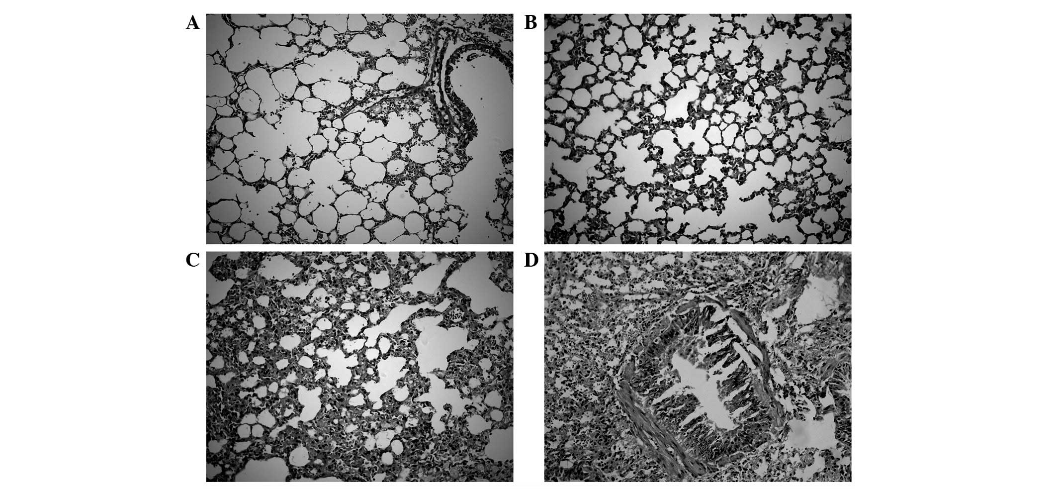
Lung tissue pathological examination
To evaluate the histopathological changes associated
with heavy ion radiation-induced lung injury and fibrosis, the
lungs of normal and 12C6+-irradiated mice
were evaluated. The results showed that there were no pathological
changes in the structure of normal lungs which consisted of a thin
alveolar wall meshwork with few cells visible inside the alveolar
spaces and no fibrosis. Significant histopathological changes and
inflammatory reactions were observed in the heavy ion
irradiated-groups, including the accumulation of numerous
inflammatory cells in the alveolar spaces, extensive collagen
deposition, heavily thickened alveolar walls and collapsed alveolar
spaces, development of fibrotic lesions in the subpleural areas and
focal honeycombing in the subpleural and peribronchial regions. The
extent of the injury was significantly increased as the radiation
dosage was increased compared with the CK mice. The injury was
gradually relieved in the 2-Gy-treated group as the time since
heavy ion irradiation increased (Fig.
2).
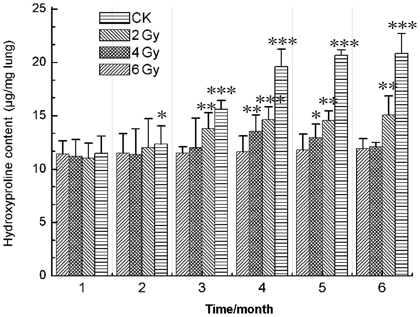
HP content of lung tissue
Pulmonary fibrosis was evaluated by measuring the HP
content of the lungs, as an index of collagen accumulation. The
results showed that the HP content of the lung tissue in the CK
group was noticeably lower than those of the experimental groups
and underwent no clear changes over the total experimental period
of 6 months. The results also demonstrated that, compared with the
CK group, the lung tissue HP content of the 4- and 6-Gy-treated
groups was increased significantly (P<0.05, P<0.01 and
P<0.001) after radiation exposure, although no statistically
significant changes were observed after month 4. In the
2-Gy-treated group, HP content was markedly increased (P<0.05
and P<0.01) between months 1 and 4, but decreased following
month 4 (Fig. 3). These results
indicated that carbon ion beam irradiation induced lung injury and
fibrosis.
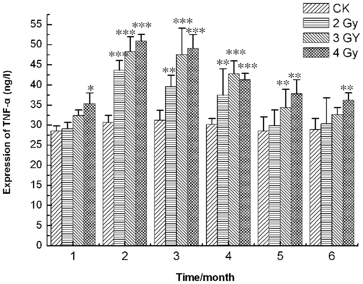
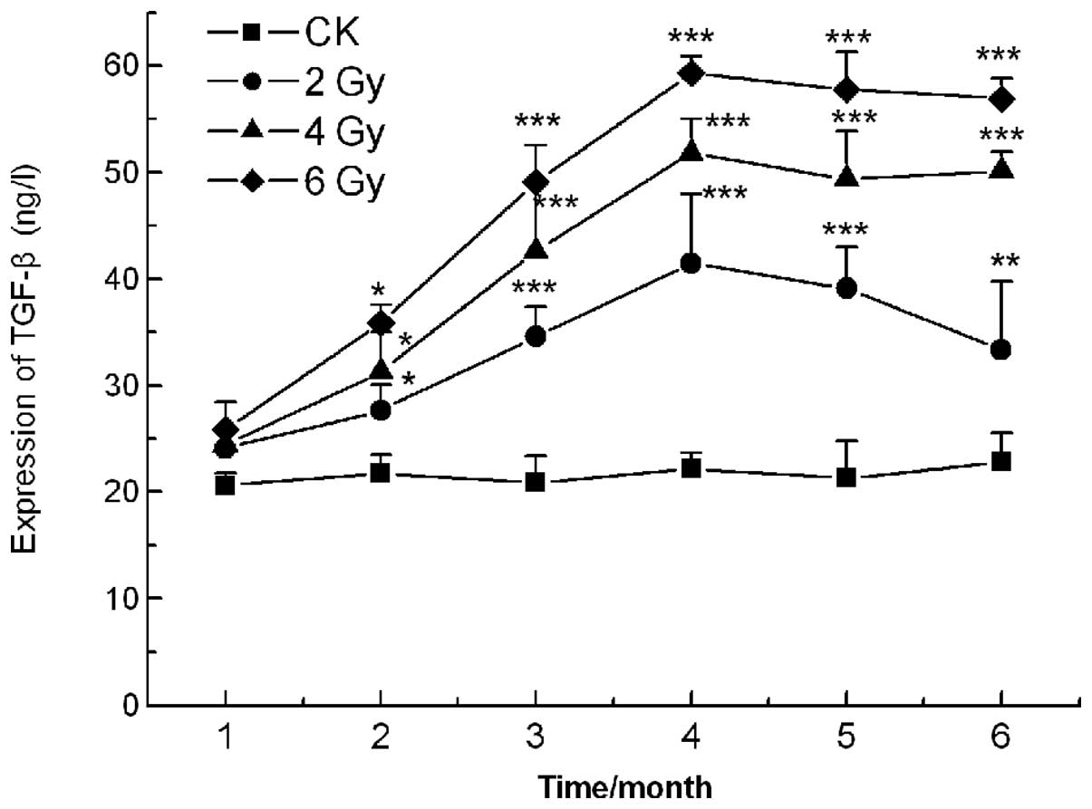
Expression of cytokines in the lung
tissues
Inflammatory cytokines are significant in the
pathogenesis and development of pulmonary fibrosis. The results
revealed that the levels of TNF-α and TGF-β in all the carbon ion
beam treatment groups were higher than those in the CK group. On
the 2nd month following irradiation, the TNF-α content reached its
maximum value, increasing to 43.7±8.6, 48.3±7.4 and 50.9±5.9 ng/l
in the 2, 4 and 6 Gy groups, respectively, compared with 30.7±3.1
ng/l (P<0.001) in the CK group. After 4 months, the levels of
TNF-α decreased noticeably in all experimental groups, but remained
higher compared with the CK group (Fig. 4). The TGF-β levels increased
markedly between months 1 and 4 in all irradiation groups, but
subsequently changed little in the 4- and 6-Gy-treated groups and
decreased sharply in the 2 Gy group (P<0.01, P<0.001;
Fig. 5).
Discussion
Heavy ion irradiation represents the best tool for
the external radiotherapy of inoperable tumors and is now
clinically used for tumor radiation therapy in Japan, America,
Germany and China due to its outstanding physical and biological
characteristics. Previous studies of heavy ions have demonstrated
that, in comparison with LET radiation, such as electrons, X-rays
and γ-rays, carbon ion beams are markedly stronger and more
effective in radiotherapy at all of levels of biological
organization (21). Heavy ions
have been used in the treatment of various tumors, particularly
radioresistant tumors mediated by hypoxia and those located near
organs at risk, such as the brain, head, lung, liver, rectum and
urogenital organs (22). However,
our previous study results also showed that carbon-ion beam
irradiation caused DNA strand breaks, cell apoptosis, lipid
peroxidation, imbalance of antioxidant status, chromosome
aberrations and inactivation of DNA repair enzymes (PARP-1) in
mouse testes (22–23). At the same time as killing
malignant cells in the treatment of cancer, carbon ion beams also
kill normal cells near the tumors.
The lung, one of the most radiosensitive organs, is
frequently irradiated as part of treatment programs for cancers of
the lung, esophagus, breast and lymphatic system. Radiation-induced
lung injury (radiation pneumonitis and radiation pulmonary
fibrosis), a well-known side-effect of radiation following the
post-operative chemotherapy or radiotherapy of breast tumors, is a
common and critical problem that limits the radiation dosage that
may be delivered in tumor therapy. At present, the etiology of this
disease remains unknown. In the present study, the results
coincided with previous studies and showed that the body weight of
the mice decreased significantly in the 1st month and slightly
increased after the 1st month following the carbon ion beam (2, 4
and 6 Gy) irradiation. This may be due to the acute radiation
injury reducing appetite and disturbing the metabolic balance. The
lung coefficient increased between the 1st and 4th months, but
slightly decreased after the 4 month in each of the treatment
groups, while no clear changes were observed in the CK group. It
appeared that the tissue edema and congestion increased in the lung
following carbon ion beam irradiation over the subsequent 4 months
but decreased after this period.
The HP content of the lung tissue homogenates was
closely associated with collagen accumulation and lung
fibrogenesis, so the HP content of the lung tissue was used as an
index of collagen accumulation and distinct marking for the
clinical diagnosis of pulmonary fibrosis. In the present study, the
lung tissue HP content increased significantly for 4 months after
radiation exposure, but decreased in the 2-Gy-treated group and did
not noticeably change in the 4- and 6 Gy-treated groups after 4
months. The histopathological changes also revealed that the extent
of the lung injury was significantly increased when the radiation
dosage was raised compared with the CK group and gradually relieved
in the 2-Gy-treated group as the time since the heavy ion
irradiation increased. A possible explanation for these findings is
that the lung injury was less severe in the low dose irradiation
group (2 Gy) than in the high dose groups and consequently easier
to repair.
Radiation-induced lung fibrosis is a dynamic process
characterized by the constant remodelling of fibrous tissue and
long-term fibroblast activation (24–27).
Previous studies have also suggested that pro-inflammatory and
pro-fibrotic cytokines such as TGF-β and TNF-α, vascular injury,
cellular adhesion molecules and oxidative stress are all vital in
the development of radiation fibrosis (28–30).
TGF-β is a well-known stimulant of extracellular matrix production
by fibroblasts and has been demonstrated to be essential in the
development of lung fibrosis (31–33).
TNF-α is a pro-inflammatory cytokine that is critical in diverse
cellular events and is also considered to be critical in the
development of lung fibrosis. In the present study, the TNF-α
content reached a maximum value in the 2nd month after radiation
and decreased noticeably in all experimental groups after month 2.
The levels of TGF-β increased markedly between months 1 and 4 in
all irradiation groups, but decreased sharply in the 2 Gy
irradiation group and changed little in the 4- and 6-Gy-treated
groups from 4 months after irradiation.
A number of studies have indicated that the
overproduction of reactive oxygen species (ROS) persisting after
irradiation may be closely associated with tissue hypoxia and
injury. ROS induce a cascade of cytokines and this is vital in the
non-healing wound response that perpetuates lung injury (34–37).
Therefore, the main task of future research is to regulate the
content and levels of TGF-β and TNF-α by molecular biology methods
such as biological modifiers, gene therapy and stem cell
transplantation, to manipulate and mini-mise radiation-induced
pulmonary fibrosis and injury.
The present study demonstrated that the carbon ion
beam radiation led to acute lung injury, inflammation and fibrosis
in mice in a time- and dose-dependent manner, similar to other
forms of radiation. However, a certain degree of repair was
observed at low doses with the lengthening of time after radiation.
These results also indicated that whole-body heavy ion beam
irradiation at lower doses (such as 2 Gy) in the normal lung is
safer. The findings of the present study may offer references and
experimental evidence for heavy ion radiotherapy.
Abbreviations:
|
TNF-α
|
tumor necrosis factor-α;
|
|
TGF-β
|
transforming growth factor-β;
|
|
H&E
|
hematoxylin and eosin;
|
|
LET
|
linear energy transfer;
|
|
RBE
|
relative biological effectiveness;
|
|
HIRFL
|
Heavy Ion Research Facility in
Lanzhou;
|
|
HP
|
hydroxyproline;
|
|
ELISA
|
enzyme-linked immunosorbent assay
|
Acknowledgements
This study was supported by grants
from the National Basic Research Program of China (973 Program;
2010CB834202), Key National Natural Science Foundation of China
(10835011), Key Scientific Technology Research Projects of Gansu
Province (0702NKDA045, O801NKDA001), Xinjiang Province Assisted by
Science and Technology project (Y160040YD0) and Western Talent
Program of Chinese Academy of Sciences (Y260230XB0). The authors
would like to thank the accelerator team at the HIRFL, National
Laboratory of Heavy Ion Accelerator in Lanzhou for supplying the
carbon beam.
References
|
1.
|
Vujaskovic Z, Marks LB and Anscher MS: The
physical parameters and molecular events associated with
radiation-induced lung toxicity. Semin Radiat Oncol. 10:296–307.
2000. View Article : Google Scholar : PubMed/NCBI
|
|
2.
|
Noth I and Martinez FJ: Recent advances in
idiopathic pulmonary fibrosis. Chest. 132:637–650. 2007. View Article : Google Scholar
|
|
3.
|
Du Bois RM: Idiopathic pulmonary fibrosis.
Annu Rev Med. 44:441–450. 1993.
|
|
4.
|
Lingos TI, Recht A, Vicini F, Abner A,
Silver B and Harris JR: Radiation pneumonitis in breast cancer
patients treated with conservative surgery and radiation therapy.
Int J Radiat Oncol Biol Phys. 21:355–360. 1991. View Article : Google Scholar : PubMed/NCBI
|
|
5.
|
Wagner GR: Asbestosis and silicosis.
Lancet. 349:1311–1315. 1997. View Article : Google Scholar : PubMed/NCBI
|
|
6.
|
Tyurina YY, Tyurin VA, Kapralova VI,
Wasserloos K, et al: Oxidative lipidomics of γ-radiation-induced
lung injury: Mass spectrometric characterization of cardiolipin and
phosphatidylserine peroxidation. Radiat Res. 175:610–621. 2011.
|
|
7.
|
Margetts PJ, Bonniaud P, Liu L, et al:
Transient over-expression of TGF-β1 induces epithelial mesenchymal
transition in the rodent peritoneum. J Am Soc Nephrol. 16:425–436.
2005.
|
|
8.
|
Martinez Flórez S, Gutiérrez Fernandéz B,
Sánchez-Campos S, González Gallego J and Tuñón MJ: Quercetin
attenuates nuclear factor-kappa activation and nitric oxide
production in interleukin-1 beta-activated rat hepatocytes. J Nutr.
135:1359–1365. 2005.PubMed/NCBI
|
|
9.
|
Mu E, Liu XJ, Chen S, et al: Changes in
factor VII-activating protease in a bleomycin-induced lung injury
rat model and its influence on human pulmonary fibroblasts in
vitro. Int J Mol Med. 26:549–555. 2010.PubMed/NCBI
|
|
10.
|
Yara S, Kawakami K, Kudeken N, et al: FTS
reduces bleomycin-induced cytokine and chemokine production and
inhibits pulmonary fibrosis in mice. Clin Exp Immunol. 124:77–85.
2001. View Article : Google Scholar : PubMed/NCBI
|
|
11.
|
Liu R, Ahmed KM, Nantajit D, et al:
Therapeutic effects of α-lipoic acid on bleomycin induced pulmonary
fibrosis in rats. Int J Mol Med. 19:865–873. 2007.
|
|
12.
|
Armutcu F, Çabuk M, Gurel A, Atmaca H and
Kart L: Caffeic acid phenethyl ester and vitamin E moderates IL-1β
and IL-6 in bleomycin-induced pulmonary fibrosis in rats. Pestic
Biochem Physiol. 88:209–212. 2007.
|
|
13.
|
Liu JF, Wang X, Wang F, Teng L and Cao J:
Attenuation effects of heparin superoxide dismutase conjugate on
bleomycin-induced lung fibrosis in vivo and radiation-induced
inflammatory cytokine expression in vitro. Biomed Pharmacother.
63:484–491. 2009. View Article : Google Scholar
|
|
14.
|
Tomita M, Okuyama T, Katsuyama H, et al:
Gene expression in rat lungs during early response to
paraquat-induced oxidative stress. Int J Mol Med. 17:37–44.
2006.PubMed/NCBI
|
|
15.
|
Hagimoto N, Kuwano K, Inoshima I, et al:
TGF-beta 1 as an enhancer of Fas-mediated apoptosis of lung
epithelial cells. J Immunol. 168:6470–6478. 2002. View Article : Google Scholar : PubMed/NCBI
|
|
16.
|
Dong XC, Li WJ, Liu QF, et al: The
influence of carbon ion irradiation on sweet sorghum seeds. Nucl
Instrum Methods Phys Res B. 266:123–126. 2008. View Article : Google Scholar
|
|
17.
|
Ritter MA, Cleaver JE and Tobias CA:
High-LET radiations induce a large proportion of non-rejoining DNA
breaks. Nature. 266:653–655. 1977. View
Article : Google Scholar : PubMed/NCBI
|
|
18.
|
Sekine E, Okada M, Matsufuji N, Yu D,
Furusawa Y and Okayasu R: High LET heavy ion radiation induces
lower numbers of initial chromosome breaks with minimal repair than
low LET radiation in normal human cells. Mutat Res. 652:95–101.
2008. View Article : Google Scholar
|
|
19.
|
Zhang H, Li S, Wang XH, et al: Results of
carbon ion radiotherapy for skin carcinomas in 45 patients. Br J
Dermatol. 166:1100–1106. 2012. View Article : Google Scholar : PubMed/NCBI
|
|
20.
|
Wu ZH, Zhang H, Wang XY, et al: Protective
effects of melatonin against 12C6+ beam
irradiation-induced oxidative stress and DNA injury in the mouse
brain. Adv Space Res. 49:196–203. 2012.
|
|
21.
|
Zhou G, Kawata T, Furusawa Y, et al:
Protective effects of melatonin against low- and high-LET
irradiation. J Radiat Res. 47:175–181. 2006. View Article : Google Scholar : PubMed/NCBI
|
|
22.
|
Zhang H, Zhao W, Wang Y, et al: Induction
of cytogenetic adaptive response in spermatogonia and spermatocytes
by pre-exposure of mouse testis to low-dose
12C6+ ions. Mutat Res. 653:109–112. 2008.
View Article : Google Scholar : PubMed/NCBI
|
|
23.
|
Liu Y, Zhang H, Zhang L, Zhang X, Xie Y
and Zhao W: Melatonin modulates acute testicular damage induced by
carbon ions in mice. Pharmazie. 64:685–689. 2009.PubMed/NCBI
|
|
24.
|
Stone HB, Coleman CN, Anscher MS and
McBride WH: Effects of radiation on normal tissue: consequences and
mechanisms. Lancet Oncol. 4:529–536. 2003. View Article : Google Scholar : PubMed/NCBI
|
|
25.
|
Finkelstein JN, Johnston CJ, Baggs R and
Rubin P: Early alterations in extracellular matrix and transforming
growth factor beta gene expression in mouse lung indicative of late
radiation fibrosis. Int J Radiat Oncol Biol Phys. 28:621–631. 1994.
View Article : Google Scholar : PubMed/NCBI
|
|
26.
|
Rodeman HP and Bamberg M: Cellular basis
of radiation-induced fibrosis. Radiother Oncol. 35:83–90. 1995.
View Article : Google Scholar : PubMed/NCBI
|
|
27.
|
Rubin P, Johnston CJ, Williams JP,
McDonald S and Finkelstein JN: A perpetual cascade of cytokines
post irradiation leads to pulmonary fibrosis. Int J Radiat Oncol
Biol Phys. 33:99–109. 1995. View Article : Google Scholar : PubMed/NCBI
|
|
28.
|
Anscher MS, Kong FM, Andrews K, et al:
Plasma transforming growth factor beta1 as a predictor of radiation
pneumonitis. Int J Radiat Oncol Biol Phys. 41:1029–1035. 1998.
View Article : Google Scholar : PubMed/NCBI
|
|
29.
|
Fu XL, Huang H, Bentel G, et al:
Predicting the risk of symptomatic radiation-induced lung injury
using both the physical and biologic parameters V30 and
transforming growth factor β. Int J Radiat Oncol Biol Phys.
50:899–908. 2001. View Article : Google Scholar : PubMed/NCBI
|
|
30.
|
Chen Y, Williams J, Ding I, et al:
Radiation pneumonitis and early circulatory cytokine markers. Semin
Radiat Oncol. 12(Suppl 1): 26–33. 2002. View Article : Google Scholar : PubMed/NCBI
|
|
31.
|
Broekelmann TJ, Limper AH, Colby TV and
Mcdonald JA: Transforming growth factor beta 1 is present at sites
of extra-cellular matrix gene expression in human pulmonary
fibrosis. Proc Natl Acad Sci USA. 88:6642–6646. 1991. View Article : Google Scholar : PubMed/NCBI
|
|
32.
|
Lee JC, Kinniry PA, Arguiri E, Serota M,
Kanterakis S, Chatterjee S, et al: Dietary curcumin increases
antioxidant defenses in lung, ameliorates radiation-induced
pulmonary fibrosis, and improves survival in mice. Radiat Res.
173:590–601. 2010. View Article : Google Scholar
|
|
33.
|
Dotor J, López-Vázquez AB, Lasarte JJ, et
al: Identification of peptide inhibitors of transforming growth
factor beta 1 using a phage-displayed peptide library. Cytokine.
39:106–115. 2007. View Article : Google Scholar
|
|
34.
|
Poli G and Parola M: Oxidative damage and
fibrogenesis. Free Radic Biol Med. 22:287–305. 1997. View Article : Google Scholar : PubMed/NCBI
|
|
35.
|
Harroon ZA, Raleigh JA, Greenberg CS and
Dewhirst MW: Early wound healing exhibits cytokine surge without
evidence of hypoxia. Ann Surg. 231:137–147. 2000. View Article : Google Scholar : PubMed/NCBI
|
|
36.
|
Vujaskovic Z, Anscher MS, Feng QF, et al:
Radiation-induced hypoxia may perpetuate late normal tissue injury.
Int J Radiat Onco Biol Phys. 50:851–855. 2001. View Article : Google Scholar : PubMed/NCBI
|
|
37.
|
Kang SK, Rabbani ZN, Folz RJ, et al:
Overexpression of extracellular superoxide dismutase protects mice
from radiation-induced lung injury. Int J Radiat Oncol Biol Phys.
57:1056–1066. 2003. View Article : Google Scholar : PubMed/NCBI
|