Introduction
The incidence of spinal fractures accounts for 5–6%
of whole-body fractures and the majority of these are thoracolumbar
fractures (1). With the high-speed
development of society, the number of high-energy injuries is
increasing, including falling from high places, traffic accidents
and bruises. Thoracolumbar spine injuries often involve the spinal
column and fragment retrusion causes spinal canal stenosis or
spinal cord and cauda equina injuries. Surgeons must
determine whether the spine is able to withstand the physiological
load and stress, and if not, spinal osseous stability must be
rebuilt. Biomechanical studies on spinal stability and innovation
of spinal fixation methods created the foundations for achieving
the therapeutic goal (2). With the
wide application of pedicle screw technology, posterior
short-segmental fixation has become a reliable method in the
treatment of thoracolumbar fractures. In thoracolumbar burst
fractures, pedicle screw placement between the adjacent upper and
lower vertebral bodies is normally used for vertebral reduction and
fixation (dual-plane fixation). This method opens the fractured
vertebra and restores the height of the vertebral bodies. However,
there are risks of postoperative kyphosis and failed internal
fixation as a result of intervertebral indirect reduction and
fixation. Postoperative failure of internal fixation and the loss
of the corrective angle have become important factors affecting
treatment efficacy. Theoretical and experimental studies of
short-segmental pedicle screw placement in the treatment of
fractured vertebrae in thoracolumbar fractures began in 1994 when
Dick et al(3) produced a
biomechanical study with this method. Since then, few in-depth
studies on this technology have been produced and clinical reports
on the treatment of thoracolumbar fractures using this technology
are rare. In this study, the admitted patients with thoracolumbar
fractures were retrospectively studied to assess the value of
applying vertical stress pedicle screws in the fractured
vertebra.
Materials and methods
General data
From March 2008 to January 2010, a total of 30
patients with single thoracolumbar fractures, receiving vertical
stress pedicle screw fixation of fractured vertebrae (group A) or
traditional double-plate fixation (group B), were retrospectively
reviewed. In group A, 11 men and 3 women, aged 33–59 years (average
46.4 years) were enrolled and 12 men and 4 women, aged 34–63 years
(average 47.2 years) were enrolled into group B. The injured
segments were T12 in 3 patients, L1 in 6
patients, L2 in 3 patients and L3 in 2
patients in group A and T12 in 3 patients, L1
in 7 patients, L2 in 4 patients and L3 in 2
patients in group B. All patients had fresh fractures with a
complete unilateral or bilateral pedicle and no explosion of the
inferior half of the vertebral body or inferior endplate. According
to load sharing classification put forward by McCormack et
al, a score <7 was an indication of simple posterior
internal fixation (4). All the
cases selected in this study had a score <7, with the exception
of 2 patients in group A that had a score >7, who received
posterior surgery for economic reasons. Preoperative and
postoperative lateral X-ray film, pedicle computerized tomography
(CT) scan and two- and three-dimensional reconstruction were used
to evaluate the fractures and the postoperative stability of the
internal fixation. In group A, patients received conventional
posterior distraction and lumbar lordosis restoration, as well as
alteration of ventral pressures using pedicle screws in the
fractured vertebra in a vertical direction to relieve stress,
achieving a local stress balance. In group B, the cephalad and
caudal area of the fractured vertebra was fixed according to the
traditional method, without applying vertical stress pedicle
screws. This study was conducted in accordance with the declaration
of Helsinki. This study was conducted with approval from the Ethics
Committee of Shanghai Punan Hospital of Pudong New Area. Written
informed consent was obtained from all participants.
Surgical method
After receiving general anesthesia, patients were
placed in the prone position with U-shaped pillows under their
chests and bilateral iliac to impend their abdomens in order to
reduce intraoperative bleeding. Following intraoperative X-ray
fracture location with a C-arm fluoroscopic device, a posterior
median incision was made with the fractured vertebra at the center
to expose the vertebral plate and the articular process layers. Two
pedicle screws were implanted into the upper vertebral body of the
fractured vertebra by the Magerl method and the lower vertebral
body by the Krag method, respectively. Two short nails or universal
nails were implanted into the fractured vertebra and the pre-bent
connection rod was attached. The upper and lower pedicle screws
were distracted and the upper and median pedicle screws were locked
to the connection rod. Then, the lower and median pedicle screws
were longitudinally distracted to recover the fractured vertebral
body height and the connection was locked. The dual-plane fixation
group received longitudinal distraction and reduction following
implantation of the pedicle screws into the upper and lower
vertebral bodies of the fractured vertebra and attachment of the
pre-bent connection rod. According to the location of the vertebral
canal blockage, patients received total laminectomy decompression
to remove the dural sac oppression and promote the bilateral
intertransverse bone graft fusion if necessary.
Postoperative treatment
The drainage tubes were kept in place for 24–48 h
after surgery. Patients were encouraged to exercise their lumbar
muscles in postoperative week 3 and perform out-of-bed activities
with a brace in postoperative months 1–2. An X-ray and CT scan were
taken regularly. The internal fixation was kept in place for one
year after surgery.
Statistical analysis
Contrast analysis was performed on Cobb’s angle
changes and vertebral body height in postoperative two weeks with
internal fixation. The vertebral body height was determined by the
ratio of the mean of the fractured vertebral body and anterior
heights of the adjacent two vertebral bodies. Cobb’s angle was
determined by the angle formed by the perpendiculars of the
extension lines of the upper endplate of the upper fractured
vertebra and the lower endplate of the lower fractured vertebra.
The changes to the preoperative and postoperative parameters were
analyzed by independent samples t-test with SPSS 13.0 software
package (SPSS Inc., Chicago, IL, USA) and all tests were two-sided.
P<0.05 was considered to indicate a statistically significant
difference.
Results
There were no significant differences in Cobb’s
angle and vertebral body height between the two groups
preoperatively, which may rule out the bias of therapeutic effect
caused by the varying degrees of instability of the fractured
vertebra. There were also no significant differences in Cobb’s
angle and vertebral body height between the two groups
postoperatively. There was a significant difference in Cobb’s
angle, but not in vertebral body height between the two groups one
year after surgery (Table I).
 | Table I.Comparison of relevant parameters in
the two groups. |
Table I.
Comparison of relevant parameters in
the two groups.
| Group A | Group B | t-value | P-value |
|---|
| Preoperative Cobb’s
angle (°) | 9.63±2.01 | 9.07±1.87 | 0.780 | 0.442 |
| Preoperative
vertebral body height (%) | 50.68±5.89 | 50.8±8.35 | −0.073 | 0.942 |
| Postoperative Cobb’s
angle (°) | 2.51±1.14 | 3.26±1.91 | −1.263 | 0.217 |
| Postoperative
vertebral body height (%) | 92.92±5.14 | 90.9±4.99 | 1.073 | 0.293 |
| Cobb’s angle in
postoperative one year (°) | 2.51±1.25 | 5.12±1.07 | −6.512 | 0.001a |
| Vertebral body height
in postoperative one year (%) | 91.43±4.99 | 89.1±2.74 | 1.552 | 0.132 |
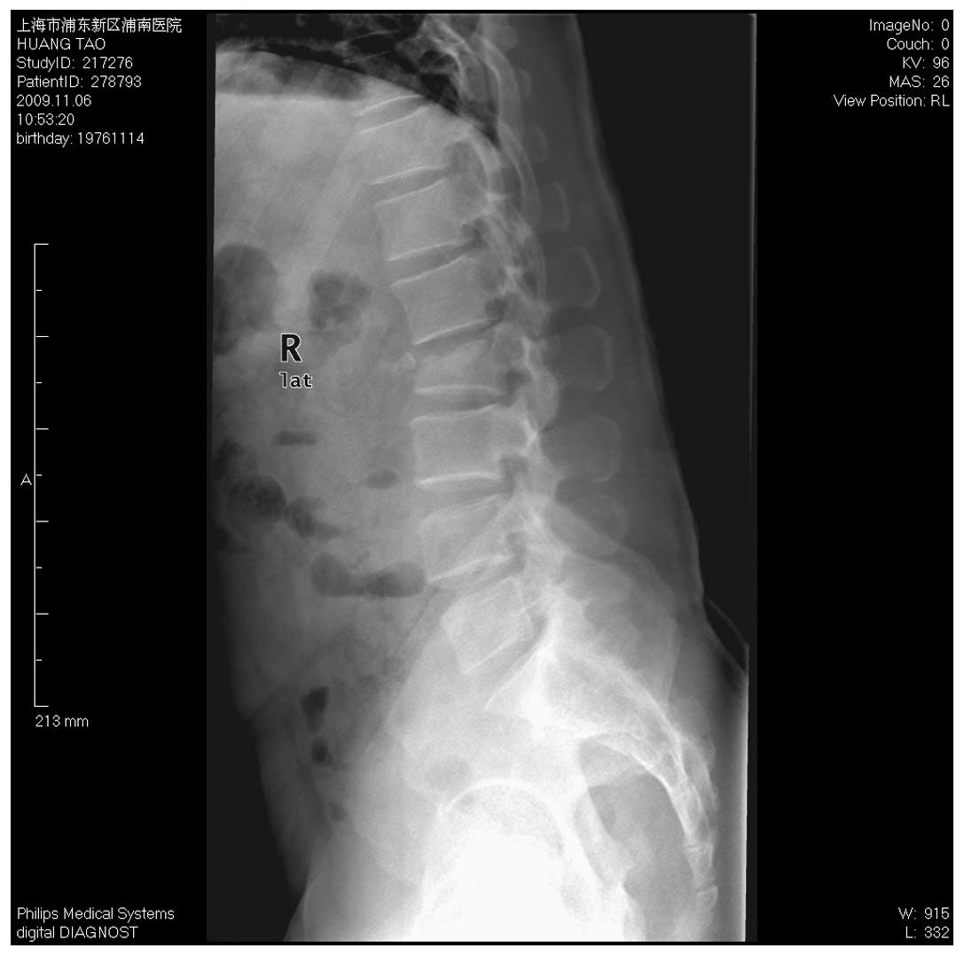
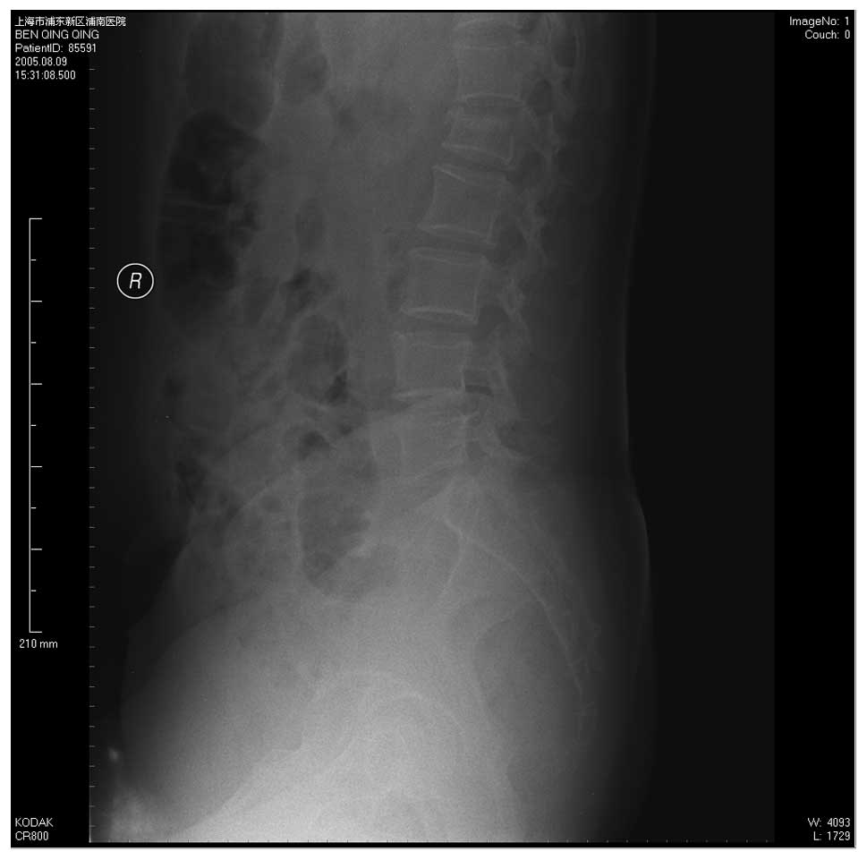
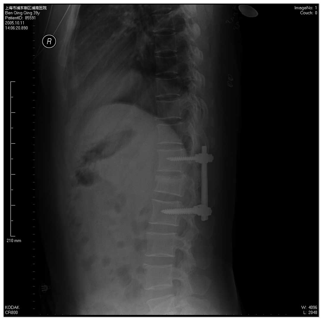
The results of the follow-up demonstrated whether
traditional dual-plane fixation or triplane fixation with stress
screw fixation through the fractured vertebra obtain a satisfactory
reduction of vertebral burst fracture (Figs. 1–3), recovery of fractured vertebral height
and correction of the forward bending of the spine (Fig. 3). However, the long-term follow-up
revealed that the two methods effectively maintain vertebral body
height and the vertical stress screw method maintained the
physiological curvature of the spine (Figs. 4–6).
Discussion
Spinal fractures cause damage to the normal
structure of the spine and affect nerve function. Therefore,
treatment of spinal fractures aims to recover the normal anatomical
structure, remove nerve oppression and promote the recovery of
nerve function. There are a number of conflicting ideas on specific
treatment, including surgical or non-surgical treatment, anterior
fixation or posterior fixation, long-segment or short-segment
fixation and fusion or non-fusion (5–8).
Spinal fractures are divided into stable and unstable fractures.
The diagnostic standards of unstable fractures include: i) anterior
vertebral compression >50%; ii) Cobb’s angle for kyphosis
>20°; and iii) a vertebral canal blockage area larger than 50%
(9,10).
Anterior surgery has the advantage of thorough
decompression, which effectively corrects the kyphosis and receives
good bone graft fusion, in order to establish the fused segment.
However, this surgery may cause large traumas that easily damage
the large blood vessels and other organs, as well as complications
caused by thoracotomy and laparotomy, including intestinal adhesion
and pneumothorax. It is recommended that compressed fractures and
mild violence fractures receive posterior surgeries. The posterior
fixation fusion through the use of pedicle screws is a simple
surgery, resulting in little injury and has a rapid recovery;
therefore, it is widely applied in the clinic (11).
McCormack et al studied various load
distributions in the reconstruction of spinal fractured implants
(4). It was considered that the
anatomical characteristics of the fracture itself are more
important than the types of implants used. In the fixation of long
fractured bones, the internal fixation system and load distribution
between the host bones are the basis of fracture healing and
internal fixation failure. The treatment of thoracolumbar fractures
should also consider the load distribution. Without reasonable load
sharing, the risks of internal fixation failure and fracture
healing failure significantly increase. Based on this, McCormack
et al presented a load sharing classification of spinal
fractures, which aids accurate assessment of the stability of the
spine after being fractured and guides method selection for
internal fixation. Additionally, this reduces the risk of internal
fixation failure and improves the effects of surgery. Scoring is
performed according to the vertebral body involved in the fracture,
the displacement of the fracture parts and the kyphotic deformity
(4). Parker et al
considered that a score of <6 points presents a good load
sharing capability and simple posterior pedicle fixation could
achieve a good stability. A score >7 points presents a poor load
sharing capability and in simple posterior pedicle fixation, there
is a risk of internal fixation failure, including a broken nail. In
these cases, the fixation should be replaced with anterior bone
graft fusion or two-stage anterior surgery (12).
Whether fractured vertebrae require vertical stress
screw fixation to assist and maintain the reduction remains
unclear. Traditional dual-plane fixation that fixes the normal
upper and lower vertebral bodies of fractured vertebrae has the
following problems: distraction of the fractured vertebral height
results in poor recovery and the intervertebral height increases,
particularly the non-affected ones; the fixation has a
parallelogram effect and a lateral instability so it often requires
the addition of a transverse connection fixation; and finally the
fixation has a suspension effect, whereby decreasing the distance
between the upper and lower anterior vertebral bodies and
increasing recession of intermediate fractured vertebrae.
The pedicle internal fixation of fractured vertebrae
is technically feasible and effective in restoring vertebral body
height and correcting dislocation (13,14).
The screw implantation through fractured vertebrae significantly
improves the stress distribution of screws, reduces screw load and
provides a fulcrum for the reduction to make it coincide with the
mechanical mechanism, so as to significantly improve its
anti-stress ability and significantly enhance the stability of the
fixation (15). In the stress
screw fixation through fractured vertebrae, an appropriate amount
of ventral pressure overcomes the kyphosis stress caused by
fractures, which is helpful for maintaining the physiological
curvature of fixed parts postoperatively to prevent the screws from
loosening. This may also benefit and reduce dislocation for
patients with fracture dislocation. The stress added between the
screw and pedicle is a ventral pressure stress but not a pullout
force. Complete pedicle fixation is enough to guarantee the
stability of the vertical stress screw (16).
Dick et al(3) reported the comparison of
biomechanical experiments of 6-screw and 4-screw fixation in a
cattle lumbar model and identified that the 6-screw fixation has
clear advantages. The axial load capacity increases 160%, the
bending resistance capacity increases 48% and the torsional
rigidity increases by 38%. The authors demonstrated that screw
implantation through the fractured vertebra increases the effect of
resistance to stress. Conversely, Hakalo and Wronski (17) considered the there is no basis for
fractured vertebrae fixation. Following spinal fracture, the
reduction of the axial ligament could rearrange the bones connected
to the ligament and restore the shape of injured vertebrae;
however, a ‘shell effect’ still existed in fractured vertebrae
following reduction. The fractured vertebra and its upper and lower
clearances did not have weight-bearing capacity and the load was
mainly conducted through the internal fixation. Therefore, pedicle
screw implantation through the fractured vertebra did not
effectively increase the spinal axial bearing capacity and initial
stability, thus it does not reduce the postoperative corrective
loss and failure rate of internal fixation. Thoracolumbar fractures
not accompanied with anterior and posterior longitudinal ligament
fracture are given inter-segmental pedicle screw fixation and
distraction. The stability is enough to meet the clinical needs and
patients do not require re-fixation of the fractured vertebra.
Fractured vertebrae fixation not only failed to clearly increase
the stability of the fixed segment, but also increased the surgery
time, as well as surgery risk and economic burden of patients.
However, the in vitro study did not consider the role of the
neuromuscular system and other stable structures on the stability
of the spine. The early postoperative evaluation lacked long-term
effect.
Maintenance of fractured vertebral height and spinal
curvature are important in the treatment of vertebral fractures.
With the distraction and reduction of the posterior pedicle screw,
seriously collapsed vertebral fractures demonstrate clear recovery
of vertebral body height intraoperatively under a C-arm
fluoroscopic device or in an X-ray film. However, the ‘shell
effect’ of fractured vertebrae still exists postoperatively in
internal fixation. The compressed trabeculae in the vertebral body
do not achieve complete reduction. The residual interspace is
difficult to heal. Therefore, if the stability of the anterior and
median column are not reestablished quickly, the posterior internal
fixation takes continuous and excessive loads, increasing the risk
of corrective angle loss and internal fixation failure (18,19).
In this study, we observed satisfactory
postoperative recovery of vertebral body height in the triplane
fixation group and dual-plane fixation group, with 92.9 and 90.9%
recovery, respectively. There was also relatively good
postoperative correction of spinal kyphosis, with a postoperative
Cobb’s angle of 2.51 and 3.26°, respectively. There were no
significant differences between the two groups. One year after
surgery, the vertebral body height in the two groups was 91.4 and
89.1%, respectively, without a significant difference. However, the
Cobb’s angles were 2.51 and 5.12°, respectively, with a significant
difference. This indicates that triplane fixation and dual-plane
fixation maintain the vertebral body height and that triplane
fixation is more effective at maintaining the Cobb’s angle and
preventing kyphosis of the spine.
The advantages of using pedicle screws to treat
fractured vertebrae include the following: i) it provides a good
three-point fixation to reduce the suspension effect of the
internal fixation system; ii) it reduces the parallelogram effect
to increase the stability; iii) it avoids stretching the normal
intervertebral disc, which is beneficial to the recovery of the
vertebral fracture form and iv) it dispenses the stress of the
pedicle screw connection. Therefore, conditional application of
vertical stress screw fixation of fractured vertebrae enhances the
stability of the posterior short-segment internal fixation system
for thoracolumbar fractures and facilitates the correction of
kyphosis and maintenance of the corrective effect (20,21).
In our comparative study of the two groups, we
demonstrate that the two internal fixation methods maintain
vertebral body height and that vertical stress screw fixation of
fractured vertebrae is more effective at maintaining spinal
postoperative physiological curvature of the spine and reducing the
angle loss. Conditional application of vertical stress screw
fixation of fractured vertebrae enhances the stability of the
posterior short-segment internal fixation system for thoracolumbar
fractures and facilitates the correction of kyphosis. The
limitations of this study are that it is a retrospective study with
a small sample size and short follow-up period. A multicenter,
large sample study with a long follow-up is required in order to
achieve a more definite conclusion.
References
|
1.
|
Define HL and Canto FR: Low thoracic and
lumbar burst fractures: radiographic and functional outcomes. Eur
Spine J. 16:1934–1943. 2007. View Article : Google Scholar : PubMed/NCBI
|
|
2.
|
Sim HB, Murovic JA, Cho BY, Lim TJ and
Park J: Biomechanical comparison of single-level posterior versus
transforaminal lumbar interbody fusions with bilateral pedicle
screw fixation: segmental stability and the effects on adjacent
motion segments. J Nerosurg Spine. 12:700–708. 2010. View Article : Google Scholar
|
|
3.
|
Dick JC, Jones MP, Zdeblick TA, Kunz DN
and Horton WC: A biomechanical comparison evaluating the use of
intermediate screws and cross-linkage in lumbar pedicle fixation. J
Spinal Disord. 7:402–407. 1994.PubMed/NCBI
|
|
4.
|
McCormack T, Karaikovic E and Gaines RW:
The load-sharing classification of spine fractures. Spine.
19:1741–1744. 1994. View Article : Google Scholar : PubMed/NCBI
|
|
5.
|
Shen WJ, Liu TJ and Shen YS: Nonoperative
treatment versus posterior fixation for thoracolumbar junction
burst fractures without neurologic deficit. Spine. 26:1038–1045.
2001. View Article : Google Scholar : PubMed/NCBI
|
|
6.
|
McLain RF: The biomechanics of long versus
short fixation for thoracolumbar spine fractures. Spine.
31:S70–S79. 2006. View Article : Google Scholar : PubMed/NCBI
|
|
7.
|
Oken F, Yildirim O, Oken O, Gulcek M,
Yavuzer G and Ucaner A: Short or long fusion after thoracolumbar
burst fractures does not alter selected gait parameters: a
preliminary study. J Orthop Res. 29:915–918. 2011. View Article : Google Scholar
|
|
8.
|
Dai LY, Jiang LS and Jiang SD: Posterior
short-segment fixation with or without fusion for thoracolumbar
burst fractures: a five to seven-year prospective randomized study.
J Bone Joint Surg Am. 91:1033–1041. 2009.PubMed/NCBI
|
|
9.
|
Umehara S, Zindrick MR, Patwardhan AG, et
al: The biomechanical effect of postoperative hypolordosis in
instrumented lumbar fusion on instrumented and adjacent spinal
segments. Spine. 25:1617–1624. 2000. View Article : Google Scholar : PubMed/NCBI
|
|
10.
|
Cho DY, Lee WY and Sheu PC: Treatment of
thoracolumbar burst fractures with polymethyl mechacrylate
vertebroplasty and short-segment pedicle screw fixation.
Neurosurgery. 53:1354–1360. 2003. View Article : Google Scholar : PubMed/NCBI
|
|
11.
|
Siebenga J, Leferink VJ, Segers MJ, et al:
Treatment of traumatic thoracolumbar spine fractures: a multicenter
prospective randomized study of operative versus nonsurgical
treatment. Spine. 31:2881–2890. 2006. View Article : Google Scholar
|
|
12.
|
Parker JW, Lane JR, Karaikovic EE and
Gaines RW: Successful short-segment instrumentation and fusion for
thoracolumbar spine fracture: a consecutive 4 1/2-year series.
Spine. 26:1157–1170. 2000.PubMed/NCBI
|
|
13.
|
Mahar A, Kim C, Wedemeyer M, et al:
Short-segment fixation of lumbar burst fractures using pedicle
fixation at the level of the fracture. Spine. 32:1503–1507. 2007.
View Article : Google Scholar
|
|
14.
|
Defino HL and Scarparo P: Fractures of
thoracolumbar spine: monosegmental fixation. Injury. 36(Suppl 2):
B90–B97. 2005. View Article : Google Scholar : PubMed/NCBI
|
|
15.
|
Lehman RA Jr, Lenke LG, Keeker KA, Kim YJ
and Cheh G: Computed tomography evaluation of pedicle screws placed
in the pediatric deformed spine over an 8-year period. Spine.
32:2679–2684. 2007.PubMed/NCBI
|
|
16.
|
Dai LY: Principles of management of
thoracolumbar fractures. Orthop Surg. 4:67–70. 2012. View Article : Google Scholar : PubMed/NCBI
|
|
17.
|
Hakalo J and Wronski J: Complications of a
transpedicular stabilization of thoraco-lumbar burst fractures.
Neural Neurolchir Pol. 40:134–139. 2006.PubMed/NCBI
|
|
18.
|
Boos N and Webb JK: Pedicle screw fixation
in spinal disorders: a European view. Eur Spine J. 6:2–18. 1997.
View Article : Google Scholar : PubMed/NCBI
|
|
19.
|
Hicks JM, Singla A, Shen FH and Arlet V:
Complications of pedicle screw fixation in scoliosis surgery: a
systematic rewiew. Spine. 35:E465–E470. 2010. View Article : Google Scholar : PubMed/NCBI
|
|
20.
|
Gaines RW Jr: The use of pedicle-screw
internal fixation for the operative treatment of spinal disorders.
J Bone Jiont Surg Am. 82-A:1458–1476. 2000.PubMed/NCBI
|
|
21.
|
Guven O, Kocaoglu B, Bezer M, Aydin N and
Nalbantoglu U: The use of screw at the fracture level in the
treatment of thoracolumbar burst fractures. J Spinal Disord Tech.
22:417–421. 2009. View Article : Google Scholar : PubMed/NCBI
|