Introduction
Breast cancer is the most common type of malignant
tumor and a leading cause of cancer mortality. At present it has
the highest incidence of malignant tumors in females (1). Approximately 200,000 females per year
are diagnosed with breast cancer and breast cancer results in
40,610 mortalities in the USA (2).
The current treatment may involve lumpectomy (surgical removal of
the tumor with clear margins) or mastectomy (surgical removal of
the breast), as well as chemotherapy, radiotherapy and other
therapeutic treatments. Among these, chemotherapy has the most
important role. Adriamycin is commonly used since it has a
broad-spectrum antitumor effect. However, it also has a strong
cytotoxic effect, including bone marrow suppression.
Heparin has long been known to possess biological
effects that are not associated with its anticoagulant activity,
and it is the first choice for the prevention and treatment of
venous thromboembolism for patients with cancer. In particular,
heparin and novel agents based upon the heparin template have been
investigated as potential antitumor agents. Previous studies have
suggested that heparin, as well as having direct effects on blood
coagulation, also has a role in the treatment of cancer by
inhibiting tumor cell proliferation, angiogenesis, invasion and
migration, and enhancing the sensitivity of tumor cells to
chemotherapeutic drugs (3–5). The possible specific mechanisms
thought to be involved in the antitumor effect of heparin include
inhibition of heparanase activity and inhibition of the tissue
factor pathway (6).
It was hypothesized in the present study that
combination treatment with small doses of Adriamycin and heparin
would lead to reduced tumor angiogenesis, and in turn metastasis,
without major adverse effects. In this study, a breast cancer model
in mice was established and the mice were randomly divided into
four groups, treated with Adriamycin or heparin alone, a
combination of Adriamycin and heparin or saline. All mice were
analyzed for tumor weight, metastasis, vascular endothelial growth
factor (VEGF), microvascular density (MVD) and overall quality of
life. The tumor metastasis rate and occurrence of adverse effects
in the various groups were compared, and the mechanism of action
was investigated. Analysis of the results was conducted to evaluate
whether heparin and Adriamycin combination treatment may be a novel
strategy for the treatment of breast cancer.
Materials and methods
Cell line and culture
C3H mouse autologous breast cancer cells
were obtained from Bengbu Medical College (Bengbu, China). Cells
were grown in Dulbecco’s modified Eagle’s medium (DMEM),
supplemented with 10% fetal calf serum, penicillin (10 U/ml),
streptomycin (100 U/ml) and HEPES (25 mM), and maintained at 37°C
in a humidified atmosphere of 5% CO2.
Experimental animals and grouping
A total of 40 healthy, female C3H mice,
weighing 17–22 g, were provided by Silaike Experimental Animals
Co., Ltd. (Shanghai, China). The mice were randomly divided into
four groups (n=10/group): normal saline group, low molecular weight
heparin (LMWH) group, Adriamycin positive control group and a
combination group (LMWH combined with Adriamycin). All animals were
examined prior to the start of the study and any animal that did
not meet the health and weight criteria was excluded from the
study. This study was performed in strict accordance with the
recommendations in the Guide for the Care and Use of Laboratory
Animals (8th edition, 2011) of the National Institutes of Health
(Bethesda, MD, USA). The study was approved by the Ethics Committee
of Bengbu Medical College.
Establishment of a breast cancer model in
C3H mice
Exponentially growing tumor cells (0.2 ml;
1×10⁷/ml) were subcutaneously injected into the right
axillary region of C3H mice. Tumor lines were achieved
by serial subcutaneous passages of tumor fragments (~3×3×3 mm) from
growing tumors into C3H mice, as previously described
(7).
When the tumor volume was the size of a millet seed,
this indicated that a breast cancer model was successfully
established in the C3H mice. The normal saline group was
administered saline intraperitoneally, once daily (1 ml/20 g). The
Adriamycin group was administered Adriamycin (Xing Jia Biological
Medicine Co., Ltd., Wuhan, China) intraperitoneally, once weekly (4
mg/kg/dose). The LMWH group was administered LMWH (Wang Bang
Biosciences Co., Ltd., Xuzhou, China) subcutaneously, once daily
(1,500 U/kg/day). The combination group was treated with Adriamycin
and LMWH, at the same doses as for the Adriamycin and LMWH groups.
Treatment was administered for one month. Tumor growth was followed
by biweekly measurements of tumor diameters using a Vernier caliper
12 days following inoculation. Tumor volume (TV) was calculated
according to the following formula: TV (mm3) =
d2xD/2, where d and D are the shortest and the longest
diameter, respectively. A tumor growth curve was then obtained.
Hematoxylin and eosin (H&E)
staining
After one month of drug administration, the mice
were decapitated and the solid tumor tissue and the lungs were
removed. The distribution of tumor cells following drug
administration was observed and the tumor metastasis was observed
in the lung. Sections of tissue were placed in phosphate-buffered
formaldehyde (4%) overnight, then stored in ethanol and embedded in
paraffin. Cross sections (4 μm) were stained with H&E.
Terminal-deoxynucleotidyl
transferase-mediated nick end labeling (TUNEL)
Tumor tissue sections fixed in 4% paraformaldehyde
accorded to the standard procedure. The slides were placed in a
plastic jar containing 200 ml citrate buffer (0.1 M, pH 6.0).
Microwave irradiation (750 W) was applied for 1 min. The slides
were cooled rapidly by immediately adding 80 ml double distilled
water (20–25°C). The slides were transferred into
phosphate-buffered saline (PBS; 20–25°C). The slides were then
immersed in Tris-HCl (0.1 M; pH 7.5), containing 3% BSA and 20%
normal bovine serum for 30 min at 15–25°C. The slides were rinsed
twice with PBS at 15–25°C and the excess fluid was drained off.
TUNEL reaction mixture (50 ml; Beyotime Institute of Biotechnology,
Nantong, China) was added to the sections. For the negative control
50 ml label solution was added. The slides were incubated for 60
min at 37°C in a humidified atmosphere in the dark and then rinsed
three times in PBS for 5 min. The sections were analyzed under a
fluorescence microscope (model BSF-60; Ba Tuo Instrument Co., Ltd.,
Shanghai, China) with blue-black particles in the nucleus
indicating apoptotic cells.
Immunohistochemical (IHC) staining
Tumor tissues fixed in 4% paraformaldehyde in PBS
were placed on ice for 2 h and saturated in 20% sucrose at 4°C. The
samples were embedded in paraffin and cut longitudinally to 4 μm.
The sections were washed with PBS and then subjected to staining
for VEGF using polyclonal mouse anti-mouse antibodies (BD
Biosciences, San Jose, CA, USA) at a dilution of 1:100. The primary
antibodies were applied and incubated for 30 min at room
temperature. The sections were washed again with PBS and incubated
in the biotinylated secondary antibodies (BioSharp, Hefei, China)
for 30 min. After three washes with PBS, the sections were
incubated in enzyme reagents, containing 50 μl avidin, 50 μl
biotinylated horseradish peroxidase and 2.5 ml PBS. Subsequently,
the sections were incubated in a few drops of peroxidase substrate
mixing liquid and were washed in deionized water. Finally, sections
were counterstained in hematoxylin followed by several washes with
deionized water. In negative controls, nonimmune serum was used
instead of primary antibodies.
Results
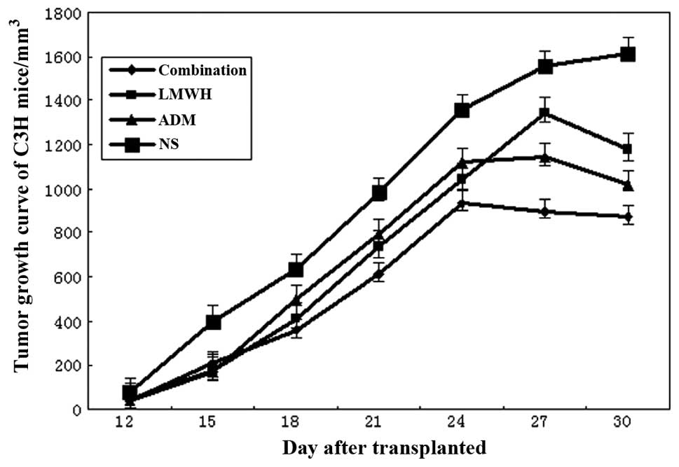
Tumor growth curve in C3H
mice
Twelve days following the inoculation, no
significant difference in tumor volume was observed between the
four groups. In the normal saline group, as time progressed, the
tumor volume increased with a linear upward trend. However, after
27 days, growth slowed. In the LMWH group, after 15 days the tumor
volume was similar to that in with the Adriamycin group and
combination group, and then increased linearly with time. After day
27, the volume began to decrease; however, it was still higher
compared with that in the Adriamycin and combination groups. In the
Adriamycin group, the tumor grew rapidly for 24 days, and then the
tumor volume gradually declined, but remained higher compared with
that in the combination group. In the combination group, after 15
days the volume growth was relatively slow compared with that in
the Adriamycin and LMWH groups. After 24 days, the tumor growth
began to decline, and the overall volume growth was less compared
with that in the other three groups. The tumor growth curves of
breast cancer in the four groups of C3H mice are shown
in Fig. 1.
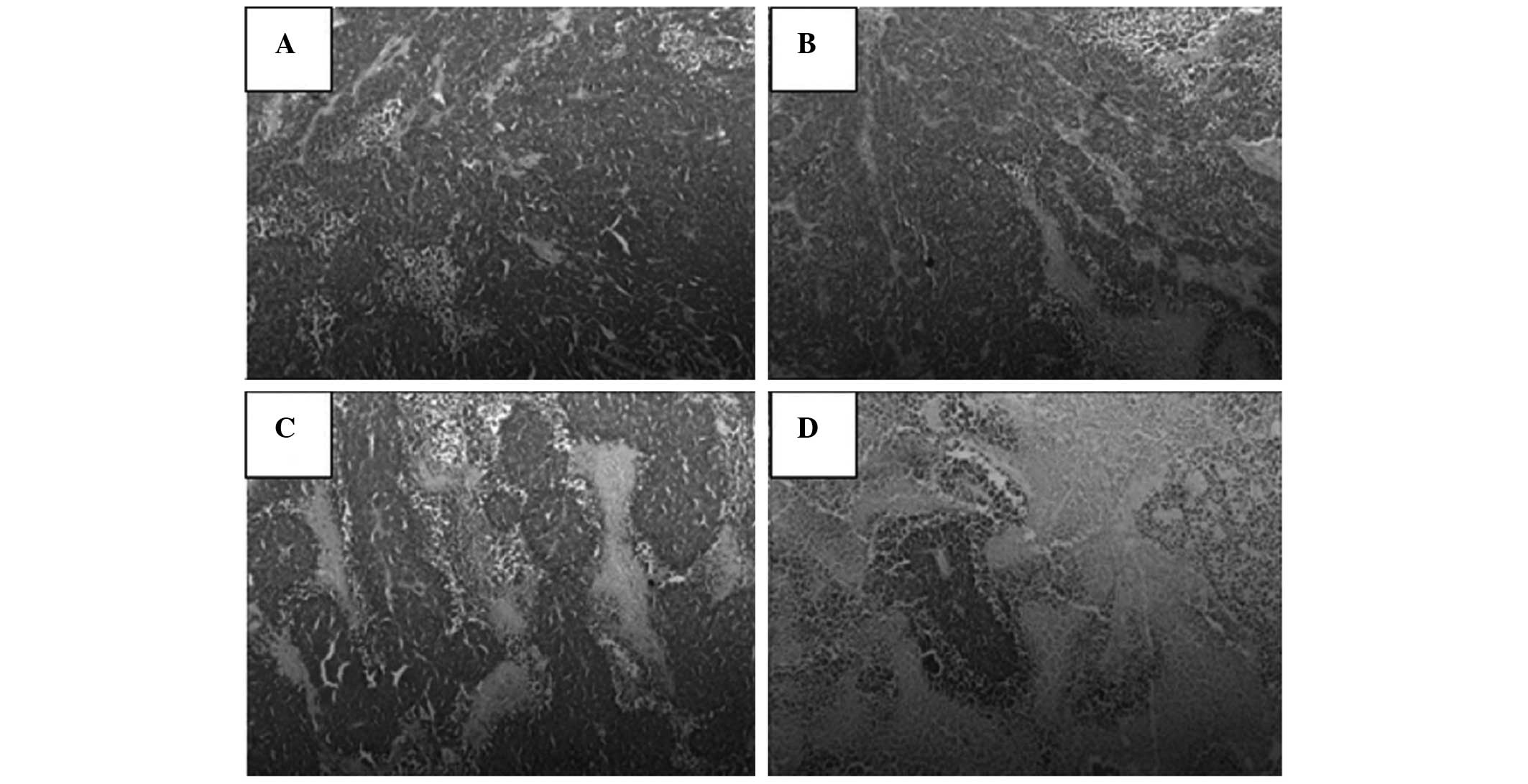
H&E staining
In the tumor tissue, the tumor cells were found to
have a disordered arrangement and a nest-like distribution. The
tumor interstitial substance and boundaries were clear, and a
slight hyperplasia of fibrous tissue was observed. The number of
tumor cells was greater than that of normal cells, and the volume
of the tumor nucleus was increased. The nuclei were found to vary
in size, shape and coloring, and a number of cases had pathological
nuclear fission. These features were most evident in the normal
saline group (Fig. 2).
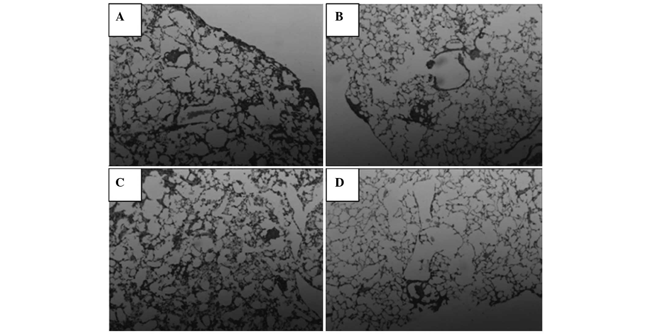
In the lung, the tumor cells were arranged in nests
and strands in the alveolar tissue, accompanied by a small amount
of adenoid material. The tumor cells showed marked pleomorphism,
and mitotic cells could be observed around the tumor mass, among
the fibrous tissue hyperplasia. The normal saline, LMWH and
adriamycin groups were observed to have less pleomorphism, mitosis
or fibrous tissue hyperplasia compared with the combination group
(Fig. 3).
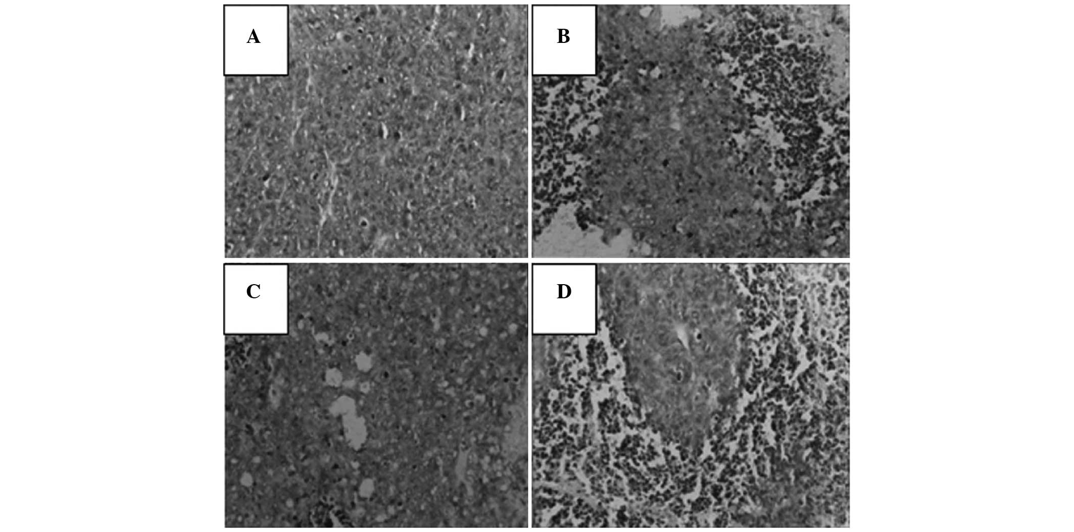
TUNEL results
TUNEL-positive apoptotic cells showed small
condensed nuclei and a circumscribed nuclear membrane, and the
nucleus was stained brown. A number of studies have demonstrated
that heparin induces the apoptosis of tumor cells (8–10),
which is in accordance with the results observed in the present
study. The TUNEL results demonstrated that following treatment, the
tumor cells became apoptotic, in particular in the combination
group (Fig. 4).
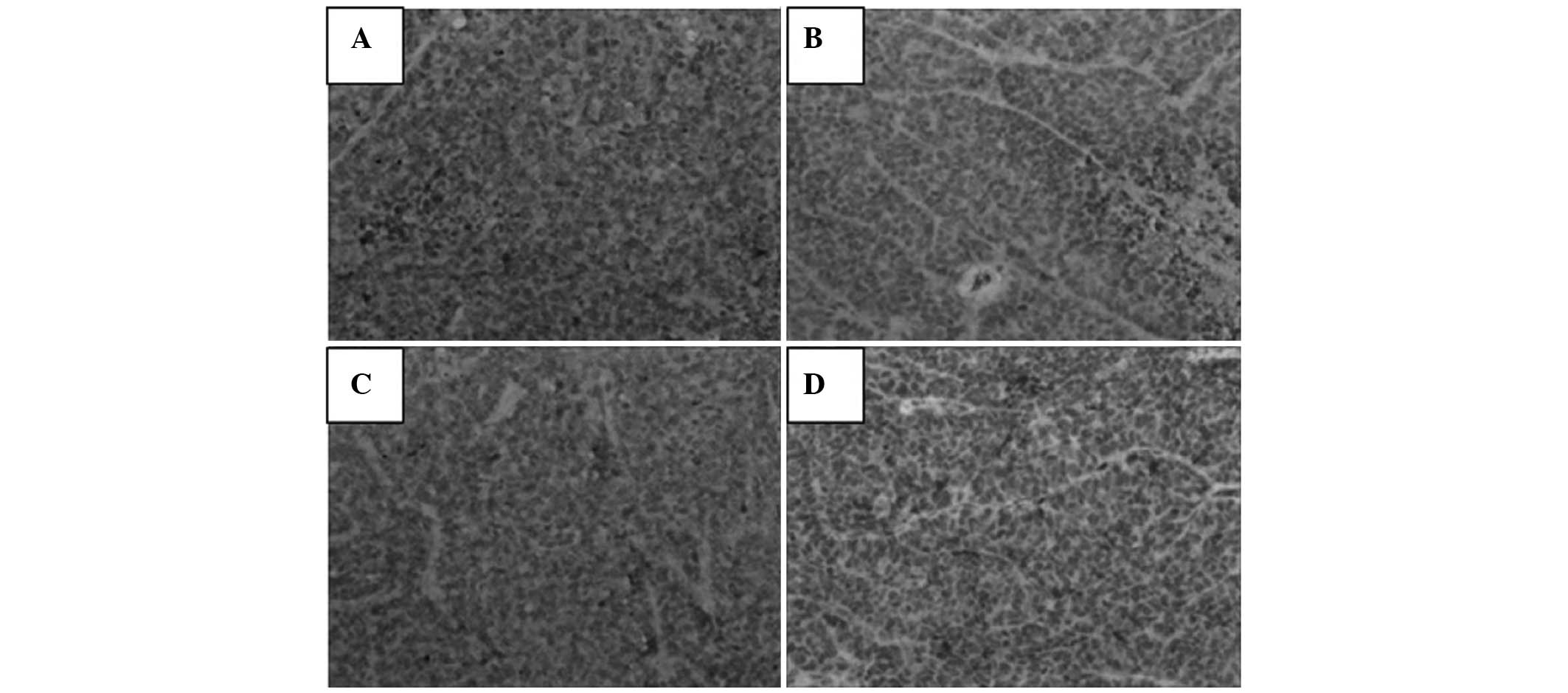
Immunohistochemical results for VEGF
The immunohistochemical results demonstrated that
cytoplasmic expression of VEGF was present in the tumor tissue.
Heparin treatment inhibited the expression of VEGF, and the lowest
levels of expression were observed in the combination group
(Fig. 5).
Discussion
Breast cancer is the most common malignant tumor in
females (11). At present,
treatments focus primarily on tumor cells. However, this approach
is changing, particularly since numerous studies over the past two
decades have demonstrated that cancer is a complex with a large
number of components affecting tumor growth, invasion and
metastasis. Previous studies have suggested that heparin, as well
as having direct effects on blood coagulation, also has a positive
effect for the treatment of cancer by inhibiting tumor cell
proliferation, angiogenesis, invasion and migration, and enhancing
the sensitivity of tumor cells to chemotherapeutic drugs (3–5).
LMWH has certain pharmacokinetic advantages over
unfractionated heparin, including a longer half-life, better
bioavailability, lower binding to plasma proteins, no requirement
for regular laboratory control and the possibility of self
treatment at home (12). The
results of preclinical and clinical studies have suggested that
LMWH inhibits cell growth, cell invasion and angiogenesis in
cancer, indicating its anticoagulant and direct antitumor effects
(13,14). However, LMWH exhibits a limited
single-agent activity, due to hemorrhage and thrombocytopenia in
the clinical setting, thus requiring combination with other agents
to achieve therapeutic effects (15). Therefore, Adriamycin was selected
since it has a broad-spectrum antitumor effect.
The extracellular matrix (ECM) and basement membrane
provides an essential physical barrier between cells and tissues,
as well as a scaffold for cell growth, migration and
differentiation. Studies of ECM molecules in cell attachment,
growth and differentiation have indicated that heparan sulfate (HS)
proteoglycans are centrally involved in embryogenesis, angiogenesis
and epithelial mesenchymal interactions (16,17).
HS chains interact with numerous proteins and ensure that a wide
variety of bioactive molecules bind to the ECM (18). Heparanase is an endoglucuronidase
that cleaves HS, and the expression levels of this enzyme correlate
with the metastatic potential of tumor cells (19). LMWH competes with heparanase for
the HS acceptor, reducing the degradation of HS and thereby
maintaining an intact ECM and inhibiting infiltration and
metastasis by the tumor. The results of H&E staining of the
lung tissue demonstrated that heparin treatment is able to reduce
tumor cell metastasis (Fig. 3) and
the TUNEL assay indicated the ability of heparin to induce
apoptosis (Fig. 4). This has
previously been demonstrated in a number of studies which have
shown that heparin reduces tumor metastasis rates by inhibiting
heparanase (20,21).
Tumor angiogenesis has an important role in tumor
growth and metastasis. The formation of tumor angiogenesis is
driven by microvascular endothelial cells (MVECs). The activation
of MVECs degrades the basement membrane and allows endothelial
cells into the interstitial matrix, where they proliferate and form
capillary-like tubular structures. VEGF has a major role in
angiogenesis by acting via tyrosine kinase receptors. VEGF
antagonists affect tumor growth and vascularization, and the
VEGF-specific antibody bevacizumab exerts antivascular effects in
patients with cancer (22). LMWH
may also inhibit the hyperplasia of endothelial cells and
competitively bind to the heparin acceptor, thus affecting
angiogenesis factors, particularly VEGF (13,23).
The efficacy of an anti-VEGF antibody to inhibit tumor angiogenesis
has been shown in lung cancer and human pediatric sarcoma (8,24,25).
The results from the present study further confirm that heparin
inhibits tumor angiogenesis by inhibiting the expression of VEGF
(Fig. 5) to reduce microvascular
density and reduce the formation of tumor blood vessels, thus
inhibiting tumor growth, invasion and migration.
In conclusion, LMWH was shown to inhibit the growth
of breast cancer tumors in C3H mice, and the mechanism
may be associated with the induction of cancer cell apoptosis and
inhibition of neovascularization. The results from the present
study indicated that LMWH, combined with a chemotherapeutic agent,
exerts an antitumor function, and may therefore provide a novel
strategy for tumor treatment. However, due to the complexity of
structure and function of heparin, further investigation is
required.
Acknowledgements
This study was supported by the Natural Science
Foundation of Anhui province in 2014 (KJ2014A165) and as a
postgraduate research and innovation project in Bengbu Medical
College (Byycx1321).
References
|
1
|
Marsden CG, Wright MJ, Carrier L, et al: A
novel in vivo model for the study of human breast cancer metastasis
using primary breast tumor-initiating cells from patient biopsies.
BMC Cancer. 12:102012. View Article : Google Scholar : PubMed/NCBI
|
|
2
|
American Cancer Society. Cancer Facts and
Figures. 2009, http://www.cancer.org/docroot/STT/content/STT_1x_Cancer_Facts__Figures_2009asp?from=fasturi.
Accessed February 5, 2010
|
|
3
|
Borsig L: Antimetastatic activities of
heparins and modified heparins. Experimental evidence. Thromb Res.
125(Suppl 2): S66–S71. 2010. View Article : Google Scholar : PubMed/NCBI
|
|
4
|
Mousa SA: Heparin and low-molecular weight
heparins in thrombosis and beyond. Methods Mol Biol. 663:109–132.
2010. View Article : Google Scholar : PubMed/NCBI
|
|
5
|
Phillips PG, Yalcin M, Cui H, et al:
Increased tumor uptake of chemotherapeutics and improved
chemoresponse by novel non-anticoagulant low molecular weight
heparin. Anticancer Res. 31:411–419. 2011.PubMed/NCBI
|
|
6
|
Teoh ML, Fitzgerald MP, Oberley LW and
Domann FE: Overexpression of extracellular superoxide dismutase
attenuates heparanase expression and inhibits breast carcinoma cell
growth and invasion. Cancer Res. 69:6355–6363. 2009. View Article : Google Scholar : PubMed/NCBI
|
|
7
|
Liu H, Tong X, Ma L, Xu Z, et al:
Establishment of a non-spontaneous breast cancer model in C3H mice.
Chinese Pharmacological Bulletin. 21:1396–1398. 2005.(In
Chinese).
|
|
8
|
Dredge K, Hammond E, Handley P, Gonda TJ,
Smith MT, Vincent C, et al: PG545, a dual heparanase and
angiogenesis inhibitor, induces potent anti-tumour and
anti-metastatic efficacy in preclinical models. Br J Cancer.
104:635–642. 2011. View Article : Google Scholar : PubMed/NCBI
|
|
9
|
Li JP: Heparin, heparin sulfate and
heparanase in cancer: remedy for metastasis? Anticancer Agents Med
Chem. 8:64–76. 2008. View Article : Google Scholar : PubMed/NCBI
|
|
10
|
Li Y, Liu H, Huang YY, Pu LJ, Zhang XD,
Jiang CC and Jiang ZW: Suppression of endoplasmic reticulum
stress-induced invasion and migration of breast cancer cells
through the downregulation of heparanase. Int J Mol Med.
31:1234–1242. 2013.PubMed/NCBI
|
|
11
|
Reisfeld RA: The tumor microenvironment: a
target for combination therapy of breast cancer. Crit Rev Oncog.
18:115–133. 2013. View Article : Google Scholar : PubMed/NCBI
|
|
12
|
Takeuchi A, Yamamoto Y, Munesue S,
Harashima A, et al: Low molecular weight heparin suppresses
receptor for advanced glycation end products-mediated expression of
malignant phenotype in human fibrosarcoma cells. Cancer Sci.
104:740–749. 2013. View Article : Google Scholar
|
|
13
|
Nagy Z, Turcsik V and Blaskó G: The effect
of LMWH (Nadroparin) on tumor progression. Pathol Oncol Res.
15:689–692. 2009. View Article : Google Scholar : PubMed/NCBI
|
|
14
|
Tan H, Yang S, Liu C, Cao J, Mu G and Wang
F: Enhanced anti-angiogenesis and anti-tumor activity of endostatin
by chemical modification with polyethylene glycol and low molecular
weight heparin. Biomed Pharmacother. 66:648–654. 2012. View Article : Google Scholar : PubMed/NCBI
|
|
15
|
Jayson GC, Hicklin DJ and Ellis LM:
Antiangiogenic therapy - evolving view based on clinical trial
results. Nature Rev Clin Oncol. 9:297–303. 2012. View Article : Google Scholar : PubMed/NCBI
|
|
16
|
Zcharia E, Jia J, Zhang X, et al: Newly
generated heparanase knock-out mice unravel co-regulation of
heparanase and matrix metalloproteinases. PLoS One. 4:e51812009.
View Article : Google Scholar : PubMed/NCBI
|
|
17
|
Kim SH, Turnbull J and Guimond S:
Extracellular matrix and cell signaling: the dynamic cooperation of
integrin, proteoglycan and growth factor receptor. J Endocrinol.
209:139–151. 2011. View Article : Google Scholar : PubMed/NCBI
|
|
18
|
Lindahl U and Li JP: Interactions between
heparan sulfate and proteins - design and functional implications.
Int Rev Cell Mol Biol. 276:105–159. 2009. View Article : Google Scholar : PubMed/NCBI
|
|
19
|
Vlodavsky I, Beckhove P, Lerner I, Pisano
C, Meirovitz A, Ilan N and Elkin M: Significance of heparanase in
cancer and inflammation. Cancer Microenviron. 5:115–132. 2011.
View Article : Google Scholar
|
|
20
|
Basappa, Sugahara K, Thimmaiah KN, Bid HK,
Houghton PJ and Rangappa KS: Anti-tumor activity of a novel
HS-mimetic-vascular endothelial growth factor binding small
molecule. PLoS One. 7:e394442012. View Article : Google Scholar : PubMed/NCBI
|
|
21
|
Battinelli EM, Markens BA, Kulenthirarajan
RA, Machlus KR, Flaumenhaft R and Italiano JE Jr: Anticoagulation
inhibits tumor cell-mediated release of platelet angiogenic
proteins and diminishes platelet angiogenic response. Blood.
123:101–112. 2014. View Article : Google Scholar : PubMed/NCBI
|
|
22
|
Jeong KW, Jeong MC, Jin B and Kim Y:
Relationship between structural flexibility and function in the
C-terminal region of the heparin-binding domain of VEGF165.
Biochemistry. 52:8823–8832. 2013. View Article : Google Scholar : PubMed/NCBI
|
|
23
|
Jia W, Feng K, Fan P, et al: Post-TACE
combination therapy of heparin and octreotide results in decreased
tumor metastasis in extrahepatic tumorigenesis. Cell Biochem
Biophys. 62:35–40. 2012. View Article : Google Scholar : PubMed/NCBI
|
|
24
|
Shafat I, Bern-Arush MW, Issakov J, Meller
I, Naroditsky I, Tortoreto M, et al: Preclinical and clinical
significance of heparanase in Ewing’s sarcoma. J Cell Mol Med.
15:1857–1864. 2011.
|
|
25
|
Judy BF, Aliperti LA, Predina JD, Levine
D, et al: Vascular endothelial-targeted therapy combined with
cytotoxic chemotherapy induces inflammatory intratumoral
infiltrates and inhibits tumor relapses after surgery. Neoplasia.
14:352–359. 2012.
|