Spandidos Publications Logo
  • About
    • About Spandidos
    • Aims and Scopes
    • Abstracting and Indexing
    • Editorial Policies
    • Reprints and Permissions
    • Job Opportunities
    • Terms and Conditions
    • Contact
  • Journals
    • All Journals
    • Oncology Letters
      • Oncology Letters
      • Information for Authors
      • Editorial Policies
      • Editorial Board
      • Aims and Scope
      • Abstracting and Indexing
      • Bibliographic Information
      • Archive
    • International Journal of Oncology
      • International Journal of Oncology
      • Information for Authors
      • Editorial Policies
      • Editorial Board
      • Aims and Scope
      • Abstracting and Indexing
      • Bibliographic Information
      • Archive
    • Molecular and Clinical Oncology
      • Molecular and Clinical Oncology
      • Information for Authors
      • Editorial Policies
      • Editorial Board
      • Aims and Scope
      • Abstracting and Indexing
      • Bibliographic Information
      • Archive
    • Experimental and Therapeutic Medicine
      • Experimental and Therapeutic Medicine
      • Information for Authors
      • Editorial Policies
      • Editorial Board
      • Aims and Scope
      • Abstracting and Indexing
      • Bibliographic Information
      • Archive
    • International Journal of Molecular Medicine
      • International Journal of Molecular Medicine
      • Information for Authors
      • Editorial Policies
      • Editorial Board
      • Aims and Scope
      • Abstracting and Indexing
      • Bibliographic Information
      • Archive
    • Biomedical Reports
      • Biomedical Reports
      • Information for Authors
      • Editorial Policies
      • Editorial Board
      • Aims and Scope
      • Abstracting and Indexing
      • Bibliographic Information
      • Archive
    • Oncology Reports
      • Oncology Reports
      • Information for Authors
      • Editorial Policies
      • Editorial Board
      • Aims and Scope
      • Abstracting and Indexing
      • Bibliographic Information
      • Archive
    • Molecular Medicine Reports
      • Molecular Medicine Reports
      • Information for Authors
      • Editorial Policies
      • Editorial Board
      • Aims and Scope
      • Abstracting and Indexing
      • Bibliographic Information
      • Archive
    • World Academy of Sciences Journal
      • World Academy of Sciences Journal
      • Information for Authors
      • Editorial Policies
      • Editorial Board
      • Aims and Scope
      • Abstracting and Indexing
      • Bibliographic Information
      • Archive
    • International Journal of Functional Nutrition
      • International Journal of Functional Nutrition
      • Information for Authors
      • Editorial Policies
      • Editorial Board
      • Aims and Scope
      • Abstracting and Indexing
      • Bibliographic Information
      • Archive
    • International Journal of Epigenetics
      • International Journal of Epigenetics
      • Information for Authors
      • Editorial Policies
      • Editorial Board
      • Aims and Scope
      • Abstracting and Indexing
      • Bibliographic Information
      • Archive
    • Medicine International
      • Medicine International
      • Information for Authors
      • Editorial Policies
      • Editorial Board
      • Aims and Scope
      • Abstracting and Indexing
      • Bibliographic Information
      • Archive
  • Articles
  • Information
    • Information for Authors
    • Information for Reviewers
    • Information for Librarians
    • Information for Advertisers
    • Conferences
  • Language Editing
Spandidos Publications Logo
  • About
    • About Spandidos
    • Aims and Scopes
    • Abstracting and Indexing
    • Editorial Policies
    • Reprints and Permissions
    • Job Opportunities
    • Terms and Conditions
    • Contact
  • Journals
    • All Journals
    • Biomedical Reports
      • Information for Authors
      • Editorial Policies
      • Editorial Board
      • Aims and Scope
      • Abstracting and Indexing
      • Bibliographic Information
      • Archive
    • Experimental and Therapeutic Medicine
      • Information for Authors
      • Editorial Policies
      • Editorial Board
      • Aims and Scope
      • Abstracting and Indexing
      • Bibliographic Information
      • Archive
    • International Journal of Epigenetics
      • Information for Authors
      • Editorial Policies
      • Editorial Board
      • Aims and Scope
      • Abstracting and Indexing
      • Bibliographic Information
      • Archive
    • International Journal of Functional Nutrition
      • Information for Authors
      • Editorial Policies
      • Editorial Board
      • Aims and Scope
      • Abstracting and Indexing
      • Bibliographic Information
      • Archive
    • International Journal of Molecular Medicine
      • Information for Authors
      • Editorial Policies
      • Editorial Board
      • Aims and Scope
      • Abstracting and Indexing
      • Bibliographic Information
      • Archive
    • International Journal of Oncology
      • Information for Authors
      • Editorial Policies
      • Editorial Board
      • Aims and Scope
      • Abstracting and Indexing
      • Bibliographic Information
      • Archive
    • Medicine International
      • Information for Authors
      • Editorial Policies
      • Editorial Board
      • Aims and Scope
      • Abstracting and Indexing
      • Bibliographic Information
      • Archive
    • Molecular and Clinical Oncology
      • Information for Authors
      • Editorial Policies
      • Editorial Board
      • Aims and Scope
      • Abstracting and Indexing
      • Bibliographic Information
      • Archive
    • Molecular Medicine Reports
      • Information for Authors
      • Editorial Policies
      • Editorial Board
      • Aims and Scope
      • Abstracting and Indexing
      • Bibliographic Information
      • Archive
    • Oncology Letters
      • Information for Authors
      • Editorial Policies
      • Editorial Board
      • Aims and Scope
      • Abstracting and Indexing
      • Bibliographic Information
      • Archive
    • Oncology Reports
      • Information for Authors
      • Editorial Policies
      • Editorial Board
      • Aims and Scope
      • Abstracting and Indexing
      • Bibliographic Information
      • Archive
    • World Academy of Sciences Journal
      • Information for Authors
      • Editorial Policies
      • Editorial Board
      • Aims and Scope
      • Abstracting and Indexing
      • Bibliographic Information
      • Archive
  • Articles
  • Information
    • For Authors
    • For Reviewers
    • For Librarians
    • For Advertisers
    • Conferences
  • Language Editing
Login Register Submit
  • This site uses cookies
  • You can change your cookie settings at any time by following the instructions in our Cookie Policy. To find out more, you may read our Privacy Policy.

    I agree
Search articles by DOI, keyword, author or affiliation
Search
Advanced Search
presentation
Experimental and Therapeutic Medicine
Join Editorial Board Propose a Special Issue
Print ISSN: 1792-0981 Online ISSN: 1792-1015
Journal Cover
January-2015 Volume 9 Issue 1

Full Size Image

Sign up for eToc alerts
Recommend to Library

Journals

International Journal of Molecular Medicine

International Journal of Molecular Medicine

International Journal of Molecular Medicine is an international journal devoted to molecular mechanisms of human disease.

International Journal of Oncology

International Journal of Oncology

International Journal of Oncology is an international journal devoted to oncology research and cancer treatment.

Molecular Medicine Reports

Molecular Medicine Reports

Covers molecular medicine topics such as pharmacology, pathology, genetics, neuroscience, infectious diseases, molecular cardiology, and molecular surgery.

Oncology Reports

Oncology Reports

Oncology Reports is an international journal devoted to fundamental and applied research in Oncology.

Experimental and Therapeutic Medicine

Experimental and Therapeutic Medicine

Experimental and Therapeutic Medicine is an international journal devoted to laboratory and clinical medicine.

Oncology Letters

Oncology Letters

Oncology Letters is an international journal devoted to Experimental and Clinical Oncology.

Biomedical Reports

Biomedical Reports

Explores a wide range of biological and medical fields, including pharmacology, genetics, microbiology, neuroscience, and molecular cardiology.

Molecular and Clinical Oncology

Molecular and Clinical Oncology

International journal addressing all aspects of oncology research, from tumorigenesis and oncogenes to chemotherapy and metastasis.

World Academy of Sciences Journal

World Academy of Sciences Journal

Multidisciplinary open-access journal spanning biochemistry, genetics, neuroscience, environmental health, and synthetic biology.

International Journal of Functional Nutrition

International Journal of Functional Nutrition

Open-access journal combining biochemistry, pharmacology, immunology, and genetics to advance health through functional nutrition.

International Journal of Epigenetics

International Journal of Epigenetics

Publishes open-access research on using epigenetics to advance understanding and treatment of human disease.

Medicine International

Medicine International

An International Open Access Journal Devoted to General Medicine.

Journal Cover
January-2015 Volume 9 Issue 1

Full Size Image

Sign up for eToc alerts
Recommend to Library

  • Article
  • Citations
    • Cite This Article
    • Download Citation
    • Create Citation Alert
    • Remove Citation Alert
    • Cited By
  • Similar Articles
    • Related Articles (in Spandidos Publications)
    • Similar Articles (Google Scholar)
    • Similar Articles (PubMed)
  • Download PDF
  • Download XML
  • View XML
Case Report Open Access

Solitary fibrous tumor of the spermatic cord: A case report and literature review

  • Authors:
    • Shanbiao Hu
    • Lu Yi
    • Luoyan Yang
    • Yinhuai Wang
  • View Affiliations / Copyright

    Affiliations: Department of Urology, Second Xiangya Hospital, Central South University, Changsha, Hunan 410011, P.R. China
  • Pages: 55-58
    |
    Published online on: November 12, 2014
       https://doi.org/10.3892/etm.2014.2066
  • Expand metrics +
Metrics: Total Views: 0 (Spandidos Publications: | PMC Statistics: )
Metrics: Total PDF Downloads: 0 (Spandidos Publications: | PMC Statistics: )
Cited By (CrossRef): 0 citations Loading Articles...

This article is mentioned in:


Abstract

A 31‑year‑old male patient with a six‑year history of left inguinoscrotal swelling was admitted to the Second Xiangya Hospital (Changsha, China). The mass was not found to be associated with intraperitoneal pressure. Ultrasonography and computed tomography examinations demonstrated several solid, botryoidal masses involving the spermatic cord, with limited capacity of mobility. The demarcation between the masses and the left testicle was clear; thus, the masses were removed by a left spermatic cord tumor resection via a left inguinal approach, under epidural anesthesia. Positive staining of the tumor markers, CD34+, CD99+ and Bcl‑2+, was confirmed by pathological examination following surgery, and a solitary fibrous tumor of the spermatic cord was diagnosed. No recurrence and metastasis were observed in the patient during the subsequent 25‑month follow‑up period.

Introduction

Solitary fibrous tumors (SFTs) are a rare type of mesenchymal tumor, which were first described by Klemperer and Rabin in 1931 (1). Although they are commonly found in the pleura, statistics show that 50–70% of all SFTs are extrapleural (2). However, few cases of SFTs in the urogenital organs have been documented. To the best of our knowledge, only a small number of these cases forming in the spermatic cord have been reported to date (3–7).

Due to the rarity of these tumors, there was no consensus on its standard treatment. However, treatment for SFTs include surgical resection, radiotherapy and chemotherapy. Resection of the tumor with a sufficient surgical margin was the mainstay of treatment. However, a number of studies have demonstrated that radiotherapy and chemotherapy, with ifosfamide and adriamycin, is effective (5,8). However, these treatments were not established yet. The present study reports a case of SFT involving the spermatic cord.

Case report

The patient was a 31-year-old male with a six-year history of left inguinoscrotal swelling, which had been gradually increasing in size for the past three years. The size of the swelling showed no association with increased intraperitoneal pressure. Physical examination revealed no evident defects in the appearance of the scrotum, with the exception of several hard, botryoidal masses, located between the left testicle and the inguinal region, with smooth surfaces and limited capacity of mobility. Ultrasound examination revealed several solid, well-demarcated, hypoechoic extratesticular masses arising from the left spermatic cord, the largest of which was ~3 × 2 cm in size. The masses were separated from the cutis and showed no connections with the left testis and epididymis, and no vascularity was observed. The patient’s family signed an informed consent prior to the treatment.

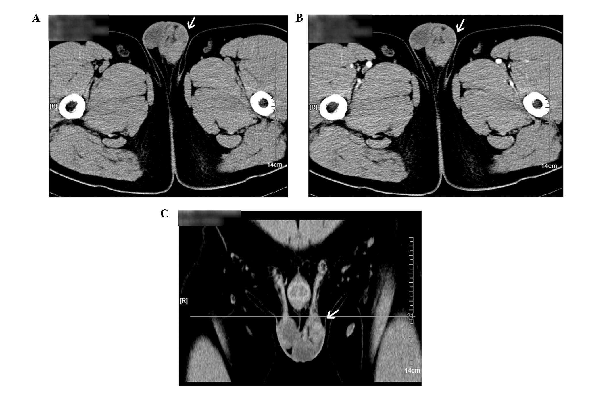
A computed tomography (CT) scan revealed multiple high-density nodules associated with the left spermatic cord, which showed partial fusion, uniform density and a clear boundary with the testis (Fig. 1). In addition, the density of the nodules was enhanced slightly and no bilateral inguinal swollen lymph nodes were observed in the enhanced scan.

Figure 1

CT images of an SFT in the spermatic cord: (A) Unenhanced CT scan; (B) enhanced CT scan (the mass density is enhanced slightly); and (C) coronal section of the enhanced CT scan. SFT, solitary fibrous tumor; CT, computed tomography.

The masses were removed by a left spermatic cord tumor resection via a left inguinal approach, under epidural 
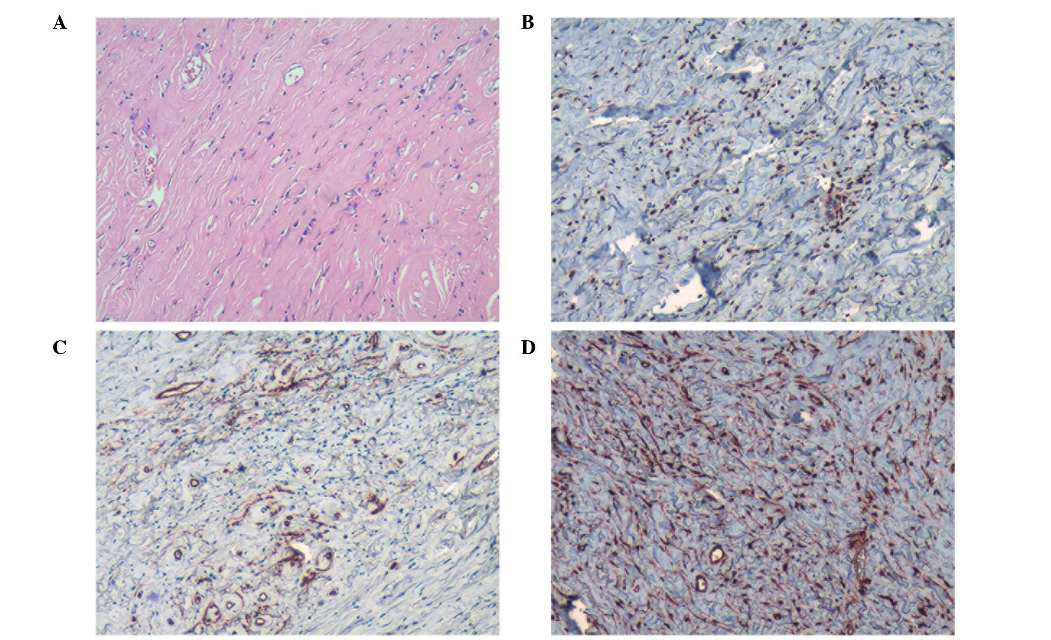
anesthesia. Benign mesenchymal tissue was diagnosed by intraoperative frozen pathological sections. Postoperative pathological examination confirmed that the masses belonged to multiple mesenchymal tumors with classic properties, including fibroplasia, small vascular hyalinization, clear boundaries, less tumor component and no nuclear division. Immunohistochemical examination revealed the presence of numerous tumor markers, including CD99+, Bcl-2+, partial CD34+, focal S-100+, SMA+ and CD68− (Fig. 2). Thus, the final diagnosis was solitary fibrous tumors of the spermatic cord. The patient remained healthy, with no local recurrence or metastasis observed during the 25-month follow-up period.

Figure 2

Immunohistochemical detection of tumor marker expression, showing (A) hematoxylin-eosin staining, (B) Bcl-2+, (C) partial CD34+ and (D) CD99+. Magnification, ×100.

Discussion

Although SFTs have been observed in male and female patients of a wide range of ages, the underlying mechanism remains unknown. Initially, SFTs were most commonly located in the pleura (9). At present, only a limited number of SFT cases have been identified in the urogenital organs, including the kidney, renal pelvis, bladder, prostate and seminal vesicles (4,6,7,9–12). Moreover, SFTs involving the spermatic cord are extremely rare and should be distinguished from other urogenital mesenchymal tumors, which include inflammatory myofibromatous tumors, hemangiopericytomas, malignant fibrohistiocytomas, fibrosarcomas, leiomyosarcomas and benign and malignant nerve sheath tumors. In addition, SFTs should be distinguished from other diseases that can result from inguinal masses, similarly to an inguinal hernia (11).

Clinical symptoms of urogenital SFTs, including a painless mass, dysuria, hematospermia and abdominal pain, are mainly determined by tumor properties, including the size, location, invasion and the degree of mucosal integrity (11).

Ultrasonography and magnetic resonance imaging (MRI), two common imaging examinations, play an important role in the diagnosis of SFTs. Ultrasound examination is more effective in the identification of solid and cystic tumors, while MRI is able to illustrate anatomical associations in the tumor surroundings more precisely (7). Isodensity with patchy hypodensity for unenhanced CT images, and mild or marked heterogeneous enhancement for contrast-enhanced CT images are the major features of pleural SFTs (13,14). A study by Zhang et al demonstrated that a well-defined, ovoid or rounded mass with low signal intensity on MRI T2-weighted images and different degrees of enhancement on CT and MRI scans may indicate the presence of an SFT (14). Positron emission tomography-CT scanning is able to preoperatively indicate malignancy and recognize local recurrence or distant metastases; however, Lococo et al deemed that it is controversial for the identification of benign and malignant SFTs for PETCT. Thus, further evaluation is required (15,16). In the present study, an unenhanced CT scan demonstrated multiple high-density nodules, and the signal only strengthened slightly in the enhanced CT scan. Therefore, the diagnosis of an SFT by imaging only is inadvisable and further pathological examinations are necessary.

Previous studies have confirmed that tumor markers, including CD34+, Bcl-2+ and CD99+, are generally positive in SFT cases, which has aided the diagnosis of SFTs (17,18). Currently, the positive staining of CD34+ is indispensable in the diagnosis of an SFT; however, the expression of CD34 is not limited to SFTs (11). In the present study, the positive staining of CD34+, Bcl-2+ and CD99+ was demonstrated by immunohistochemistry examinations, confirming the diagnosis of an SFT.

The conventional treatment for an SFT is the complete resection of the tumor with negative margins (19,20) or resection experience satisfactory health outcomes (21). However, the resection of tumors with positive margins is associated with a higher rate of local recurrence, and surgical excision remains the first choice for the treatment of local recurrence (20). With regard to urogenital SFTs, the selection of the surgical procedure should depend on the characteristics of each individual tumor, including the location, size and degree of malignancy. For tumors with infiltrative growth, of a large size or aggressive malignancy, the recommended treatment is the extended resection of the involved organs, including radical nephrectomy and cystoprostatectomy. In the present study, considering the clear demarcations between the tumors and surrounding organs, complete resection of the tumors was performed without removing the spermatic cord.

The prognosis of SFTs is obscure for rare cases. Usually, an SFT is defined as a benign form of tumor due to its low rates or even absence of local recurrence and metastasis, as demonstrated by a number of previous studies (1,2,22,23). However, an increasing number of recent studies have demonstrated that a considerable number of SFTs are malignant (11,18,24). In the case of an SFT which is unresectable, metastatic or has positive surgical margins, patients are unlikely to benefit significantly from radiotherapy or chemotherapy; the alternative treatments for an SFT (2). Therefore, a careful long-term follow-up period is advised in all SFT cases due to the possibility of a late recurrence (21). The case reported in the present study was considered to be a benign SFT due to its small size (<10 cm), lack of growth and minimal cancer component. During the 25-month follow-up period, the patient remained healthy and exhibited no tumor recurrence.

In conclusion, pathological examination remains the standard approach for the diagnosis of SFTs, while radical resection of the tumor/s is the first choice for treatment. Furthermore, patients should undergo careful follow-up examination in order to identify tumor recurrence.

References

1 

Klemperer P and Rabin LB: Primary neoplasms of the pleura. A report of five cases Arch Pathol. 11:385–412. 1931.

2 

van Houdt WJ, Westerveld CM, Vrijenhoek JE, et al: Prognosis of solitary fibrous tumors: a multicenter study. Ann Surg Oncol. 20:4090–4095. 2013. View Article : Google Scholar : PubMed/NCBI

3 

Cafiero F, Gipponi M, Peressini A, et al: Solitary fibrous tumor of the inguinal region: a clinicopathological, light-microscopic, immunohistochemical, electron microscopic and flow-cytometric DNA study. Anticancer Res. 21:4091–4094. 2001.

4 

Fisher C and Bisceglia M: Solitary fibrous tumour of the spermatic cord. Br J Urol. 74:798–799. 1994. View Article : Google Scholar : PubMed/NCBI

5 

Gold JS, Antonescu CR, Hajdu C, et al: Clinicopathologic correlates of solitary fibrous tumors. Cancer. 94:1057–1068. 2002. View Article : Google Scholar : PubMed/NCBI

6 

Márquez Moreno AJ, Vicioso Recio L, Castro Léon A, Casals Sánchez JL, Alcázar Ramírez J and Matilla Vicente A: Paratesticular solitary fibrous tumor. Arch Esp Urol. 54:716–718. 2001.(In Spanish).

7 

Ilica AT, Ors F, Bag M, Guvenc I, Duran C and Buyukbayram H: An uncommon cause of inguinoscrotal swelling: Solitary fibrous tumor of the spermatic cord. Eur J Radiol Extra. 67:25–28. 2008. View Article : Google Scholar

8 

de Perrot M, Fischer S, Brundler MA, et al: Solitary fibrous tumor of the pleura. Ann Thorac Surg. 74:285–293. 2002. View Article : Google Scholar : PubMed/NCBI

9 

Wiessner D, Dittert DD, Manseck A and Wirth MP: Large solitary fibrous tumor of the seminal vesicle. Urology. 62:9412003. View Article : Google Scholar : PubMed/NCBI

10 

Magro G, Cavallaro V, Torrisi A, Lopes M, Dell’Albani M and Lanzafame S: Intrarenal solitary fibrous tumor of the kidney report of a case with emphasis on the differential diagnosis in the wide spectrum of monomorphous spindle cell tumors of the kidney. Pathol Res Pract. 198:37–43. 2002.PubMed/NCBI

11 

Westra WH, Grenko RT and Epstein J: Solitary fibrous tumor of the lower urogenital tract: a report of five cases involving the seminal vesicles, urinary bladder, and prostate. Hum Pathol. 31:63–68. 2000. View Article : Google Scholar : PubMed/NCBI

12 

Yazaki T, Satoh S, Iizumi T, Umeda T and Yamaguchi Y: Solitary fibrous tumor of renal pelvis. Int J Urol. 8:504–508. 2001. View Article : Google Scholar : PubMed/NCBI

13 

Chong S, Kim TS, Cho EY, Kim J and Kim H: Benign localized fibrous tumour of the pleura: CT features with histopathological correlations. Clin Radiol. 61:875–882. 2006. View Article : Google Scholar : PubMed/NCBI

14 

Zhang WD, Chen JY, Cao Y, Liu QY and Luo RG: Computed tomography and magnetic resonance imaging findings of solitary fibrous tumors in the pelvis: correlation with histopathological findings. Eur J Radiol. 78:65–70. 2011. View Article : Google Scholar

15 

Lococo F, Cafarotti S and Treglia G: Is 18F-FDG-PET/CT really able to differentiate between malignant and benign solitary fibrous tumor of the pleura? Clin Imaging. 37:9762013. View Article : Google Scholar

16 

Yan J, Ahl KL, Manning KA, Mann FA and Lewis DH: Radiology-Pathology Conference: 18F FDG PET-CT imaging of solitary fibrous tumor of the pleura. Clin Imaging. 37:598–601. 2013. View Article : Google Scholar

17 

Hanau CA and Miettinen M: Solitary fibrous tumor: histological and immunohistochemical spectrum of benign and malignant variants presenting at different sites. Hum Pathol. 26:440–449. 1995. View Article : Google Scholar : PubMed/NCBI

18 

Rao N, Colby TV, Falconieri G, Cohen H, Moran CA and Suster S: Intrapulmonary solitary fibrous tumors: clinicopathologic and immunohistochemical study of 24 cases. Am J Surg Pathol. 37:155–166. 2013. View Article : Google Scholar

19 

Marak CP, Dorokhova O and Guddati AK: Solitary fibrous tumor of the pleura. Med Oncol. 30:5732013. View Article : Google Scholar : PubMed/NCBI

20 

Uemura K, Hayashi T and Matsuoka K: Solitary fibrous tumor of the greater omentum in an inguinal hernia sac. Intern Canc Conf J. 1:70–73. 2012. View Article : Google Scholar

21 

Brunnemann RB, Ro JY, Ordonez NG, Mooney J, El-Naggar AK and Ayala AG: Extrapleural solitary fibrous tumor: a clinicopatho-logic study of 24 cases. Mod Pathol. 12:1034–1042. 1999.PubMed/NCBI

22 

Lee SC, Tzao C, Ou SM, Hsu HH, Yu CP and Cheng YL: Solitary fibrous tumors of the pleura: clinical, radiological, surgical and pathological evaluation. Eur J Surg Oncol. 31:84–87. 2005. View Article : Google Scholar : PubMed/NCBI

23 

Guo W, Xiao HL, Jiang YG, Wang RW, Zhao YP, Ma Z, et al: Retrospective analysis for thirty-nine patients with solitary fibrous tumor of pleura and review of the literature. World J Surg Oncol. 9:1342011. View Article : Google Scholar : PubMed/NCBI

24 

England DM, Hochholzer L and McCarthy MJ: Localized benign and malignant fibrous tumors of the pleura. A clinicopathologic review of 223 cases. Am J Surg Pathol. 13:640–658. 1989. View Article : Google Scholar : PubMed/NCBI

Related Articles

  • Abstract
  • View
  • Download
  • Twitter
Copy and paste a formatted citation
Spandidos Publications style
Hu S, Yi L, Yang L and Wang Y: Solitary fibrous tumor of the spermatic cord: A case report and literature review. Exp Ther Med 9: 55-58, 2015.
APA
Hu, S., Yi, L., Yang, L., & Wang, Y. (2015). Solitary fibrous tumor of the spermatic cord: A case report and literature review. Experimental and Therapeutic Medicine, 9, 55-58. https://doi.org/10.3892/etm.2014.2066
MLA
Hu, S., Yi, L., Yang, L., Wang, Y."Solitary fibrous tumor of the spermatic cord: A case report and literature review". Experimental and Therapeutic Medicine 9.1 (2015): 55-58.
Chicago
Hu, S., Yi, L., Yang, L., Wang, Y."Solitary fibrous tumor of the spermatic cord: A case report and literature review". Experimental and Therapeutic Medicine 9, no. 1 (2015): 55-58. https://doi.org/10.3892/etm.2014.2066
Copy and paste a formatted citation
x
Spandidos Publications style
Hu S, Yi L, Yang L and Wang Y: Solitary fibrous tumor of the spermatic cord: A case report and literature review. Exp Ther Med 9: 55-58, 2015.
APA
Hu, S., Yi, L., Yang, L., & Wang, Y. (2015). Solitary fibrous tumor of the spermatic cord: A case report and literature review. Experimental and Therapeutic Medicine, 9, 55-58. https://doi.org/10.3892/etm.2014.2066
MLA
Hu, S., Yi, L., Yang, L., Wang, Y."Solitary fibrous tumor of the spermatic cord: A case report and literature review". Experimental and Therapeutic Medicine 9.1 (2015): 55-58.
Chicago
Hu, S., Yi, L., Yang, L., Wang, Y."Solitary fibrous tumor of the spermatic cord: A case report and literature review". Experimental and Therapeutic Medicine 9, no. 1 (2015): 55-58. https://doi.org/10.3892/etm.2014.2066
Follow us
  • Twitter
  • LinkedIn
  • Facebook
About
  • Spandidos Publications
  • Careers
  • Cookie Policy
  • Privacy Policy
How can we help?
  • Help
  • Live Chat
  • Contact
  • Email to our Support Team