Introduction
Renal ischemia and reperfusion (I/R) injury can
increase the rates of acute kidney failure, delayed graft function
and early mortality in patients undergoing kidney transplantation
(1). Picroside II is one of the
main active constituents isolated from Picrorhiza
scrophulariiflora. The roots of this plant are of benefit and
often used in traditional Chinese medicine for a number of
conditions (2). Picroside II has
been shown to possess a wide range of pharmacological effects,
including effects against oxidative stress and inflammation
(3–5). Picroside II has also been shown to
protect against I/R in other organs, including the brain (6), due to its anti-oxidative,
anti-inflammatory and anti-apoptotic properties. However, its
impact on renal I/R injury remains unknown.
Inflammation is recognized as an important component
of renal I/R injury (7,8). Leukocytes are the key mediators that
may fuel inflammatory reactions in renal I/R injury. In the early
phase of reperfusion, the levels of various pro-inflammatory
cytokines increase rapidly. Oxidative stress also plays an
important role in renal I/R injury (9,10).
The production of reactive oxygen species (ROS) in the reperfusion
period is considered to be a key reason for uncontrolled oxidative
stress (11), and the increased
amount of ROS can also drive the inflammatory cascade (12).
Toll-like receptors (TLRs) are a family of
transmembrane proteins. Their activation leads to an intracellular
cascade of events, during which nuclear factor κB (NF-κB) is
released from IκB, allowing NF-κB translocation from the cytoplasm
to the nucleus where it mediates the overexpression of inflammatory
cytokine genes leading to a pro-inflammatory response (13). The close association between the
mechanisms of renal I/R and the protection provided by picroside II
suggest that picroside II may have a beneficial effect in
protecting against renal I/R injury. Therefore, the major purpose
of the present study was to determine whether picroside II was able
to attenuate oxidative stress and inflammation following renal I/R
injury and the potential underlying mechanism.
Materials and methods
Animal model of I/R
All surgical and experimental procedures were
approved by the Institutional Animal Care and Use Committee of
Wuhan University (Wuhan, China). Adult male Sprague Dawley rats
(220–250 g) were obtained from the Center of Experimental Animals
in Wuhan University Medical College. The procedures were carried
out according to routine animal-care guidelines, and all
experimental procedures complied with the Guide for the Care and
Use of Laboratory Animals (1996). Briefly, rats were anesthetized
with pentobarbital (45 mg/kg) and placed on a homeothermic table in
order to maintain a core body temperature of 37°C. A midline
laparotomy was conducted and right nephrectomy was performed.
Subsequently, the left kidney was subjected to 45 min of ischemia
followed by 24 h of reperfusion.
The animals were divided into three groups, namely
the sham, I/R and picroside II groups. Each group contained eight
rats. In the sham group, only the right kidneys were removed. In
the I/R and picroside II groups, the left kidney vessels were
clamped for 45 min followed by 24 h of reperfusion. The
interventions were performed as described below.
Intervention study
Picroside II (CAS No: 39012-20-9, purity >98%,
molecular formula C23H28O13) was
purchased from Tianjin Kuiqing Medical Technology Co., Ltd.
(Tianjin, China). It was diluted to form a 10 g/l solution with 1
mol/l phosphate-buffered saline (PBS). Picroside II (10 mg/kg) 250
μl was administered via the tail vein to rats in the picroside II
group with a micro-syringe according to a previous study (6), at the end of the 45 min of ischemia
and prior to 24 h of reperfusion. The rats in the I/R and sham
groups were simultaneously injected with 250 μl 1 mol/l PBS.
Following the 24-h reperfusion period, the animals were sacrificed
with an overdose of pentobarbital sodium (Sigma-Aldrich, St. Louis,
MO, USA), and the left kidneys were removed for the following
experiments and blood samples were collected for the detection of
blood urea nitrogen (BUN) and creatinine (Cr) levels.
Preservation of kidneys
The left kidney was removed under fully maintained
anesthesia. After removal, the kidney was fixed in 10%
phosphate-buffered formalin or immediately frozen, and stored at
−80°C for subsequent experiments.
Serum assays
At 24 h after I/R injury in every group, 1 ml blood
samples were taken and analyzed according to the instructions of
Creatinine and Urea Assay kits (Nanjing Jiancheng Bioengineering
Institute, Nanjing, China). The absorbance was measured using a
spectrophotometer (UV-1700; Shimadzu Corporation, Tokyo, Japan) and
then the concentrations of BUN and Cr were calculated.
Histologic examination
After the kidney fixed in 10% phosphate-buffered
formalin, it was embedded with paraffin and sectioned at 4 μm
thickness. The sections were deparaffinized and hydrated gradually,
and then stained with hematoxylin and eosin (H&E). Morphologic
assessments were conducted by an experienced renal pathologist who
was unaware of the treatments. An established grading scale of 0–4,
outlined by Jablonski et al (14), was used for the histopathological
assessment of I/R-induced damage.
Assay of malondialdehyde (MDA) and
superoxide dismutase (SOD)
The frozen samples of the ischemic zone were
homogenized and centrifuged at 3,000 × g for 10 min. Subsequently,
the supernatants were collected for analysis of the MDA level and
SOD activity. The measurements were obtained spectrophotometrically
with commercial SOD and MDA Assay kits (Nanjing Jiancheng
Bioengineering Institute) according to the manufacturer’s
instructions. The MDA level was represented in nmol/mg protein. The
SOD activity was expressed as U/mg protein.
Immunohistochemistry
The expression of TLR4 and NF-κB was examined by
immunohistochemical staining. Briefly, 4-μm sections were
deparaffinized, and endogenous peroxidase activity was blocked with
3% hydrogen peroxide at 37°C for 10 min. The sections were then
treated with 10% normal goat serum in Tris-buffered saline for 30
min at 37°C. Subsequently, they were incubated overnight at 4°C
with monoclonal anti-rat anti-TLR4 (1:100; ab8376; Abcam,
Cambridge, MA, USA) and monclonal anti-rat anti-NF-κB (1:100;
sc-8008, Santa Cruz Biotechnology, Santa Cruz, CA) antibodies.
After washing three times with PBS, these sections were incubated
with the secondary antibody from the UltraVision™ Quanto Detection
System HRP DAB (Thermo Fisher Scientific, Waltham, MA, USA) for 30
min at room temperature, followed by the color reagent
3,3′-diaminobenzidine (DAB). In the negative control group, the
experiments were routinely performed.
Reverse transcription-quantitative
polymerase chain reaction (RT-qPCR)
Total RNA was isolated using TRIzol reagent
(Invitrogen Life Technologies, Carlsbad, CA, USA) and the RNA
concentration was obtained by spectrophotometry. Single-stranded
cDNA was synthesized using the cDNA synthesis kit (Takara, Kyoto,
Japan) according to the manufacturer’s instructions. qPCR was
performed with the Applied Biosystems SYBR Green mix kit (Applied
Biosystems, CA, USA). The PCR reaction mixture contained: 2 μl
cDNA, 12.5 μl 2X SYBR Green mix, 1μl forward primer, 1μl reverse
primer and 8.5 μl ddH2O, in a final volume of 25 μl. The
primers used were as follows: Tumor necrosis factor (TNF)-α
forward, 5′-CTTCTCATTCCTGCTCGTGG-3′ and reverse,
5′-TCCGCTTGGTGGTTTGCTAC-3′ (Gen-Bank accession number NM_012675.3);
interleukin (IL)-1β forward, 5′-ACTATGGCAACTGTCCCTGAAC-3′ and
reverse, 5′-GTGCTTGGGTCCTCATCCTG-3′ (Gen-Bank accession number
NM_031512.2); intercellular adhesion molecule (ICAM)-1 forward,
5′-GGGATGGTGAAGTCTGTCAA-3′, and reverse,
5′-GGCGGTAATAGGTGTAAATGG-3′ (Gen-Bank accession number NM_012967).
β-actin was used as a reference gene. The data are presented as a
ratio of the mRNA of the gene of interest to β-actin mRNA (sense:
5′-TGCTATGTTGCCCTAGACTTCG-3′ and antisense:
5′-GTTGGCATAGAGGTCTTTACGG-3′ and NM_031144).
Western blot analysis
Total proteins were extracted, and quantified using
the bicinchoninic acid method. Then, equivalent weights of protein
(40 μg/lane) were separated on 10% SDS-PAGE gels and transferred to
a nitrocellulose membrane. The membranes were blocked with 5%
non-fat milk in Tris-buffered saline and Tween 20 buffer and then
incubated with the following primary antibodies: mouse monclonal
NF-κB (p65; 1:500 dilution; sc-8008, Santa Cruz Biotechnology) and
rabbit polyclonal TLR4 (1:500 dilution; sc-10741, Santa Cruz
Biotechnology) at 4°C overnight. Subsequently, after being washed
twice with PBS, the membranes were incubated with horseradish
peroxidase (HRP)-conjugated goat anti-rabbit (ZDR-5306) and goat
anti-mouse (ZDR-5307) secondary antibodies (1:2,000; ZSGB-BIO,
Beijing, China). Specific bands were visualized using an enhanced
chemiluminescence detection kit (Immobilon Western
Chemiluminescence HRP Substrate; Merck Millipore, Darmstadt,
Germany). Optical densities were detected using Quantity One
software (Bio-Rad Laboratories, Hercules, CA, USA).
Statistical analysis
Data are presented as mean ± standard error of the
mean. The means of the different groups were compared using one-way
analysis of variance and Student-Newman-Keuls test. Statistical
analyses were conducted using SPSS version 17.0 (SPSS Inc.,
Chicago, IL, USA). Differences were considered statistically
significant when P<0.05.
Results
Renal function
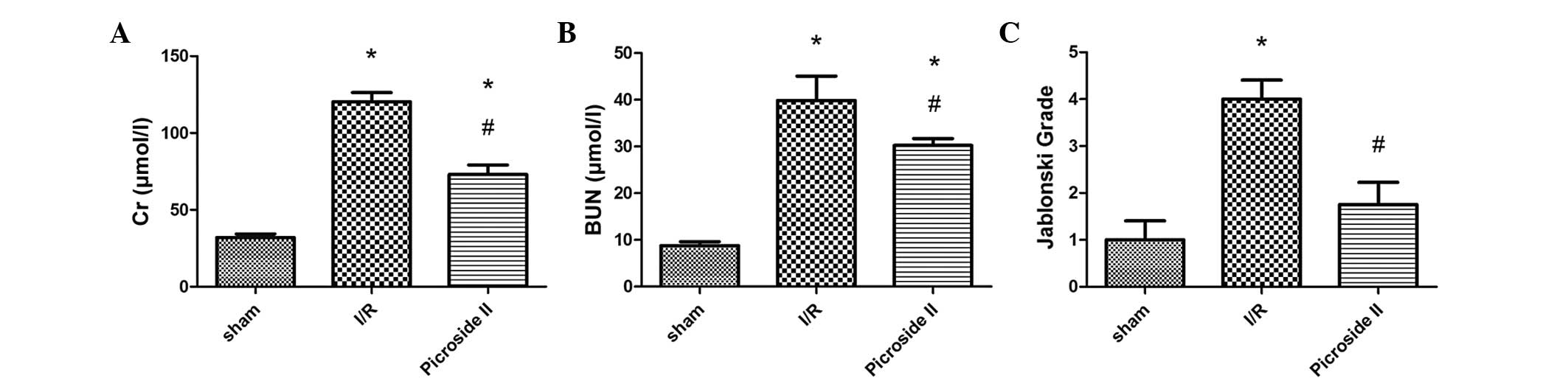
It was evident from the results of the serum assays
that the rats subjected to I/R injury exhibited significant
increases in BUN and Cr levels compared with rats in the sham
group. The renal function changes induced by I/R were significantly
ameliorated by treatment with picroside II (Fig. 1A and B).
Histopathology
Renal I/R resulted in significant renal injury, as
evidenced by tubular necrosis, medullary hemorrhage and congestion.
However, treatment with picroside II reduced the severity of the
renal damage (Fig. 2A–C).
According to Jablonski scores, 45 min of renal ischemia followed by
24 h of reperfusion resulted in severe acute tubular necrosis.
Quantitative analysis showed a significantly decreased score in the
picroside II group compared with the I/R group (Fig. 1C).
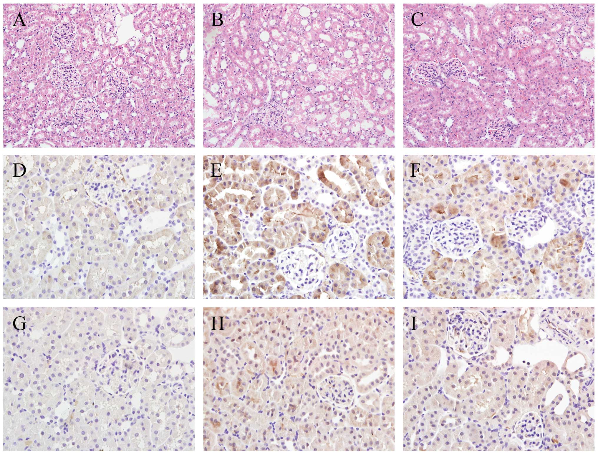 | Figure 2Histologic features were evaluated by
H&E and immunohistochemical staining. (A–C) Representative
kidney sections stained with H&E. (D–F) TLR4 expression in the
kidneys following 24 h of reperfusion. (G–I) NF-κB expression in
the kidneys following 24 h of reperfusion. (A, D, G) Sham group:
Sections from a sham-operated rat. (B, E, H) I/R group: Section
from rat subjected to I/R treatment. (C, F, I) Picroside II group:
Section from rat subjected to I/R and treated with picroside II.
H&E staining, original magnification ×200; Immunohistochemical
staining, original magnification ×400. H&E, hematoxylin and
eosin. TLR, toll-like receptor; NF-κB, nuclear factor-κB; I/R,
ischemia and reperfusion. |
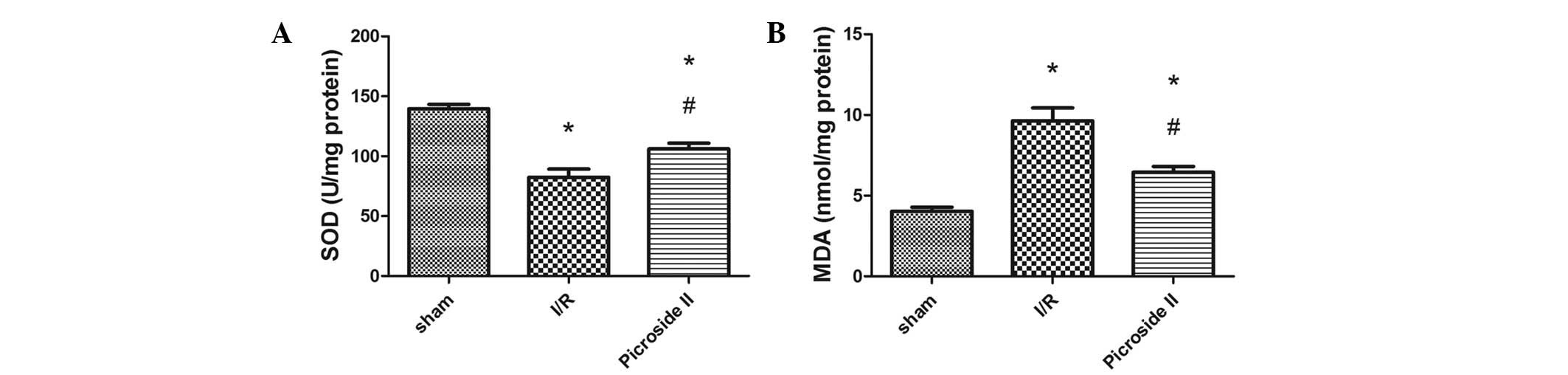
MDA and SOD analysis
As shown in Fig.
3A, the SOD activity decreased markedly in the I/R group
compared with that in the sham group; however, picroside II
inhibited the reduction of SOD activity that was induced by I/R
injury. The MDA content in the I/R group was significantly higher
than that in the sham group, whereas this increase induced by I/R
injury was significantly attenuated following treatment with
picroside II.
Immunohistochemistry
The expression of TLR4 (Fig. 2D–F) and NF-κB (Fig. 2G–I) was detected by
immunohistochemical staining. The results revealed that TLR4 and
NF-κB positive cells were rarely found in the sham group. However,
in the I/R group, renal tissues were strongly positive for TLR4 and
NF-κB expression. These expression spots were reduced in the
picroside II group compared with those in the I/R group.
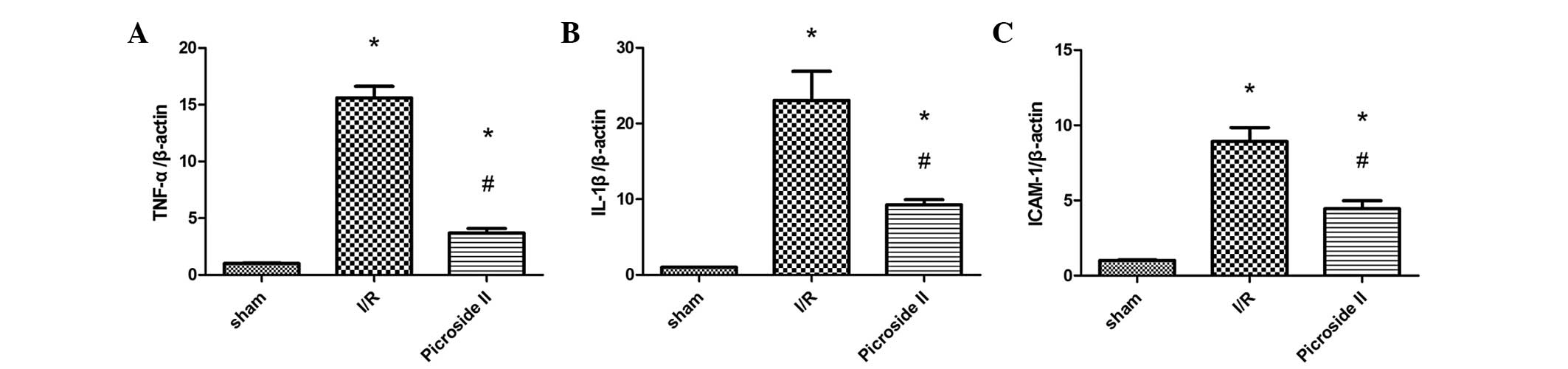
RT-qPCR analysis
The relative mRNA expression levels of TNF-α, IL-1β
and ICAM-1 to β-actin were evaluated. The mRNA levels of TNF-α,
IL-1β and ICAM-1 were significantly greater in the I/R group than
in the sham group. However, treatment with picroside II
significantly reduced the mRNA expression levels of TNF-α, IL-1β
and ICAM-1 following I/R (Fig.
4).
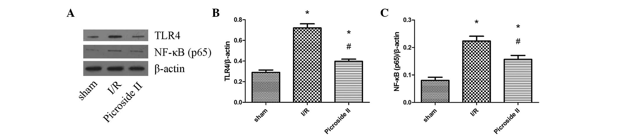
Western blot analysis
The levels of TLR4 and NF-κB (p65) protein
expression were measured by western blotting (Fig. 5). It was clear from the results
that the expression levels of TLR4 and NF-κB were upregulated in
the I/R and picroside II groups when compared with those in the
sham group. However, picroside II attenuated these I/R-induced
increases in expression.
Discussion
In this study, it was demonstrated that the
protective effects of picroside II in the kidney were associated
with a reduction of oxidative stress and inflammation. Picroside II
is one of the main active constituents of the extracts of
Picrorhiza scrophulariiflora Pennell and has been shown to
possess a wide range of pharmacological effects, including
neuroprotective, hepatoprotective, anti-apoptosis,
anti-cholestatic, anti-inflammatory and immune-modulating
activities (3–5). In a previous study, it was
demonstrated that picroside II was able to inhibit apoptosis in
rats subjected to middle cerebral I/R injury (6). Another study reported that picroside
II protected hepatocytes against injury through maintaining the
integrity of the mitochondrial membrane and enhancing the activity
of ATPase in mitochondria (15).
However, it has not previously been demonstrated whether picroside
II is able to protect tissue against renal I/R injury. In the
present study, it was demonstrated for the first time to the best
of our knowledge, that picroside II reduced the oxidative stress
and inflammation induced by renal I/R injury in rats.
Renal injury caused by I/R has a complicated
pathological course. Inflammation represents a key factor in the
occurrence and development of ischemic damage, which is considered
to occur secondary to an intense inflammatory response initiated by
the infiltration of leukocytes and the production of
pro-inflammatory cytokines following I/R (16). Within hours after ischemia, the
infiltrating leukocytes may release a large number of
pro-inflammatory mediators that contribute to the development of
tissue damage. TNF-α and IL-1β are key inflammatory cytokines
participating in the pathological process of I/R injury. Due to the
assistance of TNF-α, the infiltration of leukocytes into the kidney
may aggravate ischemic injury (17). IL-1β is an immune-derived cytokine
and can promote the secretion of itself under ischemic stimuli, the
so-called autocrine-like function (17). ICAM, as an adhesion molecule, can
facilitate leukocyte infiltration and adhesion to aggravate the
injuries caused by I/R. In the present study, it was observed that
the mRNA levels of TNF-α, IL-1β and ICAM-1 were significantly
greater in the I/R group than in the sham group. However, these
increased markers of inflammation were reduced by treatment with
picroside II (Fig. 4).
ROS play a key role in the development of renal I/R
injury. They can cause direct damage to membranes and proteins as
well as indirect damage through the activation of pro-apoptotic
pathways (18). Normally, the
generated ROS from metabolic processes can be scavenged by
endogenous antioxidant enzymes such as SOD (19), catalyzing the dismutation of the
superoxide radical to hydrogen peroxide. MDA, as a significant
product of lipid oxidation (20),
is often used to reflect the extent of cell injury by oxidative
stress. It has been reported that reducing the elevation of MDA
levels and suppressing the reduction of SOD activity has protective
effects in cardiac I/R injury (21). The present study revealed that
picroside II administration significantly reduced MDA level
elevation and attenuated the reduction of SOD activity in renal I/R
injury.
Through MyD88, TLR4 signals can lead to the
subsequent downstream activation of NF-κB (p65) and
mitogen-activated protein kinase signaling pathways (22). NF-κB, an important nuclear
transcription factor, regulates the expression of a large number of
genes, which play key roles in the regulation of apoptosis,
inflammation, viral replication and tumorigenesis (23). Numerous stimuli, including I/R
injury, can activate NF-κB signaling by the degradation of IκB and
release of the NF-κB p65-p50 dimer, which translocates to the
nucleus, binds to κB binding sites on DNA, and regulates the
transcriptional activation of target genes (24). The current study demonstrated that
renal tubular injury and inflammatory cell infiltration were
markedly alleviated in rats following picroside II treatment. The
expression levels of TLR4 and NF-κB significantly decreased in the
picroside II group compared with those in the I/R group, as did BUN
and Cr levels. This suggests that picroside II may alleviate
I/R-induced oxidative stress and inflammatory response by blocking
the TLR4/NF-κB pathway.
In conclusion, this study demonstrated for the first
time that the administration of picroside II had a protective
effect on renal I/R injury, which may be ascribed to blockade of
the TLR4/NF-κB pathway resulting in attenuation of oxidative stress
and the inflammatory response.
Acknowledgements
This study was supported by the Province Natural
Science Foundation of Hubei (grant no. 2013CFB226).
Abbreviations:
|
MDA
|
malondialdehyde
|
|
SOD
|
superoxide dismutase
|
|
I/R
|
ischemia and reperfusion
|
References
|
1
|
Kosieradzki M and Rowiński W:
Ischemia/reperfusion injury in kidney transplantation: mechanisms
and prevention. Transplant Proc. 40:3279–3288. 2008. View Article : Google Scholar : PubMed/NCBI
|
|
2
|
Li JX, Li P, Tezuka Y, Namba T and Kadota
S: Three phenylethanoid glycosides and an iridoid glycoside from
Picrorhiza scrophulariiflora. Phytochemistry. 48:537–542. 1998.
View Article : Google Scholar : PubMed/NCBI
|
|
3
|
Cao Y, Liu JW, Yu YJ, Zheng PY, Zhang XD,
Li T and Guo MC: Synergistic protective effect of picroside II and
NGF on PC12 cells against oxidative stress induced by
H2O2. Pharmacol Rep. 59:573–579.
2007.PubMed/NCBI
|
|
4
|
Smit HF, Kroes BH, van den Berg AJ, van
der Wal D, van den Worm E, Beukelman CJ, van Dijk H and Labadie RP:
Immunomodulatory and anti-inflammatory activity of Picrorhiza
scrophulariiflora. J Ethnopharmacol. 73:101–109. 2000. View Article : Google Scholar : PubMed/NCBI
|
|
5
|
He LJ, Liang M, Hou FF, Guo ZJ, Xie D and
Zhang X: Ethanol extraction of Picrorhiza scrophulariiflora
prevents renal injury in experimental diabetes via
anti-inflammation action. J Endocrinol. 200:347–355. 2009.
View Article : Google Scholar
|
|
6
|
Li Q, Li Z, Xu XY, Guo YL and Du F:
Neuroprotective properties of picroside II in a rat model of focal
cerebral ischemia. Int J Mol Sci. 11:4580–4590. 2010. View Article : Google Scholar : PubMed/NCBI
|
|
7
|
Bonventre JV and Weinberg JM: Recent
advances in the pathophysiology of ischemic acute renal failure. J
Am Soc Nephrol. 14:2199–2210. 2003. View Article : Google Scholar : PubMed/NCBI
|
|
8
|
Bonventre JV and Zuk A: Ischemic acute
renal failure: an inflammatory disease? Kidney Int. 66:480–485.
2004. View Article : Google Scholar : PubMed/NCBI
|
|
9
|
Eltzschig HK and Eckle T: Ischemia and
reperfusion - from mechanism to translation. Nat Med. 17:1391–1401.
2011. View
Article : Google Scholar : PubMed/NCBI
|
|
10
|
Brooks C, Wei Q, Cho SG and Dong Z:
Regulation of mitochondrial dynamics in acute kidney injury in cell
culture and rodent models. J Clin Invest. 119:1275–1285. 2009.
View Article : Google Scholar : PubMed/NCBI
|
|
11
|
Yang S, Chou WP and Pei L: Effects of
propofol on renal ischemia/reperfusion injury in rats. Exp Ther
Med. 6:1177–1183. 2013.PubMed/NCBI
|
|
12
|
Toufektsian MC, Boucher FR, Tanguy S,
Morel S and de Leiris JG: Cardiac toxicity of singlet oxygen:
implication in reperfusion injury. Antioxid Redox Signal. 3:63–69.
2001. View Article : Google Scholar : PubMed/NCBI
|
|
13
|
Wu H, Chen G, Wyburn KR, Yin J, Bertolino
P, Eris JM, Alexander SI, Sharland AF and Chadban SJ: TLR4
activation mediates kidney ischemia/reperfusion injury. J Clin
Invest. 117:2847–2859. 2007. View
Article : Google Scholar : PubMed/NCBI
|
|
14
|
Jablonski P, Howden BO, Rae DA, Birrell
CS, Marshall VC and Tange J: An experimental model for assessment
of renal recovery from warm ischemia. Transplantation. 35:198–204.
1983. View Article : Google Scholar : PubMed/NCBI
|
|
15
|
Gao H and Zhou YW: Anti-lipid peroxidation
and protection of liver mitochondria against injuries by picroside
II. World J Gastroenterol. 11:3671–3674. 2005. View Article : Google Scholar : PubMed/NCBI
|
|
16
|
Du X, Hu X and Wei J: Anti-inflammatory
effect of exendin-4 postconditioning during myocardial ischemia and
reperfusion. Mol Biol Rep. 41:3853–3857. 2014. View Article : Google Scholar : PubMed/NCBI
|
|
17
|
Wang Y, Ge P and Zhu Y: TLR2 and TLR4 in
the brain injury caused by cerebral ischemia and reperfusion.
Mediators Inflamm. 2013:1246142013.PubMed/NCBI
|
|
18
|
Braunersreuther V and Jaquet V: Reactive
oxygen species in myocardial reperfusion injury: from
physiopathology to therapeutic approaches. Curr Pharm Biotechnol.
13:97–114. 2012. View Article : Google Scholar
|
|
19
|
Meng FJ, Hou ZW, Li Y, Yang Y and Yu B:
The protective effect of picroside II against hypoxia/reoxygenation
injury in neonatal rat cardiomyocytes. Pharm Biol. 50:1226–1232.
2012. View Article : Google Scholar : PubMed/NCBI
|
|
20
|
Wang Y, Zhang ZZ, Wu Y, Zhan J, He XH and
Wang YL: Honokiol protects rat hearts against myocardial ischemia
reperfusion injury by reducing oxidative stress and inflammation.
Exp Ther Med. 5:315–319. 2013.
|
|
21
|
Wang X, Yu Y, Ji L, Liang X, Zhang T and
Hai CX: Alpha-lipoic acid protects against myocardial
ischemia/reperfusion injury via multiple target effects. Food Chem
Toxicol. 49:2750–2757. 2011. View Article : Google Scholar : PubMed/NCBI
|
|
22
|
Muzio M, Ni J, Feng P and Dixit VM: IRAK
(Pelle) family member IRAK-2 and MyD88 as proximal mediators of
IL-1 signaling. Science. 278:1612–1615. 1997. View Article : Google Scholar : PubMed/NCBI
|
|
23
|
Ma JQ, Liu CM, Qin ZH, Jiang JH and Sun
YZ: Ganoderma applanatum terpenes protect mouse liver against benzo
(alpha)pyren-induced oxidative stress and inflammation. Environ
Toxicol Pharmacol. 31:460–468. 2011. View Article : Google Scholar : PubMed/NCBI
|
|
24
|
Wong ET and Tergaonkar V: Roles of
NF-kappaB in health and disease: mechanisms and therapeutic
potential. Clin Sci (Lond). 116:451–465. 2009. View Article : Google Scholar
|