Introduction
Neurons in the central nervous system (CNS) of adult
mammals were not considered to be able to undergo repair or
regeneration (1). Since neural
stem cells (NSCs) were first isolated from the adult mammalian CNS,
NSCs have become increasingly studied due to their unique
self-renewal capacity and multiple differentiation potential
(2,3). Characteristics of NSCs include the
generation of new cells and the creation of an environment that is
good for axonal regeneration; thus, NSCs have become beneficial to
the repair of spinal cord injury (SCI) (4–7).
However, their potential is very limited in adult models. Firstly,
the adult CNS can only provide a small number of new NSCs. In
addition, the environment of the injured CNS is detrimental to the
survival and differentiation of NSCs (8–10).
To combat these two disadvantages, the repair strategy should be
adjusted to increase the number of NSCs through transplantation and
alter the local environment, respectively (11,12).
To date, the embryonic CNS is considered to be the
main source of NSCs. NSCs derived from the embryonic neural tube
present a unique advantage, as they have the same origin as the
cells of the SCI lesion (13–15).
Increasing the number of NSCs can be achieved via the expansion of
NSCs in vitro, of which there are two methods that are
currently used: Neurosphere culture (2) and adherent culture (16).
Buyang Huanwu decoction (BYHWD), recorded in ‘Yilin
Gaicuo’ (Correction on Errors in Medical Classics), has been used
for the treatment of stroke-induced disability and for improving
neurological functional recovery in China for hundreds of years
(17). In previous studies, BYHWD
has been widely applied for conditions of the nervous system, and
has been shown to exert neuroprotective effects on cerebral
ischemia-reperfusion following injury (18–21),
promoting peripheral nerve regeneration in vivo (22), enhancing functional recovery
following spinal cord injury (23)
and stimulating the growth and differentiation of neural progenitor
cells in vitro (24).
However, the effects of BYHWD on the differentiation of
transplanted NSCs in an injured spinal cord, as well as the
associated mechanisms underlying the improvements to the SCI
symptoms, have not yet been reported.
In the present study, the effects of this formula on
the differentiation of transplanted NSCs were investigated, with
the aim to observe the synergistical effect on the recovery of
neurological function between NSCs and BYHWD in the treatment of
SCI in rats. In addition, the present study investigated the
possible mechanisms of acquiring a higher BBB score following
combined administration of BYHWD and NSCs in a model of SCI.
Materials and methods
Preparation of embryonic neural
tube-derived NSCs
Embryonic neural tube-derived NSCs were cultured
according to previously outlined methods (25). The posterior segment of the neural
tubes from an embryonic Sprague-Dawley (SD) rat (11.5 days) were
collected. The morning of the day when a vaginal plug was found was
defined as embryonic day 0.5 post coitum. The neural tubes
were cut into sections and dissociated mechanically under an
anatomical microscope (SZ2-ILST; Olympus Corporation, Tokyo,
Japan), under sterile conditions in the tissue culture hood.
Subsequently, the samples were transferred to a centrifuge tube
that contained 1 ml Hanks balanced salt solution (HBSS; 14185-052;
Invitrogen Life Technologies, Carlsbad, CA, USA). Following
filtration with a 200-mesh sieve, the samples were centrifuged at
300 × g for 5 min and resuspended with Dulbecco’s modified Eagle’s
medium: Nutrient Mixture F/12 (DMEM/F12; 1:1; SH30126.FS, Hyclone,
Shanghai HuiYing Biotechnology Co. Ltd., Shanghai, China)
containing 2% B27 (17504-044; Invitrogen Life Technologies) and 20
ng/ml basic fibroblast growth factor (bFGF; PMG0035; Invitrogen
Life Technologies). The cell suspension was adjusted to a
concentration of 1×10⁶ cells/ml, plated and incubated at
37°C in a 5% CO2 incubator. Following culture for 3–4
days, half of the medium was replaced. After 7–9 days, the cells
were passaged. The second passage of the NSCs were collected, and
the formed neurospheres were dispersed into a single cell
suspension and transferred to a culture flask. Next,
5-bromo-2-deoxyuridine (BrdU) was added to the culture flask at a
final concentration of 10 μM (26). The newly formed neurospheres were
dispersed into a single cell suspension after BrdU labeling for 48
h. The cell suspension was centrifuged at 300 × g for 5 min, washed
with HBSS, centrifuged at 300 × g for 5 min and resuspended in
complete DMEM/F12 culture media. The cell concentration was
adjusted to a final density of 5×10⁹ cells/ml prior to
transplantation.
Composition and preparation of BYHWD
BYHWD consists of the following ingredients: Radix
Astragali (120 g), also known as huáng qí, is the root of
Astragalus membranaceus (Fisch.) Bge. var.
mongholicus (Bge.) Hsiao; Radix Angelicae Sinensis (6 g),
also known as dang gui, is the root of Angelica sinensis
(Oliv.) Diels; Radix Paeoniae Rubra (4.5 g), also known as chi
shao, is the root of Paeonia lactiflora Pall.; Rhizoma
Chuanxiong (3 g), also known as chuan xiong, is the root of
Ligusticum chuanxiong Hort.; Semen Persicae (3 g), also
known as tao ren, is the dry ripe seed of Prunus persica
(L.) Batsch; Flos Carthami (3 g), also known as hong hua, is the
flower of Carthamus tinctorius L.; and Lumbricus (3 g), also
known as di long and Pheretima aspergillum (perrier). All
the herbs were orthodox drugs that were purchased from the
Traditional Chinese Medicine Association of the Affiliated Hospital
of Binzhou Medical University (Binzhou, China), and had been
extracted according to standard methods outlined in the Chinese
Pharmacopoeia (27). The mixture
was decocted to yield a final concentration of 2 g crude drug/ml,
which was stored at 4°C for further use (28).
Animal grouping, SCI model establishment
and drug administration
In total,78 female SD rats (weight, 220–250 g) were
purchased from the Experimental Animal Center at Shandong Green
Leaf Pharmaceutical Co., Ltd. (Yantai, China). The rats were
randomly divided into five groups, which included the sham-operated
control (sham control; 6 rats), SCI (18 rats); BYHWD (18 rats), NSC
transplantation (18 rats) and NSCs transplantation combined with
BYHWD (BYHWD + NSCs; 18 rats) groups. Three time points were
selected, 7, 14 and 28 days, where each group of rats underwent
examination. Surgical procedures on the rats during the experiment
were performed following the guiding principles of care (29), and were consistent with the
National Institutes of Health Guide for the Care and Use of
Laboratory Animals (30). All
experiments involving rats in the present study were approved by
the Ethics Committee for animal care and use of Binzhou Medical
University (Yantai, China). Anesthesia was achieved by an
intraperitoneal injection of 4% chloral hydrate (0.1 ml/kg). A
laminectomy was performed for the rats in the sham control group.
The spinal cords of the rats in the remaining four groups underwent
complete transection at the T10 vertebra. During surgery, gelatin
sponges (2×2×2 mm3) containing 10 μl BrdU-labeled NSCs
(5×10⁹ cells/ml) were transplanted into the SCI site in
the NSCs and BYHWD + NSCs groups. In the SCI control group, gelatin
sponges containing 10 μl DMEM/F12 complete culture media were
transplanted into the SCI site.
The dosage of BYHWD was selected according to the
conversion factor of the dosage between humans and rats: Rat dose
(mg/kg) = W × human dose (mg/kg), where W is the conversion factor
(6.25). The BYHWD dose selected was ~14.8 g/kg/day. Thus, the rats
in the BYHWD and BYHWD + NSCs groups were administered BYHWD at
this dose by introgastric infusion once a day, while the same
volume of saline was administered to the rats in the NSC group.
Postoperative care was conducted according to the National
Institutes of Health guidelines (31).
Behavioral analysis
All the rats were evaluated by two trained examiners
via a double blind method. The Basso, Beattie and Bresnahan (BBB)
locomotor rating scale was used to determine motor function prior
to the injury and transplantation, at 24 h after the surgery, and
then weekly for four weeks. The open field locomotor activity score
was determined by observing and calculating the behaviors involving
the movement of all joints of the hindlimbs, plantar placement,
forelimb and hindlimb coordination, the trunk stability and tail
position, according to the BBB protocol. Each session lasted for 4
min. Scores from the two examiners were averaged for each rat, and
scores ranged between 0 and 21 (0, no movement; 21, normal
movement) (32).
Tissue processing
Rats were deeply anesthetized by an intraperitoneal
injection of 4% chloral hydrate. Subsequently, 0.9% saline solution
(37°C; 200 ml) was perfused through the heart, followed by 4%
paraformaldehyde in 0.1 M phosphate buffer (pH 7.4) at 4°C (200
ml). The spinal cords were dissected, post-fixed with 4%
paraformaldehyde at 4°C for 2 h, and subjected to graded
dehydration, xylene transparent clearing, wax infiltration,
paraffin embedding and sectioning.
Immunofluorescence staining
Primary antibodies were used as follows: Mouse
anti-BrdU (monoclonal IgG; 1:100; ZM-0013; Beijing Zhongshan Golden
Bridge Biotechnology Co., Ltd., Beijing, China), rabbit polyclonal
anti-nestin (1:100; Beijing Zhongshan Golden Bridge), rabbit
polyclonal anti-neuron specific enolase (NSE; 1:200; BA0535; Wuhan
Boster Biological Engineering Co., Ltd., Wuhan, China), rabbit
polyclonal anti-myelin basic protein (MBP; 1:150; BA0094; Wuhan
Boster Biological Engineering Co., Ltd.) and rabbit polyclonal
anti-glial fibrillary acidic protein (GFAP; 1:200; BA0056; Wuhan
Boster Biological Engineering Co., Ltd.). Cy3-conjugated goat
anti-rabbit IgG (1:100; BA1032; Wuhan Boster Biological Engineering
Co., Ltd) and fluorescein isothiocyanate-conjugated goat anti-mouse
IgG (1:200; ZF-0312; Beijing Zhongshan Golden Bridge, Beijing,
China) were used as secondary antibodies. Paraffin-embedded
sections were deparaffinized, rehydrated and washed three times
with 0.01 M phosphate-buffered saline (PBS). The samples were
subsequently incubated with 0.1 M HCl containing 0.4% pepsin
(Sigma-Aldrich, St. Louis, MO, USA) for 10 min at 37°C, and then
0.2 M HCl for 30 min at 37°C. After washing three times with 0.01 M
PBS, the samples were incubated with 0.01 M PBS containing 0.05%
Triton X-100 for 15 min at room temperature, washed three times
with 0.01 M PBS and incubated with goat serum working solution for
30 min at 37°C (33). Incubations
with the appropriate primary antibody were performed overnight at
4°C. Following repeated washing with 0.01 M PBS, the sections were
incubated with their respective secondary antibodies for 30 min at
37°C. Finally, the sections were washed with 0.01 M PBS, mounted
with a coverslip and examined under a confocal microscope (Leica
TCS SPE; Leica Microsystems GmbH, Wetzlar, Germany).
Cell counting
For quantitative analysis of the transplanted NSCs
in the spinal cord, beginning from the site of transplantation, the
spinal cord of the rats was sectioned continuously at a 5-μm
thickness in the transverse plane towards the rostral and caudal
end. Every fifth slice was collected, resulting in six sections in
each direction being obtained. GFAP and BrdU double positive cells
within the white matter and gray matter of every section were
counted at the same magnification (x40; Leica TCS SPE; Leica
Microsystems GmbH). Area analysis of the GFAP-positive astrocytes,
NSE-positive neurons and MBP-positive oligodendrocytes was assessed
using Image-Pro Plus 6.0 pathological image analysis software
(Media Cybernetics, Inc., Rockville, MD, USA).
Statistical analysis
Data corresponding to the BBB scores, the number of
GFAP/BrdU double positive cells and the area analysis of
astrocytes, neurons and oligodendrocytes are expressed as the mean
± standard deviation. A two-way classification analysis of variance
of the factorial experiment, followed by the Student-Newman-Keuls
test, was performed for statistical analysis. P<0.05 was
considered to indicate a statistically significant difference.
Experimental data were analyzed using the SPSS 13.0 software
package (SPSS, Inc., Chicago, IL, USA) to estimate the significance
of the difference between changes in the data.
Results
NSC culture and characterization
NSCs obtained from the embryonic neural tube at 11.5
days post coitum were cultured in DMEM/F12 medium containing
B27 and bFGF. At day 2 of in vitro culture, the NSCs
proliferated and formed neurospheres. Between days 7 and 9, the
NSCs became confluent and underwent cell passage. These
undifferentiated neurospheres expressed the primitive
neurofilament, nestin, which was used as a marker of NSCs (Fig. 1A).
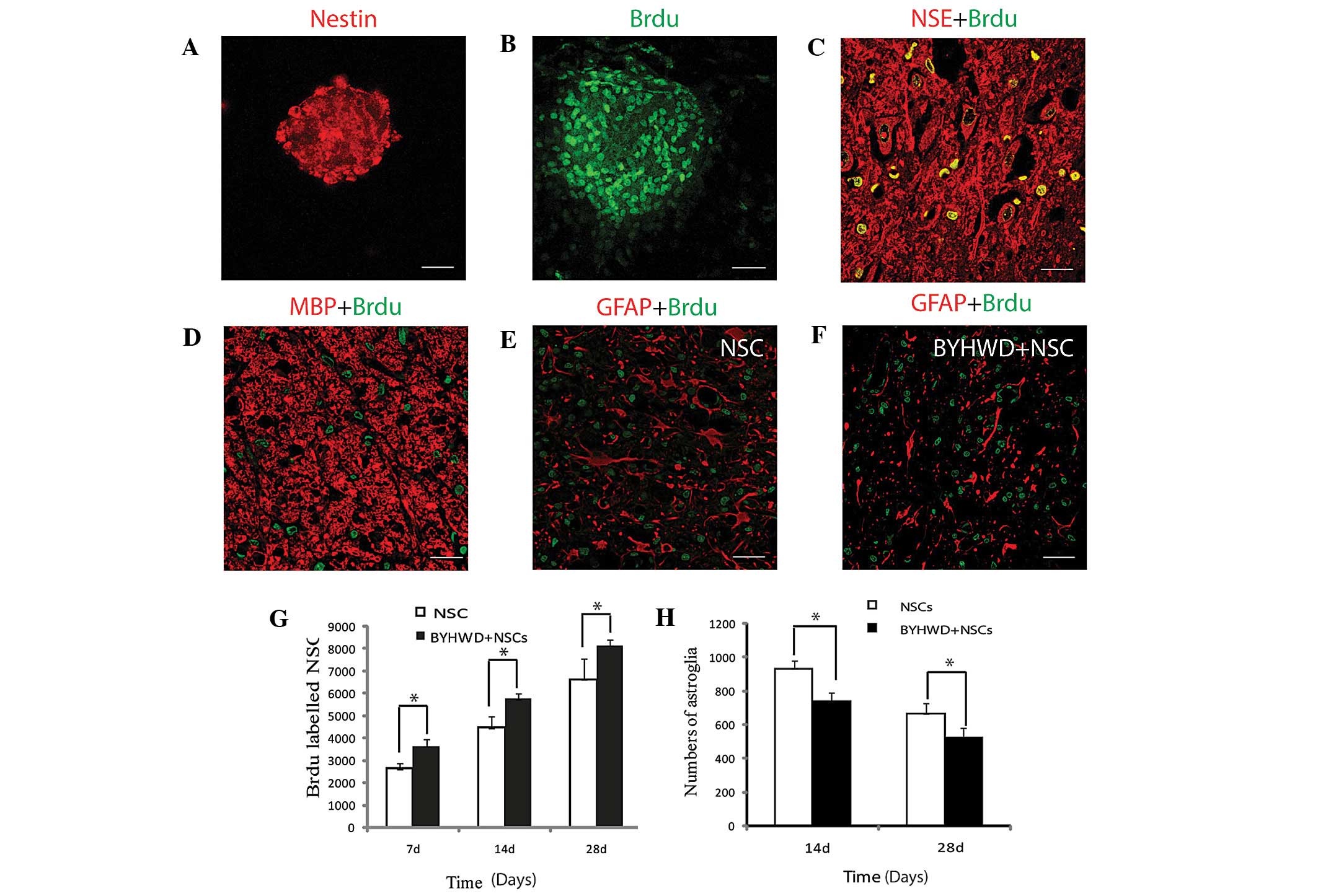 | Figure 1Characterization and differentiation
of the cultured isolated NSCs from the rat embryo neural tube. (A)
Cultured NSCs grew into neurospheres in the culture dish,
expressing the neural stem cell marker, nestin. (B) NSCs continued
to proliferate in the in vitro culture, as shown by the
incorporation of BrdU (green). (C) Transplanted NSCs in the injured
spinal cord differentiated into neurons, labeled with the neuron
marker, NSE (red) and BrdU (green). (D) Transplanted NSCs
differentiated into oligodendrocytes, expressing MBP and BrdU, and
incorporated into neural networks. (E and F) BYHWD treatment
exerted a suppressive effect on astrogliosis in the spinal cord
injury (SCI) site, as compared with NSC treatment alone. (G) BYHWD
treatment was shown to promote NSC survival at the SCI site at the
three examined time points (*P<0.05; n=36). (H) BYHWD
treatment was shown to inhibit astrogliogenesis in the surrounding
SCI site at the two examined time points (*P<0.05;
n=36). BYHWD, Buyang Huanwu decoction; NSC, neural stem cells;
BrdU, 5-bromo-2-deoxyuridine; GFAP, glial fibrillary acidic
protein; MBP, myelin basic protein; NSE, neuron specific enolase.
Scale bars, 50 μm (A and C), 30 μm (B) and 100 μm (D, E and F). |
To trace the NSCs and their fate following
transplantation adjacent to the injured spinal cord site, as well
as the response to the treatment of BYHWD, BrdU was supplemented
into the culture media. Immunocytochemistry revealed that the BrdU
was efficiently incorporated into the proliferating NSCs (Fig. 1B)
Survival and proliferation of NSCs in the
transected spinal cord
BYHWD was shown to promote the survival and
proliferation of NSCs in the SCI site. Following transplantation,
the grafted NSCs were easily identified primarily around the lesion
site of the host spinal cord, and had integrated well with the host
tissue in the NSCs only and BYHWD + NSCs groups. The total counted
number of BrdU-labeled NSCs in the NSCs only transplanted group at
day 7 was 2,627±259.435, while at day 14, the number was
4,460.33±508.723 and at day 28, the total number was
6,631.83±909.948. The number of BrdU-labeled NSCs in the NSC and
BYHWD + NSC groups at day 28 was significantly increased when
compared with the numbers at days 7 and 14; however, the difference
between the numbers at days 7 and 14 were not statistically
significant. In the BYHWD + NSCs group, the total counted number of
BrdU-labeled NSCs at days 7, 14 and 28 were 3,643.67±281.378,
5,755.50±242.561 and 8,143.50±267.644, respectively. The number of
BrdU-labeled NSCs significantly increased with time when comparing
the numbers at days 7, 14 and 28. In addition, the number of
BrdU-positive cells at each of the three time points in the BYHWD +
NSCs group was greater compared with the number at the
corresponding time point in the NSCs group (n=18; P<0.05). The
number of BrdU-positive cells at day 28 was the highest among the
three time points in the BYHWD + NSCs group (n=18; P<0.05;
Fig. 1G).
In the control experiment, a gelatin sponge injected
with 10 μl culture medium was transplanted into the SCI site. There
were no BrdU-positive cells observed in the sham-operated control,
SCI and BYHWD groups (data not shown).
NSC differentiation and migration into
the SCI site
To determine the fate of the transplanted NSCs and
their differentiation status, immunohistochemical staining of the
neuron marker, NSE, was performed. NSE/BrdU double labeling
revealed that the majority of the transplanted NSCs had
differentiated into neurons (Fig.
1C); however, there was little differentiation into astroglial
cells (Fig. 1E and F). To assess
whether the NSCs had differentiated into oligodendrocytes and were
able to express MBP to protectively wrap the axons for electrical
signal conduction, immunohistochemical staining of MBP was
performed. The results revealed that the BrdU-labeled NSCs were
closely associated with the myelin-wrapped axons (Fig. 1D).
The effect of BYHWD on NSC differentiation into
neurons was analyzed at three time points (days 7, 14 and 28). The
number of NSE/BrdU double labeled neurons in the three
corresponding time points did not exhibit a statistically
significant difference between the NSC and BYHWD + NSC groups (data
not shown).
To examine the effect of BYHWD on NSC
differentiation into astroglia, or the inhibition of gliosis in the
SCI site, GFAP/BrdU double labeling immunofluorescence was
performed. The results revealed few double labeled cells were
observed in the SCI site (Fig. 1E and
F). The number of GFAP-labeled cells in the BYHWD-treated group
was significantly decreased when compared with the number in the
NSC transplantation only group (Fig.
1H). The average optical density of GFAP immunofluorescence in
the NSC only and BYHWD + NSC groups was significantly lower when
compared with the BYHWD group (data not shown).
Since oligodendrocytes are the predominant
myelinating cell type in the CNS, whether the transplanted NSCs
were able to differentiate into oligodendrocytes in the SCI site
and integrate into the nerve fiber network was investigated.
MBP/BrdU double labeling immunofluorescence revealed that the
BrdU-labeled cells were closely incorporated inside the network of
MBP-positive nerve fibers (Fig.
1D).
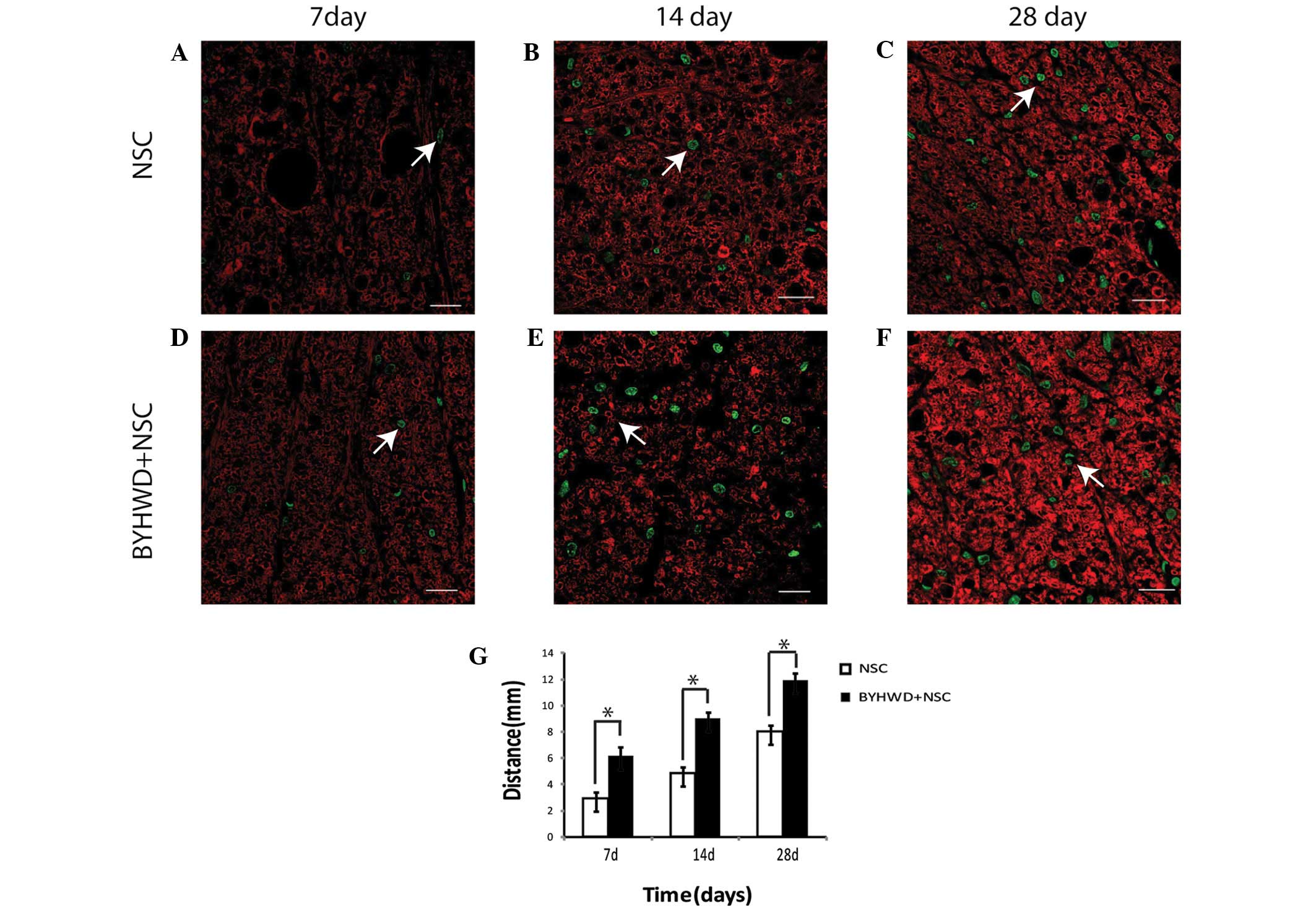
NSC migration
The distance of NSC migration into the surrounding
SCI undamaged tissue is associated with improvements to the SCI
symptoms. To further investigate the possible mechanisms underlying
the improvements in motor behavior following NSC transplantation
and treatment with BYHWD, the total distance migrated by the
BrdU-labeled cells appearing in the rostral and distal sections of
the SCI site was determined. In the BYHWD + NSC group, the total
distance traveled at day 7 was 6.15±0.49 mm, while at day 14, the
distance was 9.02±0.45 mm. By day 28, the total migration distance
was 11.95±0.50 mm. In the NSC only group, at days 7, 14 and 28, the
total distances traveled were 2.92±0.66, 4.85±0.46 and 8.01±0.51
mm, respectively. All the distances in the BYHWD + NSC group were
significantly greater when compared with the distances in the NSC
group at the respective time points (n=18; P<0.05; Fig. 2A–G).
Ultrastructure of the SCI prior to and
following the treatment
Following induction of the injury, apparent changes
were observed around the injury site of the spinal cord. Firstly,
the myelin sheath underwent loosely onion-like demyelination, while
the wrapped axons exhibited degeneration. In addition, a vacuole
change was observed inside the myelinated axon (Fig. 3A and B). In the SCI models treated
with BYHWD and NSC transplantation separately for four weeks,
improvements were observed with regard to the tightness of the
myelin sheath to varying degrees (Fig.
3C–F). However, the combined treatment of BYHWD + NSCs improved
the myelination to the greatest degree when compared with the
single treatments of BYHWD or NSCs (Fig. 3G and H).
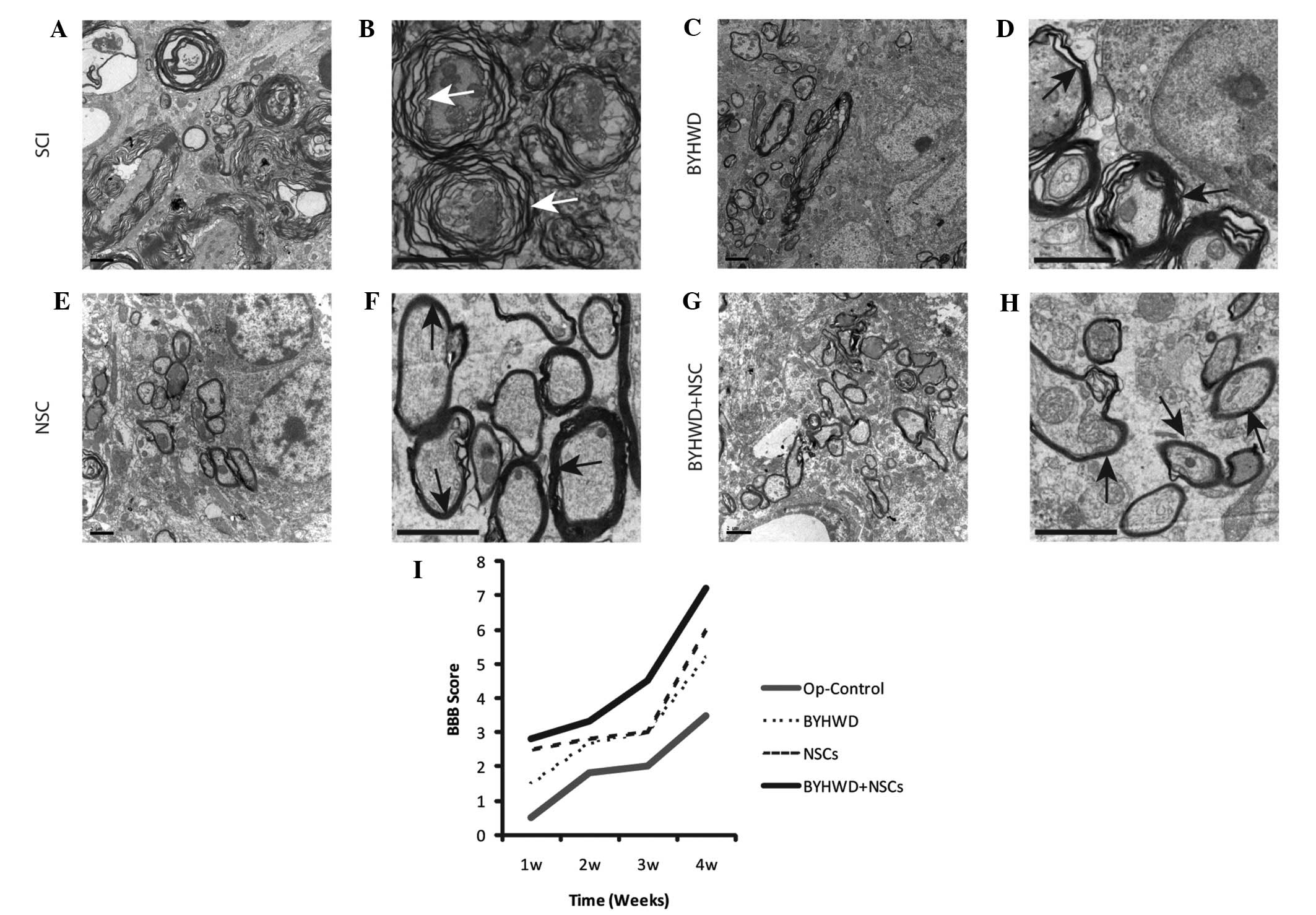 | Figure 3Ultrastructure of the spinal cord at
the SCI site and the effect of NSCs and BYHWD treatment on SCI
rehabilitation, as measured by the BBB locomotor rating scale.
Ultrastructure examination revealed the effect of the different
treatments on the SCI after 28 days. (A and B) No treatment
resulted in onion-like demyelination (white arrow). (C and D)
Treatment with BYHWD only mitigated the demyelination (black
arrow). (E and F) Treatment with NSCs only further promoted the
mitigation of demyelination when compared with the BYHWD and
control group (black arrows). (G and H) Combination treatment with
BYHWD and NSCs notably alleviated the demyelination, and the
myelination wrapping the axons was shown to recover to relative
normality. B, D, F and H are higher magnification compared with A,
C, E and G, respectively. (I) Motor rehabilitation, as measured
using the BBB locomotor rating scale. Treatment with BYHWD + NSCs
demonstrated a synergistic effect on locomotion recovery. BYHWD,
Buyang Huanwu decoction; NSC, neural stem cells; SCI, spinal cord
injury; BBB, Basso, Beattie and Brasnahan. Scale bar, 2 μm. |
Synergy between BYHWD and NSCs leads to
the recovery of neurological function
Following spinal cord surgery, the rats in the
sham-operated group exhibited normal motor function, while all the
rats in the four remaining groups were paralyzed, unable to stand
independently and moved by pulling themselves forward with their
forelimbs. Urinary incontinence was also later observed in these
rats. The hindlimb locomotor activities of all the rats were
evaluated using the BBB locomotor rating scale at four time points
between one and four weeks after surgery. The BBB scores were shown
to gradually increase over time in each group throughout the entire
follow-up period, which represented gradual improvements to the
hindlimb locomotion activity. In addition, BBB scores were
significantly higher in the three treatment groups when compared
with the SCI group in the period between two and four weeks after
surgery. Compared with the BYHWD and NSCs only groups, the BYHWD +
NSCs group exhibited a significantly higher score starting at the
second week, while the most notable difference was observed at the
fourth week following the surgery. However, the differences in the
BBB scores between the BYHWD and NSCs groups were not statistically
significant during the same time period (Fig. 3I).
Discussion
In a previous study, embryonic neural tube-derived
NSCs were demonstrated to have multiple potentiality to
differentiate into neurons, oligodendrocytes and astrocytes in
vitro (25). Using a
pharmacological serum testing method, Sun et al found that
BYHWD may directly promote the differentiation of neural progenitor
cells (24). In the current study,
the transplantation of embryonic neural tube-derived NSCs was
employed in conjunction with BYHWD administration for the treatment
of rats that had undergone transection of the spinal cord. The
efficacy of BYHWD treatment on the survival and differentiation of
the transplanted NSCs was further confirmed. The results revealed a
synergy between BYHWD and NSCs in improving the neurological
behavior performance, as measured using the BBB locomotor rating
scale.
Previous studies have shown that NSCs exert a
protective effect at the lesion site by means of multiple
mechanisms, including secreting diverse neurotrophic factors and
cytokines, mobilizing endogenous stem cells and replacing lost
cells (11,34,35).
To achieve all these protective effects, a sufficient number of
surviving NSCs are required to differentiate into neural cells
following transplantation. Compared with the NSCs only group, a
higher number of BrdU-positive cells were observed in the NSCs +
BYHWD group. Thus, BYHWD treatment was found to improve the
survival rate of the NSCs. In addition, a greater area of
MBP-positive oligodendrocytes was present in the rats of the NSCs +
BYHWD group, which was an important indicator for remyelination at
the lesion site. By contrast, a smaller number of GFAP/BrdU double
positive astrocytes were present within the injured spinal cords of
the NSCs + BYHWD group, indicating that BYHWD treatment suppresses
injury-induced astrogliosis in the spinal cord. This may further
limit scar formation or improve the reestablishment of the neuronal
innervation circuit between the upper control neurons and the
targets effect organs. Electron microscopy results clearly revealed
that the administration of NSCs and BYHWD improved the tightness of
the myelin sheath in the neural tract between the upper control
neurons and the targets effect organs. However, the exact
mechanisms and factors involved in the pathway of achieving the
improvements, as well as the establishment of a synaptic
connection, remains unclear and further investigation is required.
Recent progress in self-derived induced pluripotent stem cells has
enabled the prospect of patient-derived cells in the treatment of
SCI and other stem cell-required treatment to become a prospective
strategy (36). From the results
of the present study, BYHWD was concluded to be beneficial to the
survival and differentiation of NSCs into neurons and
oligodendrocytes, while reducing the differentiation into
astrocytes.
In the present study, rats treated with BYHWD + NSCs
following SCI exhibited significantly enhanced functional recovery
of the injured forelimb. Comprised of seven Chinese plant drugs,
BYHWD has multiple chemical constituents. Although Yang et
al reported that the chemical constituents of BYHWD are
numerous and diverse, including flavonoids, alkaloids and saponins
(37), the effective constituents
of BYHWD and the mechanisms of action remain unknown. Thus, much
research required to clarify the underlying mechanisms and
subsequently develop this formula into a potential therapeutic
reagent.
In conclusion, the results of the present study
indicate that administration of BYHWD may enhance the
differentiation of NSCs into neurons and oligodendrocytes, while
reducing the differentiation into astrocytes, in the injured spinal
cord. A synergy was identified between BYHWD and NSCs that resulted
in the recovery of neurological function, which indicated that
BYHWD may aid in the treatment of SCI in humans. However, further
studies investigating the underlying mechanisms of BYHWD are
required in the future.
Acknowledgements
This study was supported by grants from the Natural
Science Foundation of Shandong Province (no. Y2007C034) and the
Science and Technology Development Planning of Shandong Province
(no. 2010GSF10254). The authors thank Dr Fuzi Jin from the
Lunenfeld-Tanenbaum Research Institute (Toronto, ON, Canada) for
thoroughly reading and revising the manuscript.
References
|
1
|
Björklund A and Lindvall O: Cell
replacement therapies for central nervous system disorders. Nat
Neurosci. 3:537–544. 2000. View
Article : Google Scholar : PubMed/NCBI
|
|
2
|
Cummings BJ, Uchida N, Tamaki SJ, et al:
Human neural stem cells differentiate and promote locomotor
recovery in spinal cord-injured mice. Proc Natl Acad Sci USA.
102:14069–14074. 2005. View Article : Google Scholar : PubMed/NCBI
|
|
3
|
Reynolds BA and Weiss S: Generation of
neurons and astrocytes from isolated cells of the adult mammalian
central nervous system. Science. 255:1707–1710. 1992. View Article : Google Scholar : PubMed/NCBI
|
|
4
|
Marques SA, Almeida FM, Fernandes AM, et
al: Predifferentiated embryonic stem cells promote functional
recovery after spinal cord compressive injury. Brain Res.
1349:115–128. 2010. View Article : Google Scholar : PubMed/NCBI
|
|
5
|
McDonald JW, Liu XZ, Qu Y, et al:
Transplanted embryonic stem cells survive, differentiate and
promote recovery in injured rat spinal cord. Nat Med. 5:1410–1412.
1999. View Article : Google Scholar : PubMed/NCBI
|
|
6
|
Teng YD, Lavik EB, Qu X, et al: Functional
recovery following traumatic spinal cord injury mediated by a
unique polymer scaffold seeded with neural stem cells. Proc Natl
Acad Sci USA. 99:3024–3029. 2002. View Article : Google Scholar : PubMed/NCBI
|
|
7
|
Vroemen M, Aigner L, Winkler J and Weidner
N: Adult neural progenitor cell grafts survive after acute spinal
cord injury and integrate along axonal pathways. Eur J Neurosci.
18:743–751. 2003. View Article : Google Scholar : PubMed/NCBI
|
|
8
|
Namiki J and Tator CH: Cell proliferation
and nestin expression in the ependyma of the adult rat spinal cord
after injury. J Neuropathol Exp Neurol. 58:489–498. 1999.
View Article : Google Scholar : PubMed/NCBI
|
|
9
|
Rietze R, Poulin P and Weiss S:
Mitotically active cells that generate neurons and astrocytes are
present in multiple regions of the adult mouse hippocampus. J Comp
Neurol. 424:397–408. 2000. View Article : Google Scholar : PubMed/NCBI
|
|
10
|
Yamanaka H, Obata K, Kobayashi K, Dai Y,
Fukuoka T and Noguchi K: Alteration of the cell adhesion molecule
L1 expression in a specific subset of primary afferent neurons
contributes to neuropathic pain. Eur J Neurosci. 25:1097–1111.
2007. View Article : Google Scholar : PubMed/NCBI
|
|
11
|
Kamei N, Tanaka N, Oishi Y, et al: BDNF,
NT-3, and NGF released from transplanted neural progenitor cells
promote corticospinal axon growth in organotypic cocultures. Spine
(Phila Pa 1976). 32:1272–1278. 2007. View Article : Google Scholar
|
|
12
|
Pearse DD, Sanchez AR, Pereira FC, et al:
Transplantation of Schwann cells and/or olfactory ensheathing glia
into the contused spinal cord: Survival, migration, axon
association, and functional recovery. Glia. 55:976–1000. 2007.
View Article : Google Scholar : PubMed/NCBI
|
|
13
|
Hu YF, Zhang ZJ and Sieber-Blum M: An
epidermal neural crest stem cell (EPI-NCSC) molecular signature.
Stem Cells. 24:2692–2702. 2006. View Article : Google Scholar : PubMed/NCBI
|
|
14
|
Lepore AC, Walczak P, Rao MS, Fischer I
and Bulte JW: MR imaging of lineage-restricted neural precursors
following transplantation into the adult spinal cord. Exp Neurol.
201:49–59. 2006. View Article : Google Scholar : PubMed/NCBI
|
|
15
|
Reier PJ: Cellular transplantation
strategies for spinal cord injury and translational neurobiology.
NeuroRx. 1:424–451. 2004. View Article : Google Scholar
|
|
16
|
Conti L, Pollard SM, Gorba T, et al:
Niche-independent symmetrical self-renewal of a mammalian tissue
stem cell. PLoS Biol. 3:e2832005. View Article : Google Scholar : PubMed/NCBI
|
|
17
|
Wang QR: Yilin Gaicuo (Correction on
Errors in Medical Classics). 2. 1st edition. People’s Medical
Publishing House; Beijing: pp. 362005
|
|
18
|
Fan L, Wang K and Cheng B: Effects of
buyang huanwu decoction on apoptosis of nervous cells and
expressions of Bcl-2 and bax in the spinal cord of
ischemia-reperfusion injury in rabbits. J Tradit Chin Med.
26:153–156. 2006.PubMed/NCBI
|
|
19
|
Li XM, Bai XC, Qin LN, Huang H, Xiao ZJ
and Gao TM: Neuroprotective effects of Buyang Huanwu Decoction on
neuronal injury in hippocampus after transient forebrain ischemia
in rats. Neurosci Lett. 346:29–32. 2003. View Article : Google Scholar : PubMed/NCBI
|
|
20
|
Qu HD, Tong L and Shen JG: Effect of
buyang huanwu decoction drug serum on expression of p53 and p21
genes in cultured rat’s cerebral cortical neuron after hypoxia in
vitro. Zhongguo Zhong Xi Yi Jie He Za Zhi. 24:133–135. 2004.(In
Chinese). PubMed/NCBI
|
|
21
|
Tang YH, Li H and Chen BY: Effect of
active fraction of buyang huanwu decoction on caspase expression in
rats after focal cerebral ischemic reperfusion. Zhongguo Zhong Xi
Yi Jie He Za Zhi. 26:533–537. 2006.(In Chinese). PubMed/NCBI
|
|
22
|
Cheng YS, Cheng WC, Yao CH, et al: Effects
of buyang huanwu decoction on peripheral nerve regeneration using
silicone rubber chambers. Am J Chin Med. 29:423–432. 2001.
View Article : Google Scholar
|
|
23
|
Chen A, Wang H, Zhang J, et al: BYHWD
rescues axotomized neurons and promotes functional recovery after
spinal cord injury in rats. J Ethnopharmacol. 117:451–456. 2008.
View Article : Google Scholar : PubMed/NCBI
|
|
24
|
Sun J, Bi Y, Guo L, et al: Buyang Huanwu
Decoction promotes growth and differentiation of neural progenitor
cells: using a serum pharmacological method. J Ethnopharmacol.
113:199–203. 2007. View Article : Google Scholar : PubMed/NCBI
|
|
25
|
Chai Y, Yang C, Shi ZG, Zhang LX and Zhang
M: The cultivation and identification of neural stem cells from
spinal cord of embryonic rat. Shen Jing Jie Pou Xue Za Zhi.
25:339–342. 2009.(In Chinese).
|
|
26
|
Zhang XR, Guo GH, Liu DW and Peng Y:
Separation, culture and BrdU labeling of human bone marrow
mesenchymal stem cells. Zhongguo Zu Zhi Gong Cheng Yan Jiu Yu Lin
Chuang Kang Fu. 1319:3618–3622. 2009.(In Chinese).
|
|
27
|
China Pharmacopoeia Commission.
Pharmacopoeia of the People’s Republic of China (Part I).
Supplement 8:1. 1st edition. Chemical Industry Press; Beijing: pp.
2672000
|
|
28
|
Zhang J, Li C, Guo X and Wang G: Effect of
buyang huanwu decoction on platelet activating factor content in
arterial blood pre- and post-arterial thrombosis in rats. J Tradit
Chin Med. 21:299–302. 2001.
|
|
29
|
Ministry of Science and Technology of the
People’s Republic of China. Guiding principles of care. http://www.most.gov.cn/fggw/zfwj/zfwj2006/200609/t20060930_54389.htmuri.
Accessed July 3, 2012
|
|
30
|
National Institutes of Health Guide for
the Care and Use of Laboratory Animals. 8th edition. http://grants.nih.gov/grants/olaw/Guide-for-the-care-and-use-of-laboratory-animals.pdfuri.
Accessed July 3, 2012
|
|
31
|
Chen BY, You SW and Wang Y: Post-operative
care measures of transected rat spinal cord. Shiyan Dongwu Kexue yu
Guanli. 17:55–56. 2000.(In Chinese).
|
|
32
|
Basso DM, Beattie MS and Bresnahan JC: A
sensitive and reliable locomotor rating scale for open field
testing in rats. J Neurotrauma. 12:1–21. 1995. View Article : Google Scholar : PubMed/NCBI
|
|
33
|
Lu J, Sun WY and Chen DY: Preparation of
immunofluorescence double staining sample for laser confocal
microscope. Mian Yi Xue Za Zhi. 23:344–345. 3502007.(In
Chinese).
|
|
34
|
Hamasaki T, Tanaka N, Kamei N, et al:
Magnetically labeled neural progenitor cells, which are localized
by magnetic force, promote axon growth in organotypic cocultures.
Spine (Phila Pa 1976). 32:2300–2305. 2007. View Article : Google Scholar
|
|
35
|
Karimi-Abdolrezaee S, Eftekharpour E, Wang
J, Morshead CM and Fehlings MG: Delayed transplantation of adult
neural precursor cells promotes remyelination and functional
neurological recovery after spinal cord injury. J Neurosci.
26:3377–3389. 2006. View Article : Google Scholar : PubMed/NCBI
|
|
36
|
Kang E, Wu G, Ma H, Li Y, Tippner-Hedges
R, Tachibana M, Sparman M, Wolf DP, Schöler HR and Mitalipov S:
Nuclear reprogramming by interphase cytoplasm of two-cell mouse
embryos. Nature. 509:101–104. 2014. View Article : Google Scholar : PubMed/NCBI
|
|
37
|
Yang D, Cai S, Liu H, Guo X, Li C, Shang
M, Wang X and Zhao Y: On-line identification of the constituents of
Buyang Huanwu decoction in pig serum using combined HPLC-DAD-MS
techniques. J Chromatogr B Analyt Technol Biomed Life Sci.
831:288–302. 2006. View Article : Google Scholar : PubMed/NCBI
|