Introduction
Large bone defects constitute a major challenge for
reconstructive surgery. Currently, clinically established
therapeutic approaches for critically-sized bone defects mainly
include autografts, allografts and artificial materials alone or in
combination with bone grafts (1).
However, a limited source of graft material is available for
autogenous bone grafting, resulting in surgical morbidity of the
donor site. In addition, allografts pose a risk of immunogenic
reactions, disease transmission and inflammation. In recent years,
the treatment of bone defects using tissue engineering technology
has attracted increasing attention (2). The key elements of tissue engineering
technology are the seed cells, growth factors and scaffolds. In
addition, the culture environment of the tissue-engineered bones
plays a significant role. Bone marrow mesenchymal stem cells
(BMSCs) are multipotent cells that are able to differentiate into
osteoblasts, adipocytes, tenocytes and marrow stroma.
BMSCs can be extracted from bone marrow and rapidly
expanded in vitro; therefore, the cells have been widely
used as seed cells (3).
Platelet-rich plasma (PRP) is rich in growth factors that influence
bone regeneration. When activated by an agonist, such as thrombin,
PRP releases factors, including platelet-derived growth factor,
epidermal growth factor, basic fibroblast growth factor,
fibronectin and insulin-like growth factor-I. These factors have
been demonstrated to be effective in the osteogenetic induction of
BMSCs (4,5). Scaffolds play a critical role in the
practical realization of bone tissue engineering. Various
three-dimensional (3D) scaffolds are available, including ceramics,
synthetic β-tricalcium phosphates (β-TCP) and calcium phosphates
extracted from corals. In the present study, β-TCP was selected due
to its high biocompatibility, good biological absorbability and
ability to induce spontaneous osteoblastic differentiation and
amplification (6). A perfusion
bioreactor was used to simulate in vivo conditions and
create a 3D environment, promoting cell adhesion, proliferation and
differentiation. The effect of the bioreactor used in the present
study was also assessed.
Materials and methods
Animal model and protocol
In total, 10 adult male New Zealand white rabbits
(age, 2–3 months; weight, 1.7–2.3 kg) obtained from the
Experimental Animal Center of Shandong Province (Jinan, China) were
used in the study. All animal experimental protocols were approved
by the Institutional Animal Care and Use Committee of Shandong
University (Jinan, China), complying with the ‘Guide for the Care
and Use of Laboratory Animals’ published by the National Academy
Press (NIH publication no. 85-23, revised 1996). The animals were
housed in separate cages at an ambient temperature of 24°C, and
followed a standard diet. The condition, activities and excretion
of the rabbits were monitored daily. Food and water were withheld
at 6 and 1 h prior to surgery, respectively.
3D scaffold
A porous bioceramic 3D scaffold consisting of β-TCP
(Shanghai Bio-lu Biomaterials Co., Ltd., Shanghai, China) was used.
The β-TCP scaffold had an irregular cellular structure which
provided a high porosity. The scaffold porosity was 75±10%, and
>80% of the pores were spherical with a diameter of 500–600 μm.
The acquired mechanical strength of the scaffold was high due to
the spherical pores and smooth walls. The surface pores were
continuous with the external environment and were connected to
adjacent pores. A cylinder with a diameter and height of 5 mm was
constructed using a mold. The cylindrical surface was smooth and
was sterilized using ethylene oxide (7).
Isolation and cultivation of rabbit
BMSCs
The rabbits were anesthetized with 3% pentobarbital
sodium (1 ml/kg) injected into the ear vein. With the rabbits under
anesthesia, bone marrow aspirate (5 ml) was aspirated through the
tibial tuberosity using a sterile bone marrow aspiration needle
containing 1 ml heparin. The bone marrow was mixed with 1 ml sodium
citrate (5%) prior to placing in an ice tray. BMSCs were isolated
using the Percoll separation method. After mixing with an equal
volume of D-Hank’s solution (Gibco®, Invirogen Life
Technologies, Carlsbad, CA, USA) and homogenizing, the aspirate
solution was centrifuged at 1,000 × g for 6 min. The supernatant
was discarded and the remaining solution was mixed and homogenized
with an equal volume of D-Hank’s solution. Next, an equal volume of
Percoll separating medium (Solarbio Science & Technology Co.,
Ltd., Beijing, China) was added to the sample. Following
centrifugation at 2,500 × g for 20 min, the cloudy solution in the
middle of the centrifuge tube was harvested. After the addition of
5 ml D-Hank’s solution to the centrifuge tube, the sample was
centrifuged at 1,000 × g for 6 min. Next, the BMSCs were harvested
from the bottom of the centrifuge tube, and subsequently cultured
in Dulbecco’s modified Eagle’s medium (Gibco®,
Invitrogen Life Technologies), containing 10% fetal bovine serum
(Gibco® Life Technologies), and identified using flow
cytometry (BD Biosciences, Frankin Lakes, NJ, USA) with
CD34-fluorescein isothiocyanate (FITC) and CD44-FITC purchased from
eBioscience Inc. (San Diego, CA, USA). Previous studies revealed
that BMSCs are positive for CD44 and negative for CD34 (8,9).
Cell growth was observed under an inverted phase-contrast
microscope (CKX31; Olympus Corporation, Tokyo, Japan). After three
passages, the BMSCs were used to build cell scaffold
composites.
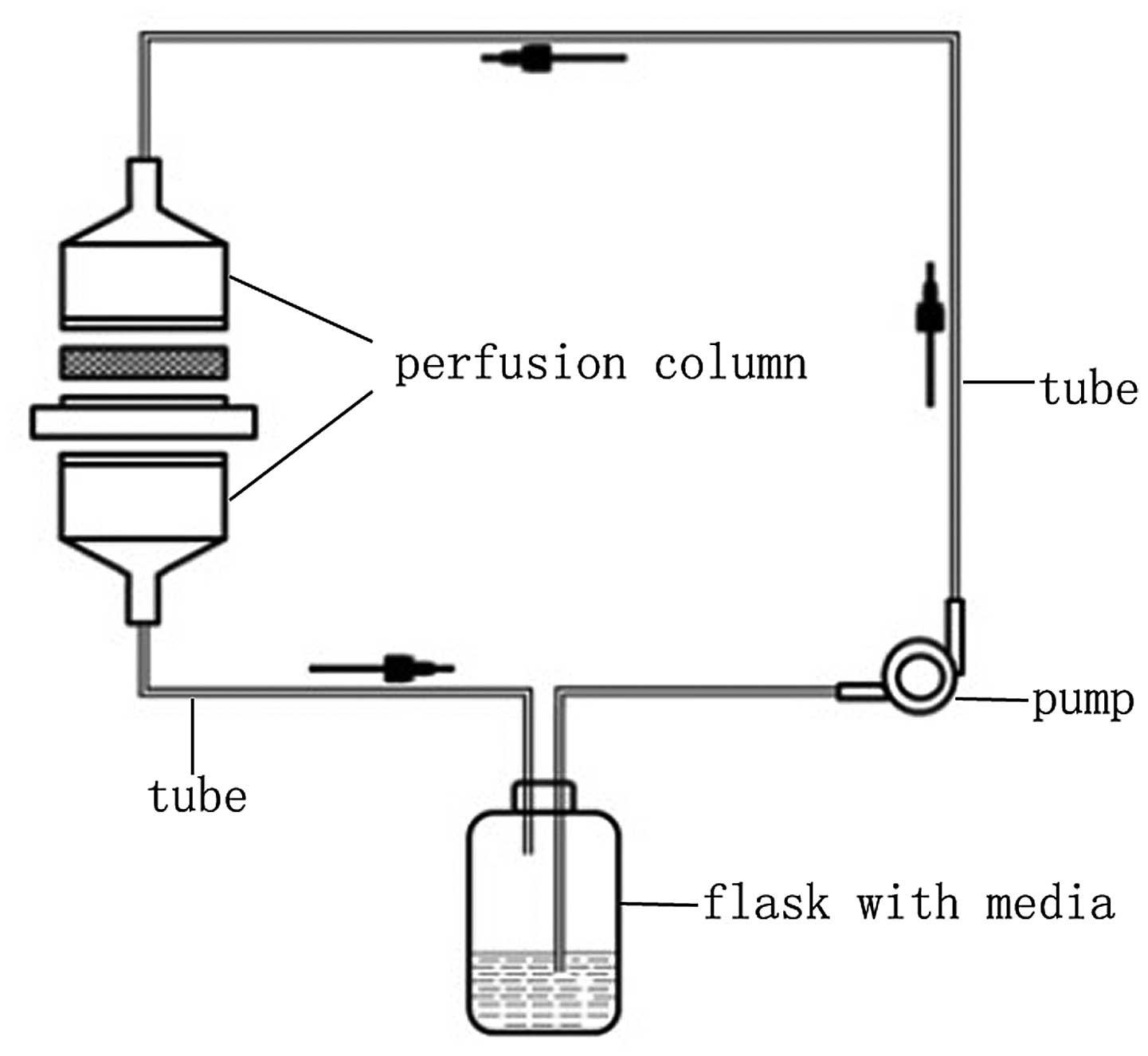
Perfusion bioreactor
The perfusion bioreactor (Fig. 1) was designed at the East China
University of Science and Technology (Shanghai, China). The system
consisted of a peristaltic pump (Masterflex peristaltic pump;
Cole-Parmer, Vernon Hills, IL, USA), a perfusion column (including
the ring and sample tank), an air filter, two silicone tubes, a 3D
interlinked connector and cell culture media. Two silicone tubes
were used to transport culture media between the perfusion column
and the flask, creating a circulation that simulated the internal
body environment (10).
Preparation and evaluation of the
PRP
PRP was prepared according to a two-step
centrifugation method. While the animals were under general
anesthesia, 5 ml blood was drawn from the central ear artery with a
10-ml sterilized syringe containing 1 ml sodium citrate, which was
used as an anticoagulant. Following homogenization, the mixture was
centrifuged at 3,500 × g for 6 min to separate the cells from the
serum components. Subsequently, the plasma and buffy layers were
collected and subjected to further centrifugation at 3,000 × g for
6 min. Following removal of the top layer, the lower part of the
solution was resuspended and designated as PRP. Next, the PRP was
mixed and homogenized with 1 ml coagulant, consisting of 1 ml
CaCl2 (10%) and 1,000 units thrombin. PRP gel, which had
a jelly-like composition, was subsequently harvested.
Tissue engineering bone building
Third generation BMSCs were prepared into a
suspension prior to addition into the β-TCP scaffold. Upon addition
of the suspension, the bracket was turned upside down to ensure
adhesion in all parts of the bracket surface. Next, 3 ml cell
culture media was added to each cultivation orifice plate and the
scaffold was placed in a 37°C and 5% CO2 incubator
overnight. The constructed bracket composites were then placed in
the bioreactor perfusion column and cultured for three weeks in
culture media with or without PRP, depending on the group
allocation. The entire system was placed in a 37°C and 5%
CO2 incubator for these three weeks, maintaining the
CO2 flow rate at 3.5 ml/min. The culture medium was
changed every 2–3 days, when the glucose content of the medium was
depleted. The adhesion, proliferation and growth of the BMSCs in
the β-TCP scaffold were examined using scanning electron microscopy
(SEM; JSM-T300; JEOL-Technics Co. Tokyo, Japan). The bracket
composites were divided into five groups as follows: Group A, BMSCs
cultured with PRP in the bioreactor; group B, BMSCs cultured with
PRP without the use of the bioreactor; group C, BMSCs cultured in
the bioreactor without PRP; group D, BMSCs cultured without PRP and
the bioreactor; and group E, β-TCP scaffold only (used as a
negative control).
Composite implantation
Following anesthesia with pentobarbital sodium (1
ml/kg) injected to the ear vein, the rabbits were placed in a left
lateral position with head and limbs fixed. The skin on the right
side at waist level was longitudinally incised to expose the
superficial fascia. The BMSC composites were implanted between the
shallow and deep fascia under the right side of the rabbit’s waist
skin, while the animals were under general anesthesia. Next, the
composites were fixed with sterile sutures and their positions were
recorded. In the present experiment, each group had 10 composites
and they were planted into 10 rabbits one by one. There were five
groups in total, so each rabbit was planted with five composites
from five groups. The grouping of the transplants was similar to
that of a previous study (10).
The rabbits were humanly sacrificed three months following surgery,
and the composites with the surrounding tissue cells were removed
in order to perform quantitative reverse transcription-polymerase
chain reaction (RT-PCR).
Detecting the expression levels of
alkaline phosphatase (ALP) and bone γ-carboxyglutamate protein 2
(BGLAP2) using quantitative RT-PCR
The primer sequences used in the quantitative RT-PCR
for rabbit ALP were 5′-TGT GCGGGGTCAAGGCTAAC-3′ (forward) and
5′-GGCGTC CGAGTACCAGTTGC-3′ (reverse), while for rabbit BGLAP2, the
primers used were 5′-CTCCTTACCCGGATCCCCTG-3′ (forward) and
5′-GTAGAAGCGCTGGTAGGCGT-3′ (reverse). Total RNA was isolated using
TRIzol® reagent (Ambion®, Invitrogen Life
Technologies) according to the manufacturer’s instructions. The
cDNA sample was generated using the RevertAid First Strand cDNA
Synthesis kit (Thermo Scientific, Waltham, MA, USA). Quantitative
RT-PCR was performed using SYBR Green Realtime PCR Master mix
(Toyobo, Osaka, Japan) in the Applied Biosystems AB7500 real-time
PCR system (Applied Biosystems®, Invitrogen Life
Technologies). The amplification was carried out under the
following conditions: 95°C for 60 sec followed by 40 cycles at 95°C
for 15 sec and 60°C for 60 sec.
Statistical analysis
Quantitative RT-PCR experiments were performed in
triplicate. The data are presented as the mean ± standard deviation
and were analyzed using SPSS 18.0 statistical software (SPSS, Inc.,
Chicago, IL, USA). One-way analysis of variance or t-tests were
used to compare the differences between groups, where P<0.05 was
considered to indicate a statistically significant difference.
Results
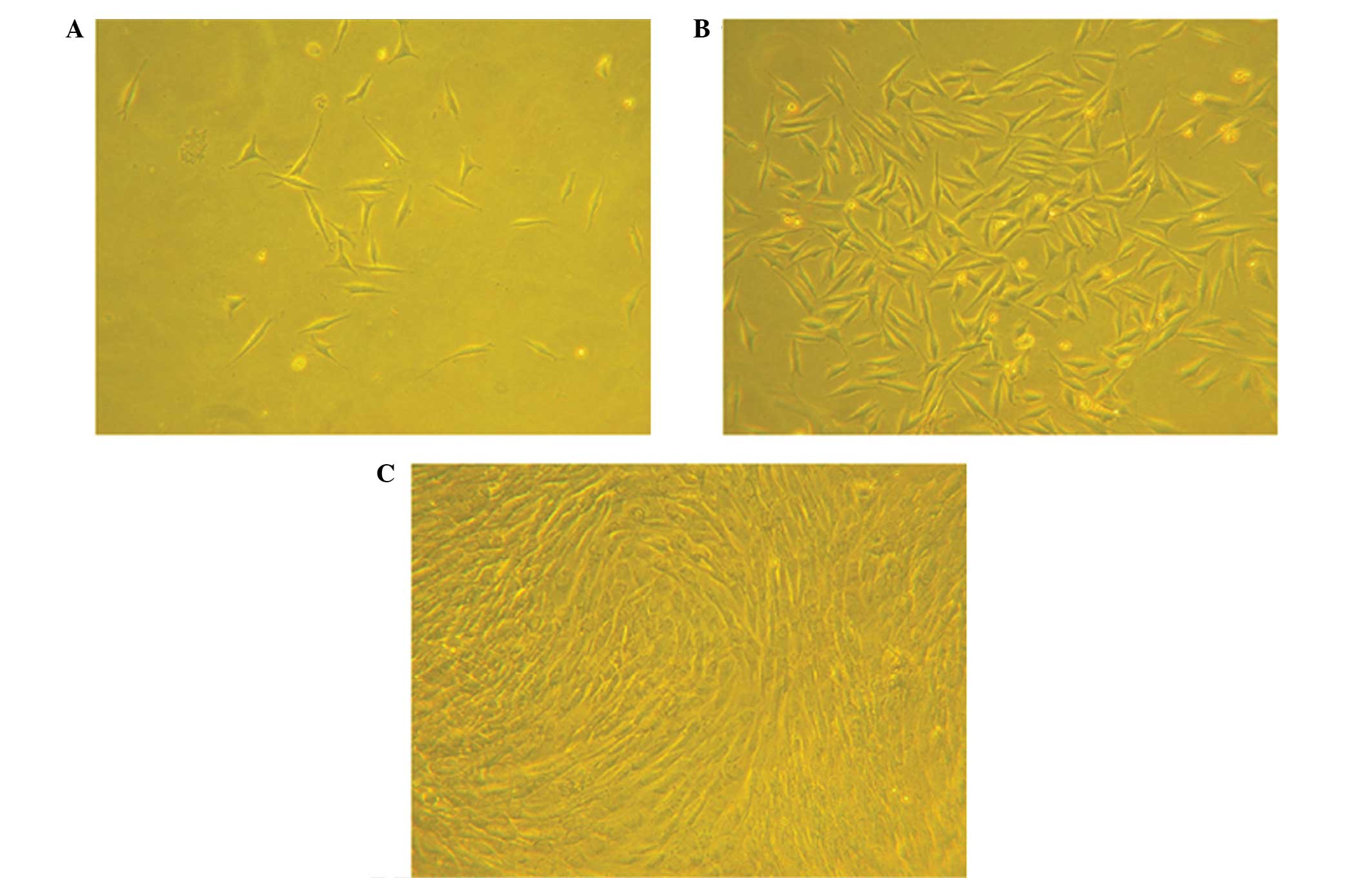
Cell culture observation
Observation of the BMSCs using an inverted
phase-contrast microscope revealed a small amount of cell adhesion
at day three following seeding. The cells were found to have a
spindle-shape at this time. Cell colonies were formed after the
primary cells had been cultured for seven days, and the cells were
gathered into swirl-shaped colonies at day seven following passage
(Fig. 2).
Identification of BMSCs
Flow cytometric analysis of BMSCs was used to
determine the gene expression profiles of cell passage 3. The
results indicated that 2% of the cells were CD34-positive and 99.8%
were CD44-positive.
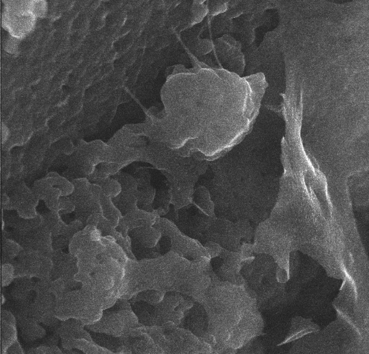
Composite examination with SEM
SEM revealed good adhesion and distribution of BMSCs
on the β-TCP scaffold. In addition, extracellular matrix secretion
was observed on the scaffold. Favorable cell proliferation and
stretch were also observed using SEM, indicating that the β-TCP
scaffold had a good affinity for the BMSCs (Fig. 3).
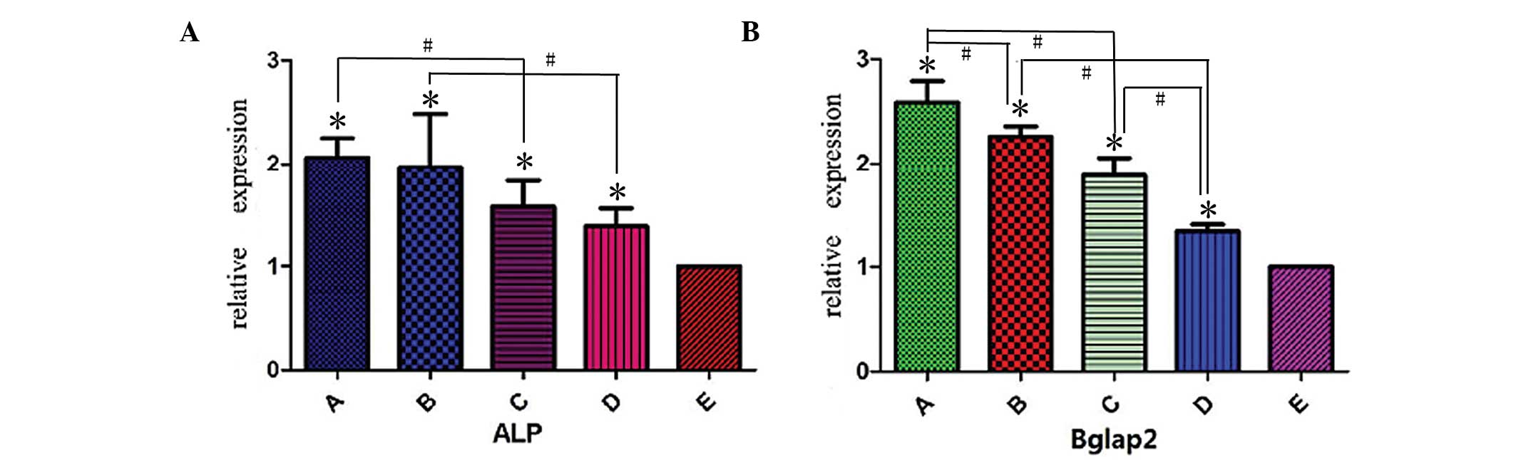
Expression levels of osteogenic
differentiation markers
The expression levels of the osteogenic
differentiation markers, ALP and BGLAP2, were detected using
quantitative RT-PCR (Fig. 4). The
expression levels of the markers were found to be much higher in
the experimental groups (groups A-D) compared with the negative
control group (group E; P<0.05). In addition, the expression
levels of ALP and BGLAP2 were higher in group A compared with group
C (P<0.05), and higher in group B compared with group D
(P<0.05), indicating the promoting effect of PRP. In addition,
the importance of BMSCs was demonstrated by the increased
expression levels of the two markers in group D, as compared with
group E (P<0.05). The expression level of BGLAP2 was found to be
higher in groups A and C compared with groups B and D (P<0.05),
respectively. Furthermore, the expression level of ALP was slightly
higher in groups A and C compared with groups B and D,
respectively; however, the differences were not found to be
statistically significant (group A vs. B, t=0.177, P>0.05; group
C vs. D, t=0.623, P>0.05). Therefore, the promoting effect of
the perfusion bioreactor was demonstrated.
Discussion
Research into the treatment of bone defects has
always received much attention; however, in recent years, the rapid
development of tissue engineering technology has provided a novel
approach for the treatment of bone defects. The concept of tissue
engineering involves the use of seed cells, growth factors and
scaffolds (11). In addition, the
culture of cells is essential in the development of
tissue-engineered bones.
In the present study, bracket composites were
constructed using BMSCs and a β-TCP scaffold. BMSCs were used as
the seed cells, while PRP was used as a growth factor source. The
composites were implanted into rabbits and the expression levels of
bone formation markers were analyzed using quantitative RT-PCR.
Alkaline phosphatase (ALP) and bone γ-carboxyglutamate protein 2
(BGLAP2) were used as indicators of cell osteogenesis
differentiation. The activity of ALP is an important index for the
evaluation of osteogenesis differentiation. In addition, ALP is an
iconic enzyme of mature osteoblasts; thus, plays a critical role in
the in vitro calcification process. Quantitative detection
of the ALP concentration is frequently used for in vitro
osteogenesis experiments as a conventional symbol of early
osteoblast differentiation (12).
BGLAP2 mainly appears in a mineralized formation of the cells as a
sign of osteoblast maturation (13). Furthermore, the expression levels
of ALP and BGLAP2 reflect the process of bone formation. As the
quantitative RT-PCR results indicated, β-TCP scaffolds, BMSCs, PRP
and the perfusion bioreactor had a significantly positive effect on
bone formation.
BMSCs are derived from the mesoderm and ectoderm at
an early developmental stage, and were first identified by
Fridenshteĭn (14). BMSCs possess
a multidirectional differentiation potential and can differentiate
into osteoblasts, adipocytes, tenocytes and marrow stroma in
vivo and in vitro, when induced under specific
conditions. In addition, BMSCs preserve a multidirectional
differentiation potential after continuous subculture and
cryopreservation; thus, are ideal as seed cells (15).
PRP consists of concentrated platelets in a small
volume of plasma. Upon activation by an agonist, the platelets in
the PRP release the inflammatory and growth factors that are
enclosed in their granules. PRP was first used in bone
reconstruction therapy in the late 1990s; however, the role of PRP
as a promoter of bone healing remains controversial (16). Previous studies have endorsed the
ability of PRP to promote bone healing (17,18).
By contrast, Kasten et al (19) investigated the effect of PRP on a
critical-size diaphyseal radius defect in a rabbit model and
observed that the combination of BMSCs and PRP had no additional
effect on bone healing. However, the bone injury resulted from
radial osteotomy, which may provide an explanation for the
aforementioned observation, since growth factors and host
precursors are known to be released following bone injury (20). The effect may have stimulated the
BMSCs, not allowing a further increase due to PRP. In the present
study, such influencing factors were excluded since the expression
levels of new bone formation markers were detected using PCR,
rather than observing the bone defect healing. Therefore, osteotomy
was not performed and growth factors were not released due to bone
injury. The data demonstrated that the addition of PRP promoted the
bone formation process.
Scaffolds play a critical role in the practical
realization of bone tissue engineering. In the current study, a
porous bioceramic 3D scaffold made of β-TCP was used due to its
favorable mechanical strength, high parity ratio, adjustable
biodegradation rate and easy process and molding. β-TCP provides a
3D space, which is more conducive for cells to adhere, adapt to
shear force and improve the unit expansion rate, promoting bone
differentiation. In addition, β-TCP gradually releases bone
induction growth factors in the process of cell culture due to the
material characteristics (21).
SEM demonstrated the good adhesion and distribution of BMSCs on the
β-TCP scaffold, as well as extracellular matrix secretion.
Favorable cell proliferation and stretch were also observed using
SEM, indicating that the β-TCP scaffold has a good affinity for
BMSCs.
Cell behavior is influenced by shear stress, tension
stimulation and hydrostatic pressure stimulation. The perfusion
bioreactor used in the present study applied continuous mechanical
stimulation to the BMSCs with mediated fluid. In addition, the
bioreactor provided the shear stress, tension and hydrostatic
pressure stimulation required to simulate a stress environment
in vivo (22,23). A previous study demonstrated that
fluid shear stress has the most significant effect on cellular
activity in the process of perfusion culture (24). Furthermore, the perfusion
bioreactor system enhanced the ability to dynamically monitor
stress. A 3D cultivation mode was established using the β-TCP
scaffold and bioreactor, which exhibited a number of advantages
over the traditional two-dimensional cultivation mode. The system
improved the distribution of nutrients within the scaffold, while
an in vivo environment was mimicked by providing mechanical
stimulation with floating culture media. In addition, a previous
study demonstrated that a contact inhibition effect appears when
cells are in close contact at a certain stage of cell proliferation
in the cell culture, which may inhibit the growth and proliferation
of cells (25). Therefore, the 3D
cultivation mode of the current study provided a 3D growth
environment that promoted cell adhesion, growth and
differentiation, reducing the inhibition of contact between the
cells. The results of the present study demonstrated that the use
of the bioreactor produced a better promotional effect on the
expression of BGLAP2, as compared with ALP, during the osteogenetic
differentiation process of the BMSCs, which should be addressed in
future studies.
In summary, BMSCs, PRP, the β-TCP scaffold and the
bioreactor had a positive effect on bone formation. BMSCs were
found to be favorable seeding cells for tissue engineering. In
addition, PRP was found to promote new bone formation through the
combined action of the growth factors released, while the β-TCP
scaffold was demonstrated to be suitable for BMSC adhesion and
proliferation. Finally, the perfusion bioreactor provided a 3D
culture mode that promoted cell adhesion, growth and
differentiation; thus, it promoted bone formation. The construction
of β-TCP scaffold composites using the 3D-bioreactor and PRP with
BMSCs is an effective method of building tissue-engineered bones
for bone formation. However, further comparative experiments are
required to investigate the best choice of cells, growth factors,
scaffolds and culture environment to build tissue-engineered
bones.
Acknowledgements
The study was supported by a grant from the National
Natural Science Foundation of China (no. 81271966).
References
|
1
|
Vacanti CA and Upton J: Tissue-engineered
morphogenesis of cartilage and bone by means of cell
transplantation using synthetic biodegradable polymer matrices.
Clin Plast Surg. 21:445–462. 1994.PubMed/NCBI
|
|
2
|
Kasten P, Vogel J, Geiger F, et al: The
effect of platelet-rich plasma on healing in critical-size
long-bone defects. Biomaterials. 29:3983–3992. 2008. View Article : Google Scholar : PubMed/NCBI
|
|
3
|
Yazdani SO, Pedram M, Hafizi M, et al: A
comparison between neurally induced bone marrow derived mesenchymal
stem cells and olfactory ensheathing glial cells to repair spinal
cord injuries in rat. Tissue Cell. 44:205–213. 2012. View Article : Google Scholar : PubMed/NCBI
|
|
4
|
Parsons P, Butcher A, Hesselden K, et al:
Platelet-rich concentrate supports human mesenchymal stem cell
proliferation, bone morphogenetic protein-2 messenger RNA
expression, alkaline phosphatase activity, and bone formation in
vitro: a mode of action to enhance bone repair. J Orthop Trauma.
22:595–604. 2008. View Article : Google Scholar : PubMed/NCBI
|
|
5
|
Park EJ, Kim ES, et al: Improved bone
healing by angiogenic factor-enriched platelet-rich plasma and its
synergistic enhancement by bone morphogenetic protein-2. Int J Oral
Maxillofac Implants. 23:818–826. 2008.PubMed/NCBI
|
|
6
|
Oreffo RO, Driessens FC, Planell JA and
Triffitt JT: Growth and differentiation of human bone marrow
osteoprogenitors on novel calcium phosphate cements. Biomaterials.
19:1845–1854. 1998. View Article : Google Scholar : PubMed/NCBI
|
|
7
|
Sun S, Ren Q, Wang D, et al: Repairing
cartilage defects using chondrocyte and osteoblast composites
developed using a bioreactor. Chin Med J (Engl). 124:758–763.
2011.
|
|
8
|
Campagnoli C, Roberts IA, Kumar S, et al:
Identification of mesenchymal stem/progenitor cells in human
first-trimester fetal blood, liver, and bone marrow. Blood.
98:2396–2402. 2001. View Article : Google Scholar : PubMed/NCBI
|
|
9
|
Mareschi K, Ferrero I, Rustichelli D, et
al: Expansion of mesenchymal stem cell isolated from pediatric and
adult donor bone marrow. J Cell Biochem. 97:744–754. 2006.
View Article : Google Scholar
|
|
10
|
Wang D, Jiang H, Wang S, et al:
Construction of tissue-engineered bone using a bioreactor and
platelet-rich plasma. Exp Ther Med. 8:413–418. 2014. View Article : Google Scholar : PubMed/NCBI
|
|
11
|
Liu Y, Zhou Y, Feng H, Ma GE and Ni Y:
Injectable tissue-engineered bone composed of human adipose-derived
stromal cells and platelet-rich plasma. Biomaterials. 29:3338–3345.
2008. View Article : Google Scholar : PubMed/NCBI
|
|
12
|
Alborzi A, Mac K, Glackin CA, Murray SS
and Zernik JH: Endochondral and intramembranous fetal bone
development: osteoblastic cell proliferation, and expression of
alkaline phosphatase, m-twist, and histone H4. J Craniofac Gent Dev
Biol. 6:94–106. 1996.
|
|
13
|
Duan Z, Zheng Q and Guo X: Dose-dependence
of bone morphogenetic protein 2-derived peptide on osteogenic
induction in marrow mesenchymal stem cells in vitro. Zhongguo Xiu
Fu Chong Jian Wai Ke Za Zhi. 21:1118–1122. 2007.(In Chinese).
PubMed/NCBI
|
|
14
|
Fridenshteĭn AIa: Stromal bone marrow
cells and the hematopoietic microenvironment. Arkh Patol. 44:3–11.
1982.(In Russian).
|
|
15
|
Van Damme A, Vanden Driessche T, Collen D
and Chuah MK: Bone marrow stromal cells as targets for gene
therapy. Curr Gene Ther. 2:195–209. 2002. View Article : Google Scholar : PubMed/NCBI
|
|
16
|
Intini G: The use of platelet-rich plasma
in bone reconstruction therapy. Biomaterials. 30:4956–4966. 2009.
View Article : Google Scholar : PubMed/NCBI
|
|
17
|
Kawase T, Okuda K, Wolff LF and Yoshie H:
Platelet-rich plasma-derived fibrin clot formation stimulates
collagen synthesis in periodontal ligament and osteoblastic cells
in vitro. J Periodontol. 74:858–864. 2003. View Article : Google Scholar : PubMed/NCBI
|
|
18
|
Yamada Y, Ueda M, Naiki T, et al:
Autogenous injectable bone for regeneration with mesenchymal stem
cells and platelet-rich plasma: tissue-engineered bone
regeneration. Tissue Eng. 10:955–964. 2004. View Article : Google Scholar : PubMed/NCBI
|
|
19
|
Kasten P, Vogel J, Geiger F, et al: The
effect of platelet-rich plasma on healing in critical-size
long-bone defects. Biomaterials. 29:3983–3992. 2008. View Article : Google Scholar : PubMed/NCBI
|
|
20
|
Lieberman JR, Daluiski A and Einhorn TA:
The role of growth factors in the repair of bone. Biology and
clinical applications. J Bone Joint Surg Am. 84-A:1032–1044.
2002.PubMed/NCBI
|
|
21
|
Santoni BG, Pluhar GE, Motta T and Wheeler
DL: Hollow calcium phosphate microcarriers for bone regeneration:
in vitro osteoproduction and ex vivo mechanical assessment. Biomed
Mater Eng. 17:277–289. 2007.PubMed/NCBI
|
|
22
|
Klein-Nulend J, van der Plas A, Semeins
CM, et al: Sensitivity of osteocytes to biomechanical stress in
vitro. FASEB J. 9:441–445. 1995.PubMed/NCBI
|
|
23
|
Owan I, Burr DB, Turner CH, et al:
Mechanotransduction in bone: osteoblasts are more responsive to
fluid forces than mechanical strain. Am J Physiol. 273:C810–C815.
1997.PubMed/NCBI
|
|
24
|
Bakker AD, Soejima K, Klein-Nulend J and
Burger EH: The production of nitric oxide and prostaglandin E(2) by
primary bone cells is shear stress dependent. J Biomech.
34:671–677. 2001. View Article : Google Scholar : PubMed/NCBI
|
|
25
|
Wang Y, Kim UJ, Blasioli DJ, et al: In
vitro cartilage tissue engineering with 3D porous aqueous-derived
silk scaffolds and mesenchymal stem cells. Biomaterials.
26:7082–7094. 2005. View Article : Google Scholar : PubMed/NCBI
|