Introduction
Hypoxia is a key adverse effect which occurs
following exposure to high altitude, leading to lung airway
inflammation and lung tissue injury. Interleukins (ILs) and
oxidative stress play an important role in this process.
IL-6 is produced by various immune and non-immune
cells, including vascular endothelial cells, monocytes/macrophages,
keratinocytes, fibroblasts, T lymphocytes and B lymphocytes. The
major biological function of IL-6 is to promote the synthesis of
acute proteins in the liver, as well as to induce the proliferation
and differentiation of, and antibody secretion from B lymphocytes.
During inflammation, IL-6 functions as an endogenous pyrogen and
enhances the tumor-killing activity of cytotoxic T cells and
natural killer (NK) cells (1).
IL-8 is a cytokine originating from various cells,
such as monocytes, fibroblasts, endothelial cells, hepatocytes,
epithelial cells and T lymphocytes. IL-8 has no species-specific
activity, and its functions include the chemotaxis and activation
of neutrophils, the chemotaxis of basophils, T lymphocytes and
other inflammatory cells, and it also plays a role in angiogenesis
(2).
IL-10 has multiple biological activities, and is
important to the functions of thymus cells, T cells, B cells, NK
cells, monocytes, macrophages, mast cells, neutrophils and
eosinophils. The physiological function of IL-10 is to inhibit
potent specific and non-specific immune reactions and subsequent
tissue injury, and to induce immune tolerance. IL-10 inhibits the
synthesis and release of pro-inflammatory mediators, as well as the
release of chemokines by neutrophils (3,4,19).
Superoxide dismutase (SOD) is an important
antioxidant that can effectively eliminate superoxide anion and
protect cells from oxidative damage. Ischemia and anoxia lead to an
imbalance in the metabolism of free radicals. With the accumulation
of free radicals, the increased production of lipid peroxides and a
significant reduction in SOD levels due to depletion, resistance to
oxidative damage is decreased, leading to cellular and even organ
damage (5). Malondialdehyde (MDA) is
the stable metabolite of lipid peroxidation. Therefore, the level
of MDA can reflect the level of the free radicals in tissue and
lipid peroxidation due to free radicals, and may indirectly reflect
cellular damage (6).
It has been reported that the expression levels of
the pro-inflammatory cytokines, IL-6, IL-8, and those of the
anti-inflammatory cytokine, IL-10, are increased following exposure
to hypoxia. The activity of the antioxidant SOD is decreased, and
the levels of the lipid peroxide, MDA, are increased (7–11).
However, the limitations of the above-mentioned is that they did
not perform combined, systemic and longitudinal observations and
comparisons.
To the best of our knowledge, research on ILs and on
the expression of oxidative stress markers in lung tissue and the
changes occurring in the levels of these markers at different
altitudes and exposure times is limited. Thus, in the present
study, we used exposure to low altitude conditions as the control
to detect the expression levels of ILs, SOD and MDA in rat lung
tissue and to observe the changes occurring in the levels of these
markers following exposure to middle and high altitude conditions
at different exposure times. We also aimed to determine the
signifiance of these changes. The findings of our study may provide
a theoretical basis for the prevention and treatment of acute and
chronic pulmonary injury in a hypoxic plateau environment.
Materials and methods
Animals
A group of 88 clean grade male Wistar rats were
randomly divided into 3 groups as follows: the control group [low
altitude (LA), Lanzhou, 1,500 m; n=8), the middle altitude group
(MA group, Xining, 2,260 m; n=40) and the high altitude group (HA
group, low pressure oxygen chamber, simulation of 5,000 m above sea
level; n=40). The MA and HA groups were further divided according
to the different observation times into the 1, 3, 7, 15, 30 day
groups (namely MA1, MA3, MA7, MA15 and MA30; HA1, HA3, HA7, HA15
and HA30), with 8 rats in each subgroup. The living conditions and
diet were basically the same for all animals apart from the
exposure to different altitudes. The control animals were
immediately collected in Lanzhou, China, while the other 2 groups
of experimental animals were transferred from Lanzhou to Xining
(China). The animals in the MA group were collected in Xining at
different exposure times and those in the HA group were placed in a
hypobaric chamber with a simulated altitude of 5,000 m above sea
level, and the animals were collected at the different specified
time points in the low pressure oxygen cabin. The animal
experimental procedures were approved by the Ethics Committee of
Qinghai Provincial People's Hospital (Xining, China).
Instruments and reagents
The low pressure oxygen chamber was obtained from
Guizhou Fenglei Aviation Armament Co., Ltd., (Anshun, China; model,
DYC-3000; volume, 8×3×3 m), the automatic blood cell analyzer was
from Shenzhen Mindray Bio-Medical Electronics Co. Ltd., (Shenzhen,
China), the microtome for paraffin embedding (Leica RM 2265) was
from (Leica Microsystems, Wetzlar, Germany), the inverted
microscope was obtained from Olympus (Tokyo, Japan), the constant
temperature water bath was purchased from (HH-S11-1 type; Beijing
Xinnuo Lihua Instrument Co., Ltd, Beijing, China), the iMark/xMark
instrument for measuring absorbance was from Bio-Rad (Hercules, CA,
USA), the IL-6, IL-8, IL-10 kits were purchased from USCN Life
Science Inc. Wuhan (Wuhan, China) and the SOD and MDA kits were
purchased from Nanjing Jiancheng Biological Co. (Jiangsu,
China).
Research methods
According to the different time points of the
experimental design, 5 ml venous blood were collected from the rats
in all the groups using EDTAK2 as an anticoagulant. An automatic
blood cell analyzer was used for the determination of the blood
hemoglobin (HGB) count, in strict accordance with the
manufacturer's instructions. The rats were anesthetized with 20%
urethane and the lung tissue was removed following dissection;
tissue from the middle lobe of the right lung was used for
pathological sections, and lung tissue morphology was observed
under a microscope. The remaining lung tissue was homogenized and
the levels of IL-6, IL-8, IL-10 levels were determined by
enzyme-linked immunosorbent assay (ELISA) in the homogenates. The
WST-1 Cell Proliferation Assay kit was used to detect SOD activity,
while the content of MDA was determined by total bile acids
colorimetric assay as described below:
Measurement of hemoglobin (HGB)
levels
Venous blood was taken from the rats and the HGB
count was measured using an automatic blood cell analyzer (BC-2300;
Mairui Biotec, Wuhan, China) following the manufacturer's
instructions.
Lung tissue morphological
analysis
The rats were anesthetized with 20% urethane and the
lung tissue was removed following dissection. A small section of
tissue from the middle lobe of the right lung was fixed in 4%
paraformaldehyde, routinely embedded in paraffin, sectioned, and
stained with hematoxylin and eosin (H&E), and lung tissue
morphology was observed under a microscope (Olympus CH-2; Olympus
Optical Co., Ltd., Tokyo, Japan).
Measurement of the levels of IL-6,
IL-8 and IL-10 by ELISA
A total of 500 µg lung tissue was homogenized by 1X
phosphate buffered saline (PBS). The total protein of rat lung
tissue was measured using a BCA Protein Assay kit (Pierce,
Springfield, IL, USA), and the levels of IL-6, IL-8 and IL-10 were
detected using a ELISA kit (USCN Life Science Inc., Los Angeles,
CA, USA) in the homogenates following the manufacturer's
instructions.
Measurement of SOD activity and MDA
content
The WST-1 Cell Proliferation Assay kit was used to
detect SOD activity, while the content of MDA was determined using
a total bile acids colorimetric assay in the homogenates according
to the manufacturer's instructions (Jiancheng Biotech Ltd.,
Nanjing, China).
H&E staining
The freshly acquired lung tissue was cut into small
sampes with a size of 1 mm3, which were then fixed in 4%
paraformaldehyde solution, dehydrated in a series concentration of
alcohol, washed with xylol and embedded in paraffin. The
paraffin-embedded tissue samples were then sectioned, and the
sections were washed with distilled water, and placed on a clean
and oil-free slide. They were then heated with a spirit lamp and
were then incubated at room temperature for 3–7 days. Subsequently,
the sections were dewaxed by dipping them first in xylol twice (for
15 min each), and then in 100% alcohol (twice), 95% alcohol, 75%
alcohol, 50% alcohol and distilled water (for 3–5 min eachy). The
sections were then stained in hematoxylin for 10–30 min and washed
with tap water for 15 min. The sections became blue in color after
washing. For color separation, the sections were decolored in 1%
HCl-alcohol (HCl: 70% alcohol = 1:100, v/v) until the sections
became light red. Subsequently, counterstaining was carried out
with 0.5% eosin-alcohol (eosin 0.5 g + 95% alcohol 100 ml) for 2–5
min. The ultrastructure (x40 and x400 magnification) of the lung
tissues was then observed under a microscope and the differences
between these samples were analyzed.
Statistical analysis
Statistical analysis was performed using SPSS 17.0
software. Data are presented as the means ± SD; comparisons between
multiple groups was made by one-way ANOVA and comparisons 2 groups
were made using the least significant difference (LSD) test. A
value of P<0.05 was considered to indicate a statistically
significant difference.
Results
Animal model
The body weight measurements of the rats and the HGB
count in the different groups are presented in Table I. The activity level and food intake
were decreased in the rats in the HA group following entry into the
low oxygen chamber. Consequently, the rate of increase in body
weight was lower than the other groups. The HGB count in the HA
group gradually increased with the passing of time, and was
significantly higher compared to the control group and MA group.
The difference was statistically significant (P<0.05),
indicating that the model of high-altitude hypoxia had been
successfully created by simulation at an altitude of 5,000 m above
sea level in a low pressure oxygen cabin. Thus, this model may be
used for future research.
 | Table I.Changes in body weight and HGB count
in the rats from the different groups. |
Table I.
Changes in body weight and HGB count
in the rats from the different groups.
| Groups | Body weight (g) | HGB (g/l) |
|---|
| LA |
159.25±3.73 |
137.25±18.22 |
| MA1 |
160.00±4.81 |
136.25±5.39 |
| MA3 |
175.63±5.95 |
138.75±9.71 |
| MA7 |
189.75±15.17 |
137.50±14.65 |
| MA15 |
228.38±7.09 |
141.13±7.85 |
| MA30 |
316.50±13.38 |
138.38±14.15 |
| HA1 |
163.50±7.27 |
141.00±8.57 |
| HA3 |
169.13±3.60 |
157.50±11.31a,b |
| HA7 |
188.00±3.02 |
187.13±16.78a–c |
| HA15 |
209.75±9.38 |
213.75±32.06a–d |
| HA30 |
237.50±12.11 |
236.00±10.86a–e |
Levels of IL-6/IL-8/IL-10 in the lung
tissue of rats in the different groups
As regards the MA group, the levels of
IL-6/IL-8/IL-10 were higher in the MA1 subgroup compared to the
control group (P<0.05), whereas no significant differences in
these levels were observed between the other MA subgroups and the
control group (P>0.05; Table
II).
| Table II.Levels of IL-6/IL-8/IL-10 in the lung
tissue of rats from the different groups. |
Table II.
Levels of IL-6/IL-8/IL-10 in the lung
tissue of rats from the different groups.
| Groups | IL-6 (pg/ml) | IL-8 (pg/ml) | IL-10 (pg/ml) |
|---|
| LA |
30.09±2.77 |
205.53±18.47 |
173.95±7.69 |
| MA1 |
53.65±3.97a |
285.10±29.36a |
199.50±38.24a |
| MA3 |
32.73±2.62 |
207.81±23.91 |
172.03±35.69 |
| MA7 |
33.04±5.40 |
205.03±28.40 |
170.90±31.28 |
| MA15 |
32.03±5.73 |
203.08±24.09 |
170.53±15.83 |
| MA30 |
31.36±2.63 |
206.85±15.41 |
174.48±27.31 |
| HA1 |
65.27±6.82a |
404.46±40.91a |
134.87±32.38a |
| HA3 |
58.76±6.91a,b |
383.69±19.68a |
113.74±19.96a |
| HA7 |
51.13±5.72a–c |
330.96±26.09a–c |
97.88±11.68a,b |
| HA15 |
43.37±5.34a–d |
284.16±18.09a–d |
99.17±3.73a,b |
| HA30 |
37.63±3.52a–e |
256.15±19.87a–e |
96.91±14.55a,b |
As regards the HA group, the levels of IL-6/IL-8 in
all the HA subgroups were higher compared to those of the control
group (P<0.05); these levels decreased with the passing of time,
but were still higher than those of the control group (P<0.05).
However, the levels of IL-10 decreased with the passing of time,
and were lower compared to those of control group and the MA group
(P<0.05; Table II).
Lung tissue SOD activity and MDA
levels in the rats in the different groups
As regards the MA group, no significant differences
in SOD activity and the MDA content were observed between all the
MA subgroups and the control group (Table III).
| Table III.Change in SOD and MDA levels in the
lung tissue of rats from the different groups. |
Table III.
Change in SOD and MDA levels in the
lung tissue of rats from the different groups.
| Groups | SOD (u/mgprot) | MDA
(nmol/mgprot) |
|---|
| LA |
439.85±33.80 |
1.808±0.240 |
| MA1 |
437.89±23.35 |
1.824±0.251 |
| MA3 |
438.86±26.74 |
1.811±0.205 |
| MA7 |
442.79±28.80 |
1.820±0.197 |
| MA15 |
441.70±15.21 |
1.816±0.206 |
| MA30 |
440.74±36.42 |
1.813±0.354 |
| HA1 |
554.07±24.59a |
2.386±0.345a |
| HA3 |
486.78±18.78a,b |
2.993±0.340a,b |
| HA7 |
403.71±16.57a–c |
3.441±0.303a–c |
| HA15 |
341.98±17.89a–d |
4.033±0.876a–d |
| HA30 |
295.98±11.08a–e |
4.661±0.742a–e |
As regards the HA group, when compared to the
control group and with the passing of time, SOD activity decreased,
and the MDA content gradually increased (P<0.05; Table III).
Pathological observation of the lung
tissue sections from the rats in the different groups
The lung tissue sections obtained from the rats in
the different groups were stained with hematoxylin and eosin, and
the morphological observations were observed under a microscope.
Images of the pathological sections are shown in Fig. 1.
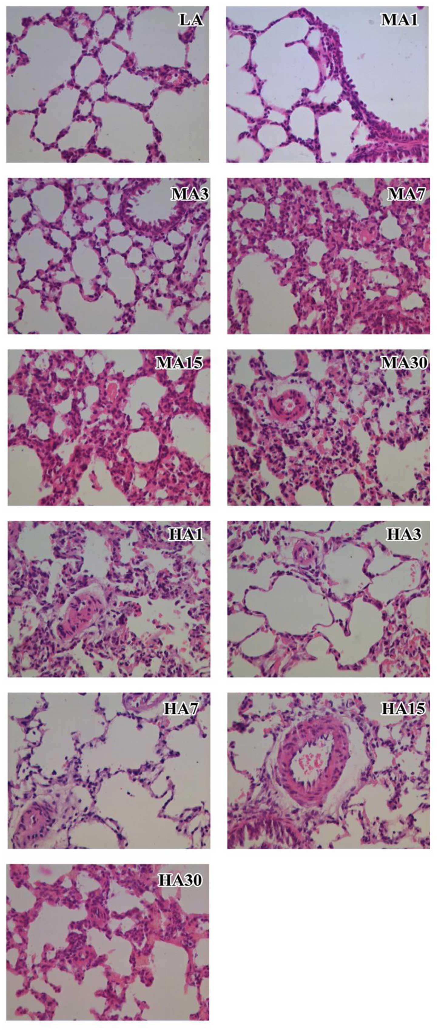 | Figure 1.Images of the pathological observation
of the lung tissues of the rats in the different groups
(magnification, x40). See text for a detail description of the
results in each group. LA, low altitude; HA, MA, middle altitude;
HA, high altitude; MA and HA1, 2, 7, 15, 30 represent the MA and HA
subgroups at 1, 3, 7, 15 and 30 days, respectively. |
As regards the control group, the longitudinal plica
mucosa in the terminal bronchioles was not obvious. In the
respiratory bronchioles, the epithelial mucosa consisted of a
simple columnar epithelium or a cuboidal epithelium, and the
alveolar wall consisted of flat type I alveolar cells and cuboidal
alveolar type II cells. The alveolar septum was rich in
capillaries, elastic fibers and macrophages, and the structure of
the large and small pulmonary arteries was normal.
As regards the MA group, the longitudinal plica
mucosa in the terminal bronchioles was evident. In the respiratory
bronchioles, the epithelial mucosa consisted of a simple columnar
epithelium or a simple cuboidal epithelium, and with the passing of
time, due to capillary dilatation and congestion, the alveolar wall
widened in part of the region, with focal alveolar hemorrhage,
light thickening of the pulmonary artery and focal emphysema being
observed.
As regards the HA group, the longitudinal plica
mucosa in the terminal bronchioles was not obvious. In the
respiratory bronchioles, the epithelial mucosa consisted of a
columnar epithelium or a simple cuboidal epithelium, and due to
capillary dilatation and congestion, the alveolar wall had widened
in part of the region, with focal alveolar hemorrhage being
observed. Focal alveolar hemorrhage and massive hemorrhage of the
individual pulmonary tissue were also observed. The pulmonary small
artery had slightly thickened. The wall of the larger arteriolar
vessels showed focal thickening and there were a few lymphocytic
infiltrations around the small blood vessels. With the passing of
the time of exposure to hypobaric hypoxic conditions, damage to the
structure of the lung tissue was observed, which gradually became
more severe, as noted under a microscope.
Discussion
The results of this study demonstrated that the
levels of IL-6/IL-8/IL-10 in the MA1 subgroup of the MA group were
higher than those of the control group (P<0.05); however, no
significant differences in these levels were observed among the
other MA subgroups and the control group (P>0.05). The levels of
IL-6/IL-8 in all the HA subgroups were higher compared to those of
the control group (P<0.05), and even though these levels
decreased with the passing of time, they were still higher than
those of the control group (P<0.05). The levels of IL-10
decreased with the passing of time in the HA group, and were lower
than those in the control group and the MA group (P<0.05).
The biological effects of IL-6 involve the promotion
of B lymphocyte proliferation and differentiation and the secretion
of antibodies, functioning as endogenous pyrogens participating in
inflammatory reactions, and enhancing the antitumor activity of
cytotoxic T cells and natural killer cells (12). As an inflammatory cytokine, IL-6 has
a number of biological effects, participating in the vascular
endothelial inflammatory response, but also stimulating the
production of inflammatory cytokines and increasing vascular
inflammatory reactions (13,14). IL-8 has strong chemotactic properties
and activates neutrophil chemotaxis, as well as the chemotaxis of
basophilic granulocytes, T lymphocytes and other inflammatory cells
for angiogenesis (15,16). On the other hand, IL-10 has an
inhibitory effect on immune cell functions, and can prevent or
inhibit specific and non-specific immune responses and the
resulting damage, thus contributing to the induction of immune
tolerance (17–19). Therefore, IL-6 and IL-8 are both
pro-inflammatory cytokines, and participate in the inflammatory
response and angiogenesis. IL-10 is an inhibitory inflammation
factor, and can reduce tissue damage and promote the process immune
tolerance.
In this study, since the levels of IL-6/IL-8/IL-10
in the MA1 group were higher than those of the control group
(P<0.05), and no significant differences in these levels were
observed among the other MA subgroups and the control group, this
indicates that exposure to mid-altitude environment increases the
inflammatory reaction in the lung tissue of rats. As the
anti-inflammatory effect took place, the levels of IL-6/IL-8/IL-10
quickly returned to levels close to the basal (control) levels
after 3 days, indicating that the altitude conditions did not have
a long-term effect on airway inflammatory reactions in the lung
tissue. In the HA group however, with the passing of time, the
IL-6/IL-8 levels gradually decreased following a sudden increase on
day 1, but were still higher than those of the control group,
indicating that the inflammatory reaction resulting from hypoxia
decreased with the passing of time; this indicates the adaptation
process to hypoxic conditions. However, the inflammation state
induced by exposure to hypoxia persisted, and injury due to
inflammation was sustained. The level of IL-10 decreased under
hypoxic conditions, indicating that hypoxia leads to a decrease in
the ability to inhibit immunity and inflammation. Therefore, IL-10
cannot prevent and suppress strong specific and non-specific immune
responses and the resulting damage, as well as reduce immune
tolerance. This explains why a high altitude hypoxic environment
causes airway inflammation of the lung tissue and injury to a
certain extent.
The results of this study demonstrated that there
were no significant differences in SOD activity and the MDA content
between all MA subgroups and the control group. With the passing of
time, SOD activity decreased and the MDA content gradually
increased in all the HA subgroups (P<0.05). SOD is an
antioxidant and can protect the cells of the body from oxidative
damage (5,20). Ischemia and hypoxia inhibit SOD
activity, weakening its antioxidant abilities.
MDA is a stable metabolite of lipid peroxidation,
and reflects the free radical content and the degree of lipid
peroxidation, and indirectly reflects the degree of cell injury
(6,21). The results of this study demonstrated
that there were no differences in SOD activity and the MDA content
between the MA group and the control group, suggesting that the
antioxidant activity and lipid peroxidation in the lung tissue were
not altered, and there is no obvious cell injury in the lung
tissue. However, in the HA group, the activity of SOD in all the
subgroups was higher than that of the control group, and with the
passing of time it gradually decreased to levels lower than those
of the control group. This indicates that following initially entry
into a high altitude environment, the lung tissue antioxidant
capacity is increased, so that the cells of the body become immune
to oxidative damage. However, with prolonged exposure to low
oxygen, the increasing consumption of SOD causes rapid oxidative
damage and an ongoing development of oxidative cellular damage. In
the HA group, since the MDA content gradually increased with the
passing of time, this further confirmed that the content of free
radicals in the lung tissue increased, resulting in a greater
degree of lipid peroxidation and severe damage to lung tissue. This
explains why a high altitude hypoxic environment causes oxidative
damage to lung tissue to a certain extent.
Subsequently, through the observation of the
pathomorphism of the lung tissue of the rats in each group, we
found that compared to the low altitude control group, there was a
gradual and mild damage to the lung tissue of the rats in the MA
group with the prolongation of the exposure time, but no
inflammatory cell infiltration. However, in the HA group, with the
extension of the time of exposure to hypoxia, lung tissue damage in
the rats was aggravated, which is consistent with the changes in
interleukin, SOD and MDA levels in lung tissue.
Therefore, the results of this study suggest that
high altitude hypoxia induces lung inflammation and progressive
oxidative damage which causes a serious degree of damage to lung
tissue and the weakening of the inhibition of the inflammatory
response.
Acknowledgements
This study was funded by Foundation Research Project
of Qinghai Science and Technology Department (no. 2013-Z-741).
References
|
1
|
Opal SM and DePalo VA: Anti-inflammatory
cytokines. Chest. 117:1162–1172. 2000. View Article : Google Scholar : PubMed/NCBI
|
|
2
|
Li A, Dubey S, Varney ML, Dave BJ and
Singh RK: IL-8 directly enhanced endothelial cell survival,
proliferation, and matrix metalloproteinases production and
regulated angiogenesis. J Immunol. 170:3369–3376. 2003. View Article : Google Scholar : PubMed/NCBI
|
|
3
|
Hilgenberg E, Shen P, Dang VD, Ries S,
Sakwa I and Fillatreau S: Interleukin-10-producing B cells and the
regulation of immunity. Curr Top Microbiol Immunol. 380:69–92.
2014.PubMed/NCBI
|
|
4
|
Hofmann SR, Rösen-Wolff A, Tsokos GC and
Hedrich CM: Biological properties and regulation of IL-10 related
cytokines and their contribution to autoimmune disease and tissue
injury. Clin Immunol. 143:116–127. 2012. View Article : Google Scholar : PubMed/NCBI
|
|
5
|
Seyhan N and Canseven AG: In vivo effects
of ELF MFs on collagen synthesis, free radical processes, natural
antioxidant system, respiratory burst system, immune system
activities, and electrolytes in the skin, plasma, spleen, lung,
kidney, and brain tissues. Electromagn Biol Med. 25:291–305. 2006.
View Article : Google Scholar : PubMed/NCBI
|
|
6
|
Niki E: Lipid peroxidation products as
oxidative stress biomarkers. Biofactors. 34:171–180. 2008.
View Article : Google Scholar : PubMed/NCBI
|
|
7
|
Sarada S, Himadri P, Mishra C, Geetali P,
Ram MS and Ilavazhagan G: Role of oxidative stress and NFkB in
hypoxia-induced pulmonary edema. Exp Biol Med (Maywood).
233:1088–1098. 2008. View Article : Google Scholar : PubMed/NCBI
|
|
8
|
Kubo K, Hanaoka M, Yamaguchi S, Hayano T,
Hayasaka M, Koizumi T, Fujimoto K, Kobayashi T and Honda T:
Cytokines in bronchoalveolar lavage fluid in patients with high
altitude pulmonary oedema at moderate altitude in Japan. Thorax.
51:739–742. 1996. View Article : Google Scholar : PubMed/NCBI
|
|
9
|
Heffner JE and Repine JE: Pulmonary
strategies of antioxidant defense. Am Rev Respir Dis. 140:531–554.
1989. View Article : Google Scholar : PubMed/NCBI
|
|
10
|
Bulger EM and Maier RV: Antioxidants in
critical illness. Arch Surg. 136:1201–1207. 2001. View Article : Google Scholar : PubMed/NCBI
|
|
11
|
Nakanishi K, Tajima F, Nakamura A, Yagura
S, Ookawara T, Yamashita H, Suzuki K, Taniguchi N and Ohno H:
Effects of hypobaric hypoxia on antioxidant enzymes in rats. J
Physiol. 489:869–876. 1995. View Article : Google Scholar : PubMed/NCBI
|
|
12
|
Diehl S and Rincón M: The two faces of
IL-6 on Th1/Th2 differentiation. Mol Immunol. 39:531–536. 2002.
View Article : Google Scholar : PubMed/NCBI
|
|
13
|
Packard RR and Libby P: Inflammation in
atherosclerosis: From vascular biology to biomarker discovery and
risk prediction. Clin Chem. 54:24–38. 2008. View Article : Google Scholar : PubMed/NCBI
|
|
14
|
Akira S, Taga T and Kishimoto T:
Interleukin-6 in biology and medicine. Adv Immunol. 54:1–78. 1993.
View Article : Google Scholar : PubMed/NCBI
|
|
15
|
Remick DG: Interleukin-8. Crit Care Med.
33 (Suppl 12):S466–S467. 2005. View Article : Google Scholar : PubMed/NCBI
|
|
16
|
Waugh DJ and Wilson C: The interleukin-8
pathway in cancer. Clin Cancer Res. 14:6735–6741. 2008. View Article : Google Scholar : PubMed/NCBI
|
|
17
|
Delogu G, Antonucci A, Signore M,
Marandola M, Tellan G and Ippoliti F: Plasma levels of IL-10 and
nitric oxide under two different anaesthesia regimens. Eur J
Anaesthesiol. 22:462–466. 2005. View Article : Google Scholar : PubMed/NCBI
|
|
18
|
Miller BJ, Buckley P, Seabolt W, Mellor A
and Kirkpatrick B: Meta-analysis of cytokine alterations in
schizophrenia: Clinical status and antipsychotic effects. Biol
Psychiatry. 70:663–671. 2011. View Article : Google Scholar : PubMed/NCBI
|
|
19
|
Moore KW, de Waal Malefyt R, Coffman RL
and O'Garra A: Interleukin-10 and the interleukin-10 receptor. Annu
Rev Immunol. 19:683–765. 2001. View Article : Google Scholar : PubMed/NCBI
|
|
20
|
Suzer T, Coskun E, Demir S and Tahta K:
Lipid peroxidation and glutathione levels after cortical injection
of ferric chloride in rats: effect of trimetazidine and
deferoxamine. Res Exp Med (Berl). 199:223–229. 2000.PubMed/NCBI
|
|
21
|
Nielsen F, Mikkelsen BB, Nielsen JB,
Andersen HR and Grandjean P: Plasma malondialdehyde as biomarker
for oxidative stress: reference interval and effects of life-style
factors. Clin Chem. 43:1209–1214. 1997.PubMed/NCBI
|


