Introduction
Experimental tests have been performed on various
bioactive compounds from marine organisms, for the purpose of
studying their biological effects and thereby identifying novel
drugs. Natural products isolated from cyanobacteria have attracted
great attention, since they comprise a valuable resource for
providing promising drugs for the prevention and treatment of
cancer (1,2). Aphanizomenon flos-aquae is a
filamentous and heterocytic cyanobacterium (3,4),
commonly found in nutrient-rich freshwaters as one of the dominant
species in cyanobacterial bloom. During the growth process of an
Aphanizomenon bloom outbreak, extracellular polymeric
substances of A. flos-aquae (EPS-A), as well as paralytic
shellfish poisons, are secreted into the surrounding environment
(5). Extracellular polymeric
substance (EPS), a high molecular weight biopolymer produced via
excretion, secretion, sorption and cell lysis, is a substance
secreted during algal growth (6).
Recent studies have shown that algal EPS has an ecological
importance and exhibits numerous biological activities (7–10). It
has been found to have anti-thrombotic, -aging, -coagulant and
-viral effects, be resistant to radiation, protect against
endothelial cell damage, decrease hematic fat and blood sugar
levels, regulate the immune response (7,8) and
induce cell apoptosis (9,10).
Aberrant regulation of apoptosis is observed in a
number of major human diseases, including cancer. Numerous
therapeutic agents inhibit tumor cell growth by inducing apoptotic
cell death. Mitochondria have also been found to play an important
role in cell apoptosis (11).
Apoptosis is a sequential process, during which unwanted cells are
eliminated in a well-organized manner, and it is characterized by
various biochemical and morphological changes, such as pyknosis,
mitochondrial membrane permeability and plasma membrane blebbing.
In apoptosis, an alteration in the permeability of the
mitochondrial membrane causes the loss of mitochondrial membrane
potential (ΔΨm) (12). Previous
studies have reported that EPS can induce cell apoptosis (9,10) and it
is likely that, in the future, marine algae-derived
materials/compounds will be used more widely in pre-clinical
studies for drug discovery.
In the present study, A431 human epidermoid
carcinoma cells were selected as the target cells, EPS-A from Lake
Dianchi (Kunming, China) was used as the treatment agent and
stabilized in vitro cultivation was conducted in order to
observe the anticancer properties of EPS-A. The cell cycle and
membrane potential of the mitochondria in the A431 cells were
analyzed using flow cytometry (FCM), in order to explore the
potential mechanism of apoptosis in A431 cells induced by EPS-A
from Lake Dianchi. In the present study, the activities of EPS-A,
including the inhibition of cell proliferation and induction of
apoptosis in A431 cell lines, were reported, and the possibility
that EPS-A could comprise the basis of an anticancer drug was
investigated.
Materials and methods
Reagents
The A431 human epidermoid carcinoma cell line was
purchased from the China Center for Type Culture Collection of
Wuhan University (Wuhan, China). Fetal bovine serum (FBS) was
purchased from Gibco-BRL (Grand Island NY, USA). Dulbecco's
modified Eagle's medium (DMEM), Rhodamine 123 (Rh123) and nitroblue
tetrazolium were purchased from Wuhan Boshide Biological Technology
Co. (Wuhan, China) and propidium iodide (PI) was purchased from
Sigma-Aldrich (St. Louis, MO, USA). All other chemicals were of the
highest grade available from commercial sources.
Culture of A. flos-aquae
A strain of A. flos-aquae, isolated from
Dianchi Lake in China, was obtained from the Freshwater Algae
Culture Collection of the Institute of Hydrobiology, Chinese
Academy of Sciences (Wuhan, China). According to methods of Zhang
(13), with minor modifications.
A. flos-aquae were cultured in 50 ml sterilized BG11 medium
for 30 days at 25±1°C, with a 12 h light/dark cycle under a photon
irradiance of 40 µE/m2/s, which was provided by daylight
fluorescent lamp. A. flos-aquae media were thoroughly shaken
2–3 times daily to prevent mat formation, then diluted into 1 L
sterilized BG11 medium and cultured under identical conditions for
a further 30 days. Large-scale culture was performed by diluting
stock cultures (1 L) into 10 L sterile BG11 medium (cell
concentration, ~1×10⁴ cells/ml). The culture media were
harvested after 100 days.
Cell culture
The A431 cells were cultured in DMEM, supplemented
with 10% FBS and 100 U/ml penicillin-streptomycin at 5%
CO2 at a temperature of 37°C. When they reached 85%
confluence, cells were harvested using 0.25% trypsin and then
subcultured in flasks measuring 75 cm2, as described in
the following experiments. Fresh conditioned medium was added every
3 days and subcultures were digested by 0.25% trypsin every 7 or 8
days.
Morphological observation
The apoptosis and cell viability of cells treated
with EPS-A were assessed by differential acridine orange/ethidium
bromide (AO/EB) staining. A431 cells treated with
phosphate-buffered saline (PBS) were also run under identical
conditions and served as controls. Cells were collected after
treatment with various concentrations of EPS-A (1, 2, 3 and 4
mg/ml) for 48 h before 20 µl AO/EB dye mix (100 µl/ml AO and 100
µl/ml EB, both prepared in PBS) was added. The suspension was
concentrated via centrifugation at 2,800 × g for 5 min at room
temperature, and the cell pellet was resuspended in 10 µl cell
suspension and plated on a clean slide; a coverslip was immediately
placed on the slide. The analysis was conducted immediately using a
fluorescence microscope (BX51TF; Olympus, Tokyo, Japan).
Cell cycle of the A431 cells
For the assessment of the effect of EPS-A on cell
cycle progression, the A431 cells were incubated with 3 mg/ml EPS-A
for 48 and 72 h. The A431 cells treated with PBS were also run
under identical conditions and served as controls. The cells were
harvested using 0.25% trypsin, washed with 0.01 mol/l PBS (pH 7.4),
counted and adjusted to 1×10⁶ cells/ml. The cell
suspension was then centrifuged at 560 × g for 5 min at room
temperature. The cells were fixed in 70% ethanol, stained with 100
µg/ml PI for 30 min and subsequently analyzed using FCM
(FACSCalibur™; BD Biosciences, Franklin Lakes, NJ, USA) at a 488-nm
wavelength.
Rh123/PI double staining and FCM
analysis
In order to further understand the ΔΨm of the cells
and the integrity of the cell membrane, as revealed by PI and Rh123
double staining, the A431 cells were incubated with 3 mg/ml EPS-A
for 48 h, and those treated with PBS were also run under identical
conditions and served as controls. The cells were harvested using
0.25% trypsin, washed with 0.01 mol/l PBS (pH 7.4), counted and
adjusted to 1×10⁶ cells/ml. The cell suspension was
centrifuged at 560 × g for 5 min at room temperature. Rh123 was
then added to a final concentration of 1 mmol/l and the sample was
incubated for 5 min at 37°C in the dark, washed with dye-free PBS,
to eliminate non-specific binding of the dye to the mitochondria,
and centrifuged again. PI was added to a final concentration of 100
µg/ml and the sample was incubated for 5 min at 37°C in the dark.
Finally, the sample was resuspended in PBS and analyzed using FCM
(FACSCalibur™) at a 488-nm wavelength.
Transmission electron microscopy
(TEM)
In order to study the EPS-A-induced apoptosis in
A431 cells, the cells that had been treated with various
concentrations of EPS-A (1, 2 and 3 mg/ml) and PBS for 48 h were
prefixed with 2.5% w/v glutaraldehyde for 2 h, rinsed three times
in 0.1 mol/l PBS (pH 7.4), and post-fixed for 2 h in 1% w/v osmium
tetroxide at a temperature of −4°C. The fixed cells were dehydrated
with a series of increasing concentrations of ethanol until they
were completely dehydrated in absolute ethanol. The cells were
detached using propylene oxide and then infiltrated with Spurr
Low-Viscosity Embedding Medium (Wuhan Boshide Biological Technology
Co., Wuhan, China). Sections were cut using an ultramicrotome with
a diamond knife and stained with uranyl acetate and lead citrate.
Observations were made using a transmission electron microscope
(JEM-1230, Olympus Corporation, Tokyo, Japan).
Results
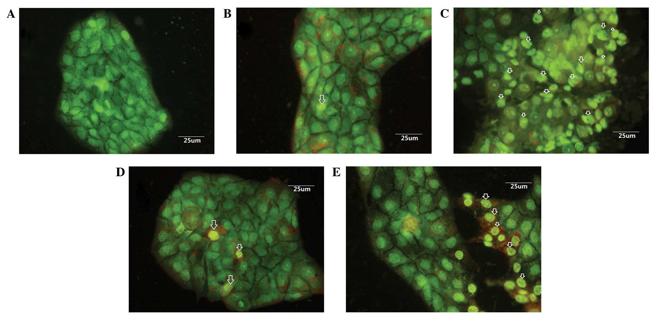
Morphological changes
In order to determine whether EPS-A induced
apoptosis in the A431 cells, morphological changes were examined
using a fluorescence microscope. It was shown that the cells
exhibited an intact morphology of the nucleus and cytoplasm in the
control group (Fig. 1A), while cells
treated with EPS-A displayed typical apoptotic features (Fig. 1B–E).
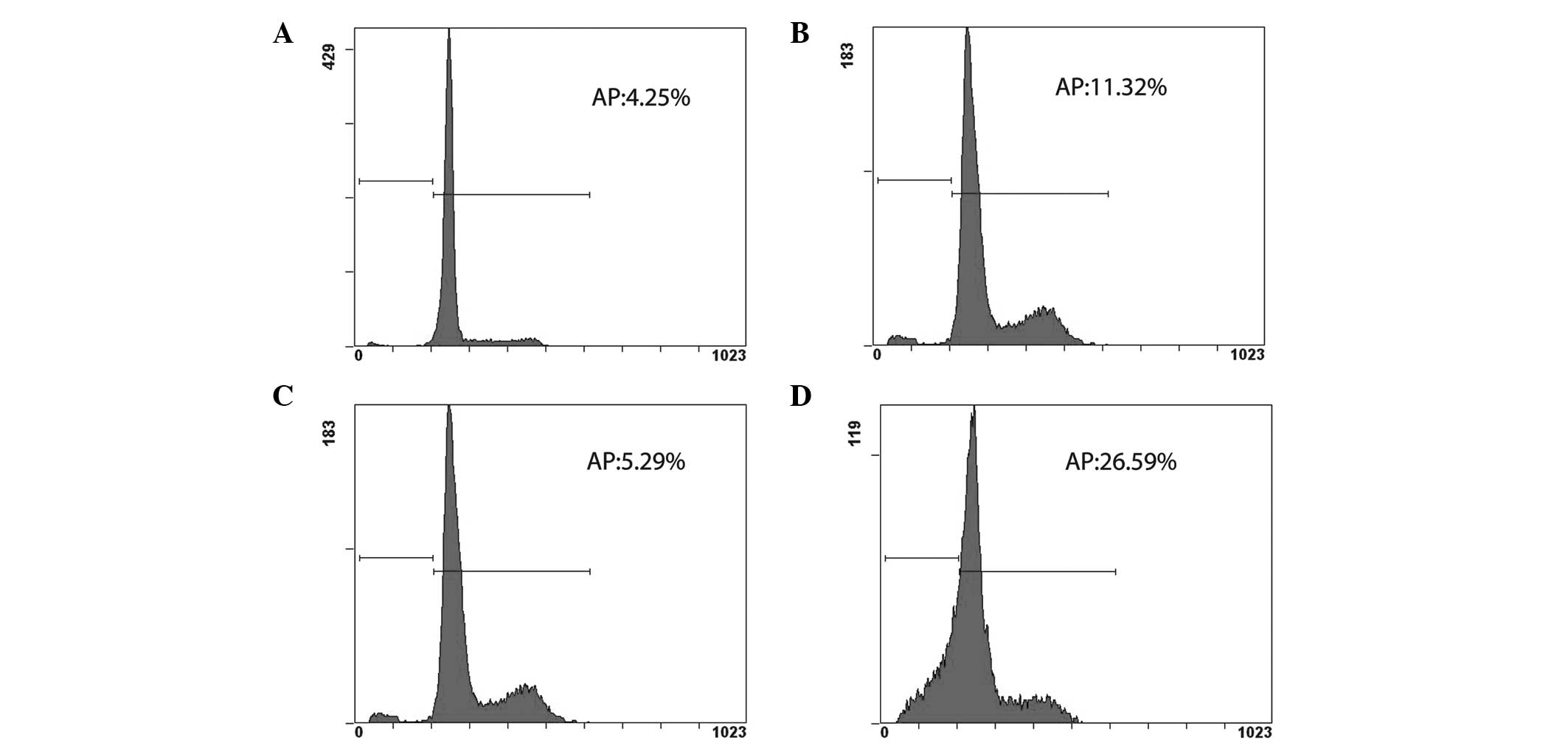
EPS-A causes cell cycle arrest and
induces apoptosis in A431 cells
The effect of EPS-A on the cell cycle progression of
A431 cells was studied both 48 and 72 h after treatment. FCM
analysis indicated that EPS-A caused cell cycle arrest and induced
apoptosis in the A431 cells, which was not obvious at 48 h after
treatment. The apoptotic rate of the control group was 4.25%
(Fig. 2A), while that of the EPS-A
group was 11.32% (Fig. 2B). At 72 h
after treatment the apoptotic rate of the EPS-A group reached
26.59% (Fig. 2D), which was 5-fold
higher than that of the control group (5.29%; Fig. 2C).
Staining of A431 cells with PI and
Rh123
Following FCM, the A431 cells that had been stained
with Rh123/PI were easily divided into four groups (Fig. 3): The lower right quadrant of each
graph, Rh123+/PI−, showed the A431 cells with
normal mitochondrial function; the lower left quadrant contained
Rh123−/PI− A431 cells that had lost their
mitochondrial function; the upper left quadrant,
Rh123−/PI+, showed necrotic A431 cells and
the upper right quadrant, Rh123+/PI+, showed
late apoptotic A431 cells. As shown in Fig. 3A, the A431 cells in the
Rh123+/PI− group were centered in the lower
right quadrant, which meant that the A431 cells in the control
group exhibited a good mitochondrial function. The A431 cells of
the EPS-A group; however, were predominantly concentrated in the
lower left and lower right quadrants, which indicated that the A431
cells in the EPA-S group had lost mitochondrial function and the
number of apoptotic A431 cells had increased (Fig. 3B).
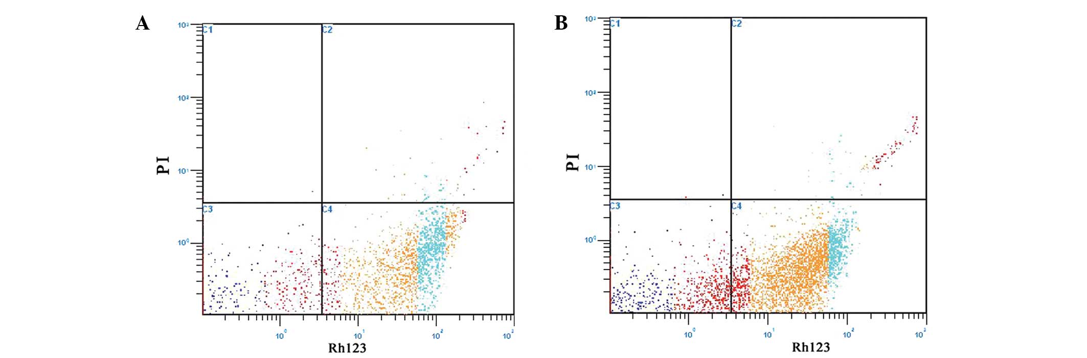 | Figure 3.Staining of A431 cells with PI and
Rh123 revealed that EPS-A induced apoptosis in A431 cells. (A) The
control cells were treated with phosphate-buffered saline. (B) The
A431 cells were treated with 3 mg/ml EPS-A for 48 h, and their
apoptotic rate was measured using flow cytometry. Cells were
stained with Rh123 and PI. Each value represents the average of
three independent experiments. Light blue represents normal cells,
red represents necrotic cells, yellow and deep blue represent
apoptotic cells. The lower right quadrant of each graph,
Rh123+/PI−, showed A431 cells with normal
mitochondrial function; the lower left quadrant,
Rh123−/PI−, showed A431 cells losing their
mitochondrial function; the upper left quadrant,
Rh123−/PI+, showed necrotic A431 cells and
the upper right quadrant, Rh123+/PI+, showed
late apoptotic A431 cells. EPS-A, extracellular polymeric
substances of Aphanizomenon flos-aquae; PI, propidium
iodide; Rh123, Rhodamine 123. |
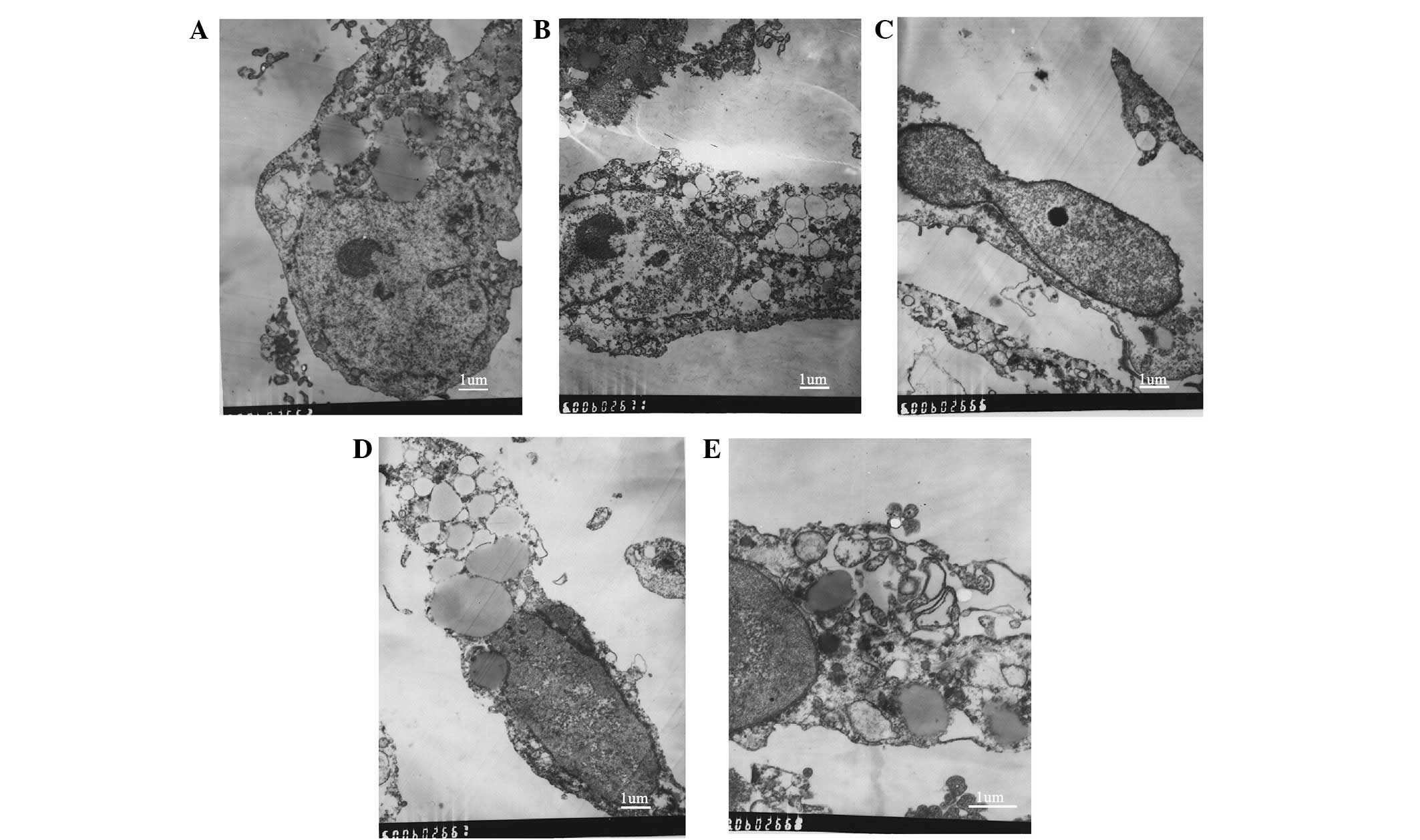
Effects of EPS-A on the ultrastructure
of the A431 cells
The ultrastructural changes of the A431 cells were
observed using TEM. The cell ultrastructure of the control group
was observed to be normal (Fig. 4A).
Following treatment with a low dose of EPS-A, nuclear
fragmentation, chromosome condensation and cell shrinkage (Fig. 4B and C) were observed in the A431
cells. Their rough-surfaced endoplasmic reticulum exhibited
enlarged cisternae (Fig. 4D).
Following treatment with a higher dose of EPS-A, apoptotic bodies
were observed (Fig. 4E).
Discussion
Numerous bioactive extracellular polymeric
substances with noteworthy functional properties have been
discovered in marine organisms, and studies that focus on marine
natural products, particularly marine algae EPS, are increasingly
attracting worldwide attention (8,13).
Natural products that have been isolated from cyanobacteria, as
well as their derivatives, have been proven to be a valuable
chemical resource for finding promising drugs that could assist the
prevention and treatment of cancer (14). The aim of the present study was to
evaluate the effect that EPS-A has on A431 cells and to explore its
anticancer activity.
Apoptosis is an important aspect of
chemotherapy-induced tumor cell death as well as the major
mechanism by which numerous anticancer drugs and natural products
induce cell death (15). Apoptosis
is also a type of programmed cell death, which, through a series of
biochemical events, leads to cellular morphological changes and
cell death. In order to explore the mechanism responsible for the
anticancer effects of EPS-A, the changes in cell morphology were
assessed. Following treatment with different concentrations of
EPS-A, morphological changes, including cell shrinkage, nuclear
fragmentation and chromatin condensation, were observed in the A431
cells. AO/EB staining of EPS-A-treated A431 cells showed that EPS-A
resulted in nuclear condensation and fragmentation, a morphological
hallmark of apoptosis (16).
In the present study, FCM analysis revealed that
EPS-A treatment resulted in an increase in the proportion of
apoptotic cells. When treated for 48 h, the proportion of apoptotic
cells in the EPS-A group was low, mainly due to the fact that there
had been no loss of DNA by fragmentation, although DNA damage
occurred at an early phase of apoptosis. The secondary reason may
be that for apoptotic cells in the S or G2/M phase, even
if the DNA content decreased, the actual DNA content was not lower
than that of diploid cells, and the proportion of cells in the S
and G2/M phases in the EPS-A group was higher than that
in the control group. This may be due to the fact that EPS-A caused
cell cycle arrest of the A431 cells in the G2/M phase,
affected spindle formation during cell division and inhibited cell
division (17,18). At 72 h the apoptosis peak
(G2/M) was not obvious. The results suggested that EPS-A
initially affected the S and G2/M phases of the A431
cells at the early treatment stage, induced cell cycle arrest in
the S and G2/M phases and then cell apoptosis, and the
proportion of apoptotic cells increased in a time-dependent manner.
Therefore, EPS-A induced apoptosis of the A431 cells by affecting
the whole cell cycle, but the exact mechanism remains unclear.
In the early phase of apoptosis, when changes in
mitochondrial morphology are undetectable, the dissipation of the
membrane potential has already occurred. The dissipation of the
membrane potential is considered to be part of a cascade reaction
that occurs early in the process of apoptosis, prior to nuclease
enzyme activation and the exposure of phosphatidylserine at the
cell surface. Once the ΔΨm is dissipated, apoptosis is irreversible
(19,20).
The fluorescent dyes Rh123 and PI can be used to
evaluate the function of the mitochondria and nucleus, respectively
(21). In order to further
understand the ΔΨm of a cell and the integrity of the cell
membrane, PI and Rh123 double staining was carried out. PI is a
fluorescent dye that is not membrane permeable and binds to DNA,
whereas Rh123 is a lipophilic dye that is absorbed by the
mitochondria. When the integrity of the cell membrane is intact, PI
is not able to enter the cell to stain the DNA; therefore, a lack
of PI staining indicates cell membrane integrity. The presence of
Rh123 staining suggests that the ΔΨm is normal. The cellular uptake
of Rh123 is positively correlated with the ΔΨm (22). FCM analysis showed that the cells
comprised two main subpopulations: PI−Rh123+
cells that retained their cell membrane integrity and had a ‘steady
state’ ΔΨm, and PI−Rh123+ cells that retained
their cell membrane integrity but had a decreased ΔΨm. In the cells
treated with EPS-A for 48 h, the number of
Rh123−PI− cells (those with neither PI nor
Rh123 staining) increased. This indicated that some of the cells
had an intact cell membrane but had lost ΔΨm. In conclusion, EPS-A
may induce the apoptosis of A431 cells by regulating the
mitochondrial membrane permeability.
To date, cell ultrastructure observation using TEM
comprises the most common and reliable method of detecting
apoptosis, and is considered as the gold standard. TEM showed
visible cell nuclei, uniform chromatin distribution, abundant
organelles and cell membrane and nuclear membrane integrity in the
control group. Following treatment with a low dose of EPS-A for 48
h, nuclear fragmentation, chromosome condensation, cell shrinkage
and expansion of the endoplasmic reticulum were observed in the
A431 cells. Following treatment with a high dose of EPS-A, the
shape of the A431 cells was markedly altered, and apoptotic bodies
were observed. The results showed that EPS-A influenced the A431
cell structure in a direct manner, disrupted cell metabolism,
damaged DNA and ultimately induced apoptosis in the A431 cells.
In conclusion, the results showed that EPS-A has
anti-cancer properties and can induce apoptosis in A431 cells via
the mitochondrial pathway. The cause of apoptosis in A431 cells may
be cell cycle arrest and collapse of the ΔΨm; therefore, EPS-A
plays an important anti-cancer role, the exact mechanism of which,
however, requires further research.
Acknowledgements
The present study was supported by the Natural
Science Foundation of Gansu Province of China (grant no.
145RJZA171) and the National Natural Science Foundation of China
(grant no. 81271390). The authors would like to thank Professor
Chunxiang Hu, Delu Zhang and Yongding Liu for their assistance in
the running of the experiments.
References
|
1
|
Tan W, Lu J, Huang M, et al: Anti-cancer
natural products isolated from chinese medicinal herbs. Chin Med.
6:272011. View Article : Google Scholar : PubMed/NCBI
|
|
2
|
Monks NR, Li B, Gunjan S, et al: Natural
products genomics: A novel approach for the discovery of
anti-cancer therapeutics. J Pharmacol Toxicol Methods. 64:217–225.
2011. View Article : Google Scholar : PubMed/NCBI
|
|
3
|
De Nobel WT, Matthijs HCP, Elert von Elert
E and Mur LR: Comparison of the light-limited growth of the
nitrogen-fixing cyanobacteria AnabaenaAphanizomenon.
New Phytologist. 138:579–587. 1998. View Article : Google Scholar
|
|
4
|
Reynolds CS, Huszar V, Kruk C, et al:
Towards a functional classification of the freshwater
phytoplankton. J Plankton Res. 24:417–428. 2002. View Article : Google Scholar
|
|
5
|
Liu Y, Chen W, Li D, et al: First report
of aphantoxins in China - waterblooms of toxigenic Aphanizomenon
flos-aquae in lake Dianchi. Ecotoxicol Environ Saf. 65:84–92.
2006. View Article : Google Scholar : PubMed/NCBI
|
|
6
|
Klock J, Wieland A, Seifert R and
Michaelis W: Extracellular polymeric substances (EPS) from
cyanobacterial mats: Characterisation and isolation method
optimisation. Marine Biol. 152:1077–1085. 2007. View Article : Google Scholar
|
|
7
|
Mooberry SL, Leal RM, Tinley TL, et al:
The molecular pharmacology of symplostatin 1: A new antimitotic
dolastatin 10 analog. Int J Cancer. 104:512–521. 2003. View Article : Google Scholar : PubMed/NCBI
|
|
8
|
Singh RK, Tiwari SP, Rai AK and Mohapatra
TM: Cyanobacteria: An emerging source for drug discovery. J
Antibiot (Tokyo). 64:401–412. 2011. View Article : Google Scholar : PubMed/NCBI
|
|
9
|
Park HK, Kim IH, Kim J and Nam TJ:
Induction of apoptosis by laminarin, regulating the insulin-like
growth factor-IR signaling pathways in HT-29 human colon cells. Int
J Mol Med. 30:734–738. 2012.PubMed/NCBI
|
|
10
|
Xue M, Ge Y, Zhang J, et al: Anticancer
properties and mechanisms of fucoidan on mouse breast cancer in
vitroin vivo. PLoS One. 7:e434832012. View Article : Google Scholar : PubMed/NCBI
|
|
11
|
Ou Y, Xu S, Zhu D, et al: Molecular
mechanisms of exopolysaccharide from Aphanothece halaphytica
(EPSAH) induced apoptosis in HeLa cells. PLoS One. 9:e872232014.
View Article : Google Scholar : PubMed/NCBI
|
|
12
|
Hou R, Zhou QL, Wang BX, et al: Diosgenin
induces apoptosis in HeLa cells via activation of caspase pathway.
Acta Pharmacol Sin. 25:1077–1082. 2004.PubMed/NCBI
|
|
13
|
Zhang D, Hu C, Wang G, Li D, Li G and Liu
Y: Zebrafish neurotoxicity from aphantoxins - cyanobacterial
paralytic shellfish poisons (PSPs) from Aphanizomenon
flos-aquae DC-1. Environ Toxicol. 28:239–254. 2013. View Article : Google Scholar : PubMed/NCBI
|
|
14
|
Yap TA and Workman P: Exploiting the
cancer genome: Strategies for the discovery and clinical
development of targeted molecular therapeutics. Annu Rev Pharmacol
Toxicol. 52:549–573. 2012. View Article : Google Scholar : PubMed/NCBI
|
|
15
|
Yang L, Wang P, Wang H, et al: Fucoidan
derived from Undaria pinnatifida induces apoptosis in human
hepatocellular carcinoma SMMC-7721 cells via the ROS-mediated
mitochondrial pathway. Mar Drugs. 11:1961–1976. 2013. View Article : Google Scholar : PubMed/NCBI
|
|
16
|
Alarifi S, Ali D, Alakhtani S, et al:
Reactive oxygen species-mediated DNA damage and apoptosis in human
skin epidermal cells after exposure to nickel nanoparticles. Biol
Trace Elem Res. 157:84–93. 2014. View Article : Google Scholar : PubMed/NCBI
|
|
17
|
Zhang X, Chen W, Guillermo R, et al:
Alpha-santalol, a chemopreventive agent against skin cancer, causes
G2/M cell cycle arrest in both p53-mutated human epidermoid
carcinoma A431 cells and p53 wild-type human melanoma UACC-62
cells. BMC Res Notes. 3:2202010. View Article : Google Scholar : PubMed/NCBI
|
|
18
|
Ma S, Shan LQ, Xiao YH, et al: The
cytotoxicity of methacryloxylethyl cetyl ammonium chloride, a
cationic antibacterial monomer, is related to oxidative stress and
the intrinsic mitochondrial apoptotic pathway. Braz J Med Biol Res.
44:1125–1133. 2011. View Article : Google Scholar : PubMed/NCBI
|
|
19
|
Lemasters JJ, Nieminen AL, Qian T, Trost
LC, Elmore SP, Nishimura Y, Crowe RA, Cascio WE, Bradham CA,
Brenner DA and Herman B: The mitochondrial permeability transition
in cell death: A common mechanism in necrosis, apoptosis and
autophagy. Biochim Biophys Acta. 1366:177–196. 1998. View Article : Google Scholar : PubMed/NCBI
|
|
20
|
Xu F, Zhang SH, Shao RG and Zhen YS:
Anticancer activity of sodium caffeate and its mechanism. Acta
Pharmacol Sin. 26:1248–1252. 2005. View Article : Google Scholar : PubMed/NCBI
|
|
21
|
Galfano A, Novara G, Iafrate M, et al:
Improvement of seminal parameters and pregnancy rates after
antegrade sclerotherapy of internal spermatic veins. Fertil Steril.
91:1085–1089. 2009. View Article : Google Scholar : PubMed/NCBI
|
|
22
|
Zou T, Liu X, Ding S and Xing J:
Evaluation of sperm mitochondrial function using rh123/PI dual
fluorescent staining in asthenospermia and oligoasthenozoospermia.
J Biomed Res. 24:404–410. 2010. View Article : Google Scholar : PubMed/NCBI
|