Introduction
Chronic hepatitis B (CHB) is caused by infection by
the hepatitis B virus (HBV) (1–3). HBV
belongs to the Hepadnaviridae family of viruses. Currently,
the Hepadnaviridae family is known to include HBV, woodchuck
hepatitis virus (WHV), ground squirrel hepatitis virus, heron
hepatitis B virus and duck hepatitis B virus (DHBV) (4). All virus types within the
Hepadnaviridae family are tiny and exhibit hepatotropism.
Hepadnaviruses are a type of DNA virus, with similar viron shape
and genome and replicate via RNA reverse transcription (5). The discovery of hepadnaviruses in
mammals and birds provided the experimental and ethical basis on
the study of HBV biological mechanisms (6,7). In a
previous study of human HBV infection mechanisms, marmots infected
with WHV (8) and the ducks infected
with DHBV are the most widely used model (9–11). Due
to the similarity between HBV-infected humans and DHBV-infected
duck, ducks infected with DHBV are an effective model for the study
of hepadnaviruses. Super spiral of covalently closed circular DNA
molecules (cccDNA) are viral genome replication intermediates in
the hepatocyte nuclei, and the key factor underlying persistent
Hepadnaviridae infection (12–14).
Currently, no methods are available for the complete inhibition of
their formation. The approved drugs for the treatment of CHB, which
are nucleotide analogs and interferons, have certain disadvantages,
such as a poor side-effect profile. The identification of novel
anti-HBV drugs has become a key focus of research in the area of
viral hepatitis (15–18). Duck hepatitis B virus polymerase
(DHBVP) is essential for duck hepatitis B virus (DHBV) replication
(19,20); therefore, the functional blockade of
DHBVP has the potential to inhibit HBV genome replication. In the
present study, phage display technology (PDT) was used to screen
for mimic peptides that specifically interact with DHBVP. The
inhibitory effect of these mimic peptides on DHBV replication in
primary duck hepatocytes was investigated in vitro in an
effort to identify novel effective drugs against HBV
infections.
Materials and methods
PDT screening test for mimic peptides
specifically targeting DHBVP and the determination of the
associated nucleotide sequences
Peptides targeting DHBVP functional sites were
dissolved in dimethyl sulfoxide at a final concentration of 100
µg/ml. These peptides were synthesized according to the DHBVP
sequence of Shaoxing duck, which surrounding the YMDD sites. Each
well of a 96-well ELISA plate (Greiner Bio-One, Frickenhausen,
Germany) was coated with peptide solution and then treated with 150
µl synthesized peptide (1 mg/ml) and incubated at 4°C overnight.
Following blocking at 4°C for ≥1 h, each ELISA plate was washed
with Tris-buffered saline with Tween-20 (TBST; Promega Corporation,
Madison, WI, USA) six times. A diluted phage peptide library (C7C
Phage Display Peptide library; New England Biolabs, Beverly, MA,
USA) was added and the plate was incubated at room temperature for
60 min. Each plate was then washed with TBST 10 times and each well
was eluted with 100 µl acidic eluent (provided with the C7C
library) at room temperature for ≤10 min. Eluents were collected in
microcentrifuge tubes and neutralized with neutralizing solutions
(provided with the C7C library). Titers were determined using 1 µl
eluents, while the remaining eluents were added to 20 ml
Escherichia (E.) coli ER2537 (New England Biolabs; early
logarithmic phase) and incubated for 4.5 h at 37°C. Samples were
precipitated and purified for further screening, following the
manufacturer instructions included with the experimental kit. Three
rounds of the above-mentioned screening process were performed.
Elutions from the third screening step were diluted and spotted
into the plates. Clear phage plaques were removed for DNA
extraction and, following DNA sequencing, the corresponding amino
acid sequences were determined and used for the mimic peptide
synthesis process. The selected mimic peptides were synthesized by
Chinese Peptide Co. (Hangzhou, China).
Serum sample and hepatocyte
preparation
Six randomly selected domestic male ducks (Shaoxing
ducks; average age, 1 year; average weight, 1 kg; Zhejiang Academy
of Agricultural Sciences, Zhejiang, China) were prepared and 1 ml
serum from each duck was isolated for analysis. Primary duck
hepatocytes were isolated from liver tissue as described previously
(21). In brief, the liver was
aseptically removed from each duck and washed twice with sterile
saline solution. The livers were then cut into pieces and digested
with trypsin in serum-free Dulbecco's modified Eagle's medium
(DMEM; Gibco Life Technologies, Carlsbad, CA, USA) for 30 min at
37°C. The resulting hepatocytes were collected by centrifugation at
1,600 × g for 12 min and then washed with sterile saline solution
and culture medium twice. Cells were counted and seeded into 60-mm
dishes at a density of 2×10⁶ per well. The cells were
cultured in a 5% CO2 incubator at 37°C. This study was
carried out in strict accordance with the recommendations in the
Guide for the Care and Use of Laboratory Animals of the National
Institutes of Health (8th edition, 2011). The protocol was approved
by the Committee on the Ethics of Animal Experiments of the First
Affiliated Hospital, Zhejiang University School of Medicine
(Hangzhou, China; Permit Number, 162). All surgery was performed
under sodium pentobarbital anesthesia, and all efforts were made to
minimize suffering.
Infection of hepatocytes with
DHBV
Blood from DHBV-positive adult ducks was aseptically
collected and incubated overnight at room temperature. The samples
were centrifuged at 3,000 × g for 10 min to isolate the serum.
Disposable filters were used to completely remove the bacteria. The
infection of hepatocytes with the serum was then performed as
follows (22): Primary duck
hepatocytes were incubated for 18 h and then washed with DMEM
containing 1% fetal bovine serum (FBS; Hyclone, Logan, UT, USA).
Viral solution (30 µl duck serum; 5×10⁹ virions/ml) was
added to the cells, followed by 3 ml serum-free DMEM, and the cells
were then incubated.
Mimic peptide treatment of
hepatocytes
Ten groups of infected hepatocytes were established,
including eight synthetic mimic peptide groups [experimental (EXP)
groups], an entecavir-treated group (positive control), and a
phosphate-buffered saline (PBS)-treated group (negative control).
In the EXP groups, 30 µl (100 µmol) mimic peptide solution was
added to each well, and 3 µl (10 mmol) entecavir stock solution or
30 µl PBS was added to the control groups, respectively. The cells
were incubated at 37°C in a 5% CO2 incubator and the
medium was changed every 48 h. Cell culture supernatants were
collected and stored at −20°C for future analysis. Following each
change of medium, 30 µl mimic peptide solution was again added to
the wells of the EXP groups and 3 µl entecavir stock solution or 30
µl PBS was added to the control groups, respectively. After a
10-day incubation, the cell supernatants were collected and the
cells were washed twice with PBS and digested with 0.25% trypsin.
Following centrifugation at 1,500 × g at 4°C for 5 min and washing
twice with PBS, the cells in each well were collected and counted.
Three replications were performed under each condition.
DHBV-DNA extraction and determination
by quantitative polymerase chain reaction (qPCR)
The DHBV-DNA extraction of cell fractions was
performed as described previously (23). Hepatocytes from the various treatment
groups were washed twice with PBS and then 0.5 ml TBS lysis buffer
was added. Following centrifugation at 1,500 × g at 4°C for 5 min,
cell nuclear fractions were sedimented and separated from the
cytoplasmic fractions in the supernatants. Nuclear fractions were
washed twice with 0.5 ml TBS and then dissolved in 1 ml
radio-immunoprecipitation assay (RIPA) solution. The cytoplasm
fractions were mixed with equivalent volumes of RIPA solution.
Equal volumes of lysis buffer were then added and the resulting
mixtures were incubated at 55°C for 2 h to digest the proteins.
Following digestion, the samples were extracted with equal volumes
of a phenol-chloroform-isoamyl alcohol (25:24:1) mixture and the
DNA in the aqueous phase was sedimented with 2.5 volumes of alcohol
and 1/10 volumes of 3 M sodium acetate. The DNA was recovered
following centrifugation at 12,000 × g at 4°C for 15 min. Following
washing with 70% alcohol, DNA was dissolved with
Tris-ethylenediaminetetraacetic acid buffer and quantified using
agarose gel and fluorescence, according to the manufacturer
instructions of a QIAquick Gel Extraction Kit (Qiagen, Hilden,
Germany).
The DHBV-DNA of the cell culture supernatant was
extracted using the QIAamp MinElute Virus Spin kit (Qiagen)
according to the manufacturer's instructions. The DNA samples were
stored at −20°C for future analysis.
The qPCR for DHBV-DNA was performed using SYBR-Green
I (Bioasia Life Technology Co. Ltd., Shanghai, China) in a
real-time PCR instrument (Mastercycler® ep realplex; Eppendorf,
Hamburg, Germany; Registration ID 2273536). The PCR program was
designed as follows: Pre-denaturation at 95°C for 5 min and 42
cycles including 15-sec denaturation at 94°C, 30-sec annealing at
56°C, and a 45-sec extension at 72°C. The DNA contents were
measured at each cycle end and calculated using a standard
curve.
Construction of mimic
peptide-expressing recombinant plasmids
Purified DHBVP PCR products (8 µl) were obtained
using a QIAquick PCR Purification Kit (Qiagen), according to the
manufacturer instructions. These products were thoroughly mixed
with 1.5 µl BamHI, 1.5 µl HindIII, 25 µl sterile
water and 4 µl 10X buffer (Qiagen). The mixtures were incubated at
37°C for 1.5 h. Purified pGEM® (4 µl; Promega) was digested
similarly. The products were purified using gel extraction methods
and the DHBVP fragment was ligated into the pGEM vector using T4
DNA ligase. The ligation products were used to transform E.
coli competent cells, and the positive clones were chosen for
sequencing.
Measurement of the inhibitory effect
on DHBV inhibition of mimic peptides expressed intracellularly
Plasmids expressing the mimic peptides were used to
transfect duck primary hepatocytes pre-infected with DHBV. The
DHBV-DNA contents of the cells were then determined. In the EXP
groups, 1.2 µg mimic peptide-expressing plasmid combined with 3 µl
liposomes were added to each well. In the control groups, 1.2 µg
pEGFP-N1 plasmid (Clontech Laboratories, Inc., Mountain View, CA,
USA) combined with 3 µl liposomes were used. The medium was changed
to serum-free DMEM following transfection and the cells were
incubated at 37°C in a 5% CO2 incubator. The medium was
changed to DMEM containing 5% FBS, 300 mg/l penicillin, 100 mg/l
streptomycin, 1 mg/l insulin, 1.5 mg/ml glucose, 10 U/ml nystatin
and 1×10⁻⁵ M hydrocortisone-hemisuccinate after 6-h
incubation. Entecavir (3 µl) was added to each well in the
entecavir-treated group while 3 µl PBS was added to the wells of
the negative control group. The medium was changed every 48 h and
the collected cell supernatants were stored at −20°C for future
analysis. After 10 days of incubation, the cell supernatants were
collected and the cells were washed twice with PBS and digested
with 0.25% trypsin. Following centrifugation at 1,500 × g at 4°C
for 5 min and washing twice with PBS, the cells in each well were
collected and counted. DHBV-DNA was then extracted as described
previously.
Inhibition rate analysis
The DHBV-DNA inhibition rate of the mimic peptides
was calculated as follows: Inhibition rate (%) = [(measured DNA
content of the negative control - measured DNA content of the
specific treatment group)/measured value of the negative control]
×100.
Statistical analysis
The data were analyzed by SPSS 17.0 for Windows
(SPSS Inc., Chicago, IL, USA). A homogeneity test of variance
revealed unequal variances between groups, so a Kruskal-Wallis test
was performed for further analysis. P<0.05 was considered to
indicate a statistically significant difference.
Results
Mimic peptides specifically targeting
DHBVP and the associated nucleotide sequences
Three screening rounds were performed and the
nucleotide sequences of eight mimic peptides were selected
(Table I; the DNA content of another
two mimic peptides were too weak to be detected). The amino acid
sequences of the selected mimic peptides were deduced from the
nucleotide sequences (Table I). The
eight mimic peptides were synthesized according to the associated
amino acid sequences.
 | Table I.Nucleotide sequences and amino acid
sequences of the selected mimic peptides. |
Table I.
Nucleotide sequences and amino acid
sequences of the selected mimic peptides.
| No. | Nucleotide
sequences | Amino acid
sequences |
|---|
| 1 |
ACCTCCACCTGTAAAAGTCCTCTGATCCTGAGAGTGAGA |
Gln-Asp-Gln-Arg-Thr-Phe-Thr |
| 2 |
ACCTCCACCAATCGGAGTATGAGTAACCAGAGAGTGAGA |
Leu-Val-Thr-His-Thr-Pro-Ile |
| 3 |
ACCTCCACCCCGAAACAGCGTAGCAGGAACAGAGTGAGA |
Val-Pro-Ala-Thr-Leu-Phe-Arg |
| 4 |
ACCTCCACCCCGCGGCGGATGAAGATGAGGAGAGTGAGA |
Pro-His-Leu-His-Pro-Pro-Arg |
| 5 |
ACCTCCACCATGCGAATGATGTAGAGACATAGAGTGAGA |
Met-Ser-Leu-His-His-Ser-His |
| 6 |
ACCTCCACCACTCGACGCAGTACGCAGAGCAGAGTGAGA |
Ala-Leu-Arg-Thr-Ala-Ser-Ser |
| 7 |
ACCTCCACCATGACGCGGATAAATAGCATGAGAGTGAGA |
His-Ala-Ile-Tyr-Pro-Arg-His |
| 8 |
ACCTCCACCAGTAGCAGTCGGAGGCGGCCTAGAGTGAGA |
Arg-Pro-Pro-Pro-Thr-Ala-Thr |
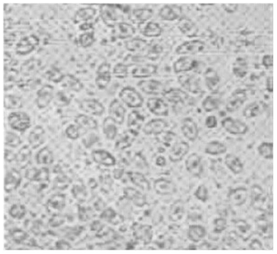
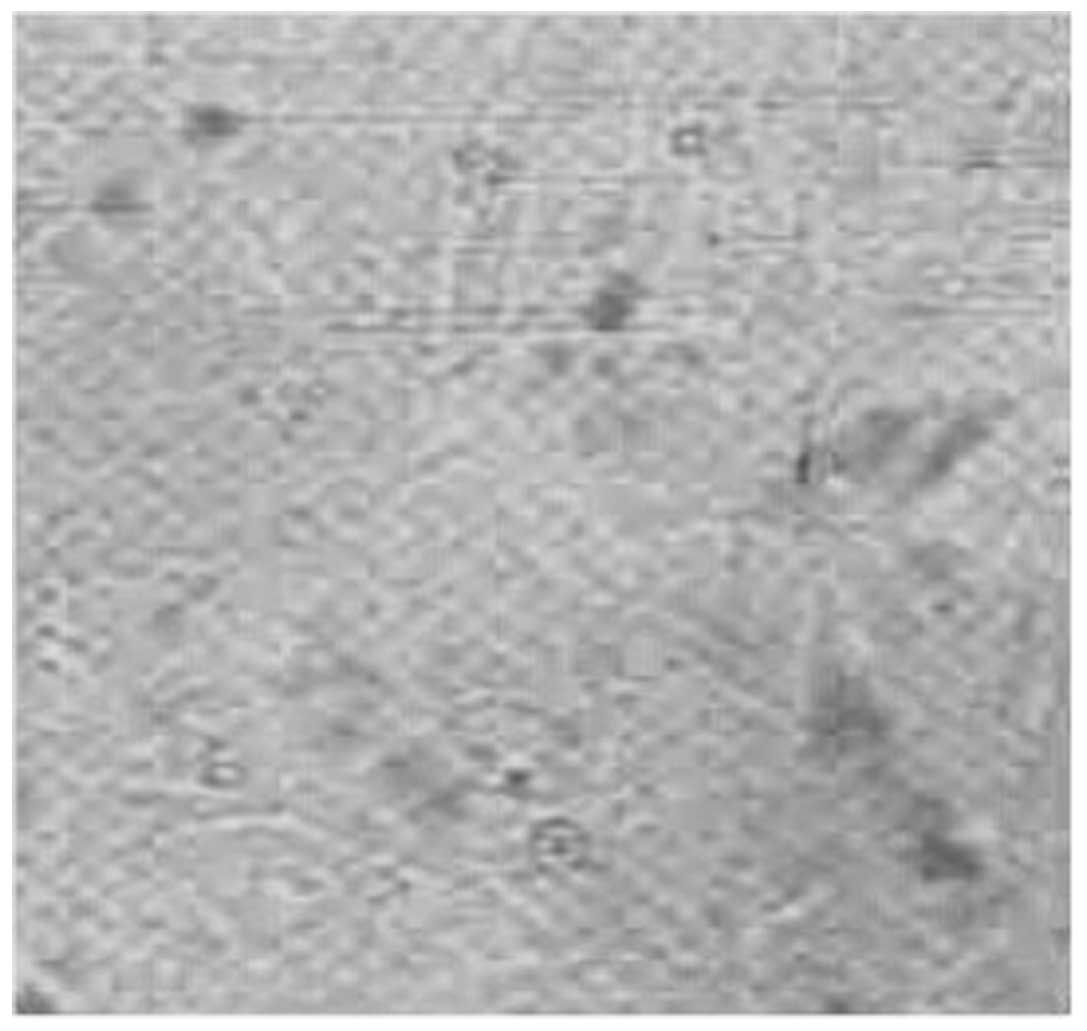
DHBV infection of primary cultured
cells
The primary duck hepatocytes were successfully
isolated and attached in culture 12 h following their isolation.
After culturing for 8 days, the proliferating hepatocytes covered
the entire surface of the dish. No significant differences were
observed between the EXP and control groups. Cell morphology at
different time-points is shown in Figs.
1–4.
Inhibition of DHBV-DNA by treatment
with mimic peptides
The DHBV-DNA levels in the cell culture
supernatants, cytoplasmic fractions and nuclear fractions are shown
in Table II.
| Table II.DHBV-DNA levels of the cell culture
supernatants, cytoplasmic fractions and nuclear fractions following
treatment with mimic peptides targeting DHBV polymerase. |
Table II.
DHBV-DNA levels of the cell culture
supernatants, cytoplasmic fractions and nuclear fractions following
treatment with mimic peptides targeting DHBV polymerase.
| No. | Cell culture
supernatants | Cytoplasmic
fractions | Nuclear
fractions |
|---|
| 1 |
1.73E5±2.16E5 |
6.53E5±6.37E3 |
1.34E5±4.27E4 |
| 2 |
5.68E4±7.07E5a,b |
5.31E4±1.59E4a,b |
4.14E5±5.17E4 |
| 3 |
6.31E5±4.18E4 |
4.83E5±3.53E4 |
6.32E5±5.14E3 |
| 4 |
1.14E5±7.35E3 |
1.79E5±2.13E5 |
7.15E5±3.82E4 |
| 5 |
1.48E5±7.39E4 |
1.63E5±6.45E4 |
4.79E5±2.24E4 |
| 6 |
2.65E5±3.24E4 |
2.43E5±2.74E4 |
6.37E5±1.31E4 |
| 7 |
2.99E4±2.41E4a,b |
3.22E4±5.23E4a,b |
3.41E5±9.47E4 |
| 8 |
6.37E5±2.14E4 |
3.01E5±5.38E4 |
4.49E5±3.25E4 |
| Positive
control |
2.47E4±3.39E4a |
4.94E4±1.47E3a |
5.72E5±1.56E4 |
| Negative
control |
9.63E5±1.59E5 |
5.25E5±1.59E5 |
7.74E5±1.63E4 |
The DHBV-DNA levels of the cell culture supernatants
and cytoplasm fractions in the EXP groups treated with mimic
peptides 2 or 7 and the positive (entecavir) group were
significantly decreased compared with those in the negative control
group (P<0.05). In addition, there were no significant
difference between the EXP groups treated with mimic peptides 2 or
7 and the positive control (P>0.05).
The DHBV-DNA inhibition rates in the cell culture
supernatants for hepatocytes treated with mimic peptides 2 and 7
were 94.1 and 96.9%, respectively, similar to the inhibition ratio
of 97.4% in the positive control (P>0.05). The DHBV-DNA
inhibition rates in the cytoplasm fractions for mimic peptides 2
and 7 were 89.9 and 93.9%, respectively, similar to the
entecavir-mediated inhibition rate of 90.6% (P>0.05).
Inhibition of DHBV-DNA by
intracellularly expressed mimic peptides
The DHBV-DNA levels of the cell culture
supernatants, cytoplasmic fractions and cell nuclear fractions from
cells intracellularly expressing mimic peptides are shown in
Table III.
| Table III.DHBV-DNA contents of the cell culture
supernatants, cytoplasmic fractions and nuclear fractions following
cellular expression of mimic peptides. |
Table III.
DHBV-DNA contents of the cell culture
supernatants, cytoplasmic fractions and nuclear fractions following
cellular expression of mimic peptides.
| No. | Culture
supernatants | Cytoplasmic
fractions | Nuclear
fractions |
|---|
| 1 |
1.80E5±3.86E4 |
2.01E5±3.40E4 |
1.67E5±2.66E4 |
| 2 |
3.51E4±1.51E4a,b |
7.09E4±5.91E3a,b |
3.89E5±4.66E4 |
| 3 |
1.96E5±3.30E4 |
2.56E5±3.74E5 |
4.02E5±2.08E4 |
| 4 |
1.94E5±3.45E4 |
2.48E5±3.53E4 |
2.05E5±7.09E4 |
| 5 |
1.85E5±3.44E4 |
2.07E5±4.10E4 |
2.01E5±6.25E4 |
| 6 |
2.22E5±1.20E4 |
4.01E5±4.48E4 |
2.82E5±6.09E4 |
| 7 |
1.14E4±8.68E3a,b |
8.50E4±5.94E4a,b |
9.83E4±6.02E3a |
| 8 |
4.74E5±3.84E4 |
2.35E5±4.63E4 |
2.37E5±6.17E4 |
| Positive
control |
1.40E4±1.18E4a |
8.79E4±8.38E3a |
4.37E5±2.55E4 |
| Negative
control |
7.09E5±6.15E4 |
5.14E5±7.95E4 |
6.94E5±1.53E4 |
The DHBV-DNA levels in the EXP groups
intracellularly expressing mimic peptides 2 or 7 and the positive
control group were significantly decreased compared with those in
the negative control group (P<0.05). Furthermore, the DHBV-DNA
levels of the nuclear fractions in the EXP group intracellularly
expressing mimic peptide 7) were significantly decreased compared
with those in the negative control group, and were the lowest among
all the groups (P<0.05).
The inhibitory effect on DHBV-DNA levels in the cell
culture supernatants was similar among the EXP groups treated with
mimic peptides 2 or 7 and the positive control, and their
respective inhibition rates were 95.0, 98.4 and 98.0% (P>0.05).
The respective inhibition rates of the cytoplasmic DHBV-DNA levels
in the EXP groups treated with mimic peptides 2 or 7 and the
positive control were 86.2, 83.5 and 82.9% (P>0.05). The EXP
group treated with mimic peptide no 7, however, had a much lower
DHBV-DNA level in the nuclear fractions than did the positive
control, and their respective inhibition rates were 85.8 and 37.0%
(P<0.05).
Discussion
Chronic HBV infection is a devastating health
problem that is closely associated with different stages of liver
injury, hepatic fibrosis and hepatocellular carcinoma (24–26). As
such, the development of an effective HBV treatment is a major task
in the medical field (27). HBV is a
member of Orthohepadnavirus belonging to the family
Hepadnaviridae. The Avihepadnavirus genus, which
includes DHBV and can infect bird species, also belongs to the same
family (28,29). Viruses of this family are quite small
and show hepatotropic characteristics. These viruses are DNA
viruses with similar virion morphology/genomes that replicate
through RNA reverse transcription. DHBV contains a relaxed circular
partially double-stranded DNA (rcDNA) genome that is 3,021 or 2,027
bp long. A minus-strand nick exists in DHBV rcDNA, whereas the
plus-strand of DHBV rcDNA remains intact (30,31).
DHBVP contains 788 amino acids, weighs ~89 kD and is composed of
four functional domains starting from the terminal protein domain
in the N terminus, spacer domain, reverse transcriptase domain, and
RNaseH domain in the C terminus (32). Since viral polymerase is essential
for the biological cycle of Hepadnaviridae, anti-HBV drugs
in development mainly focus on viral polymerase (33). In the present study, peptides
surrounding the YMDD site, which is targeted by nucleotide analogs
(34), were selected as DHBV
drug-screening targets. Current DHBV drug development is mainly
focused on nucleotide analogs, although the clinical applications
of nucleotide analogs have been limited by problems such as long
research and development cycles, high toxicity, single target sites
and acquired drug resistance (35,36).
PDT is a novel technique in which foreign proteins
or peptides are fused with phage coat proteins at the phage surface
while maintaining specific spatial conformations. It enables the
screening of proteins and peptides via specific affinity. PDT is an
efficient screening technique for biological macromolecules, which
combines physically linked genotypes and phenotypes to identify
proteins and peptides with specific molecular binding activity for
phage amplification. Specific peptides can be efficiently screened
by leveraging the affinity between peptides displayed by M13 phages
and target proteins or other biological macromolecules. Peptide
sequences can be deduced from the associated nucleotide sequences
(37–39). In the present study, mimic peptides
were screened using PDT to investigate their inhibitory effect on
DHBV functions. Mimic peptides have extensive application prospects
due to their small size, low cytotoxicity, high stability and high
membrane permeability.
The in vitro DHBV model is frequently used to
perform pharmacodynamic analyses of HBV infection. Duck primary
hepatocytes can be infected by DHBV 4 days following isolation;
such a model can be used to investigate the effects of treatments
on viral load, viral attacks and infection pathways of DHBV
infection. Although DHBV and HBV have different genetic structures
and functions, duck primary hepatocytes can be used to investigate
the early steps of the viral replication process. Hence, results
obtained from duck primary hepatocyte cultures could provide
valuable and strong evidence to support studies of HBV (22).
To investigate the anti-viral activities of the
mimic peptides in the present study, duck hepatocytes were treated
directly with synthetic mimic peptides or transfected with plasmids
expressing mimic peptides. The amino acid sequences of the mimic
peptides were deduced from nucleotide sequences and synthesized.
The synthetic mimic peptides were used to treat duck primary
hepatocytes infected with DHBV, and the DHBV-DNA content of the
nuclear fractions, cytoplasmic fractions and culture supernatants
were determined at different time-points. In this study, normal
cell morphology was found in each group, and the cell numbers of
the experimental and control groups were similar.
The DHBV-DNA contents of the cell culture
supernatants and cytoplasm fractions significantly decreased when
the cells were treated with mimic peptides 2 or 7, or these mimic
peptides were intracellularly expressed. The DHBV-DNA content of
the nuclear fractions of cells expressing mimic peptide 7 decreased
the most.
The inconsistencies observed between the synthetic
mimic peptide treatment and intracellular mimic peptide expression
may be attributable to non-specific binding since this could not be
completely ruled out by PDT. Mimic peptides with similar structures
may have different biological functions due to their distinct
functional sites. Accordingly, the present results demonstrated
that mimic peptides 2 and 7 inhibited DHBV replication when applied
directly, while the intracellular expression of mimic peptide 7
inhibited DHBV replication. These results indicate that mimic
peptide 7 may have the potential to become an anti-HBV drug.
Acknowledgements
The present study was supported by the National
Nature and Science Youth Fund of China (grant no. 81100286), the
National Key Program for Infectious Diseases of China (grant no.
2013ZX10002001), the 12th Five-Year Significant New Drugs Creation
Plan of the Ministry of Science and Technology of China (grant no.
2011ZX09302-003-03), and State Key Laboratory for Diagnosis and
Treatment of Infectious Diseases (grant no. 2013013).
Abbreviations:
|
DHBV
|
duck hepatitis B virus
|
|
DHBVP
|
duck hepatitis B virus polymerase
|
|
PDT
|
phage display technology
|
|
HBV
|
hepatitis B virus
|
References
|
1
|
Rong Y, Song H, You S, Zhu B, Zang H, Zhao
Y, Li Y, Wan Z, Liu H, Zhang A, et al: Association of Toll-like
receptor 3 polymorphisms with chronic hepatitis B and hepatitis
B-related acute-on-chronic liver failure. Inflammation. 36:413–418.
2013. View Article : Google Scholar : PubMed/NCBI
|
|
2
|
Tong MJ, Pan CQ, Hann HW, Kowdley KV, Han
SH, Min AD and Leduc TS: The management of chronic hepatitis B in
Asian Americans. Dig Dis Sci. 56:3143–3162. 2011. View Article : Google Scholar : PubMed/NCBI
|
|
3
|
Zhao Q, Peng L, Huang W, Li Q, Pei Y, Yuan
P, Zheng L, Zhang Y, Deng J, Zhong C, et al: Rare inborn errors
associated with chronic hepatitis B virus infection. Hepatology.
56:1661–1670. 2012. View Article : Google Scholar : PubMed/NCBI
|
|
4
|
Meier A: Visualization and
characterization of HBV-receptor interactions (unpublished PhD
dissertation). Heidelberg University 2010.
|
|
5
|
Köck J and Schlicht HJ: Analysis of the
earliest steps of hepadnavirus replication: genome repair after
infectious entry into hepatocytes does not depend on viral
polymerase activity. J Virol. 67:4867–4874. 1993.PubMed/NCBI
|
|
6
|
Drexler JF, Geipel A, König A, Corman VM,
van Riel D, Leijten LM, Bremer CM, Rasche A, Cottontail VM, Maganga
GD, et al: Bats carry pathogenic hepadnaviruses antigenically
related to hepatitis B virus and capable of infecting human
hepatocytes. Proc Natl Acad Sci USA. 110:16151–16156. 2013.
View Article : Google Scholar : PubMed/NCBI
|
|
7
|
Lanford RE, Chavez D, Brasky KM, Burns RB
3rd and Rico-Hesse R: Isolation of a hepadnavirus from the woolly
monkey, a New World primate. Proc Natl Acad Sci USA. 95:5757–5761.
1998. View Article : Google Scholar : PubMed/NCBI
|
|
8
|
Menne S and Cote PJ: The woodchuck as an
animal model for pathogenesis and therapy of chronic hepatitis B
virus infection. World J Gastroenterol. 13:104–124. 2007.
View Article : Google Scholar : PubMed/NCBI
|
|
9
|
Schultz U, Grgacic E and Nassal M: Duck
hepatitis B virus: An invaluable model system for HBV infection.
Adv Virus Res. 63:1–70. 2004. View Article : Google Scholar : PubMed/NCBI
|
|
10
|
Nassal M, Leifer I, Wingert I, Dallmeier
K, Prinz S and Vorreiter J: A structural model for duck hepatitis B
virus core protein derived by extensive mutagenesis. J Virol.
81:13218–13229. 2007. View Article : Google Scholar : PubMed/NCBI
|
|
11
|
Murray SM, Freiman JS, Vickery K, Lim D,
Cossart YE and Whiteley RK: Duck hepatitis B virus: A model to
assess efficacy of disinfectants against hepadnavirus infectivity.
Epidemiol Infect. 106:435–443. 1991. View Article : Google Scholar : PubMed/NCBI
|
|
12
|
Wong DK, Yuen MF, Yuan H, Sum SS, Hui CK,
Hall J and Lai CL: Quantitation of covalently closed circular
hepatitis B virus DNA in chronic hepatitis B patients. Hepatology.
40:727–737. 2004. View Article : Google Scholar : PubMed/NCBI
|
|
13
|
Levrero M, Pollicino T, Petersen J,
Belloni L, Raimondo G and Dandri M: Control of cccDNA function in
hepatitis B virus infection. J Hepatol. 51:581–592. 2009.
View Article : Google Scholar : PubMed/NCBI
|
|
14
|
Yang HC and Kao JH: Persistence of
hepatitis B virus covalently closed circular DNA in hepatocytes:
Molecular mechanisms and clinical significance. Emerg Microbes
Infect. 3:e642014. View Article : Google Scholar : PubMed/NCBI
|
|
15
|
Li G, Fu L, Jiang J, Ping Y, Huang Y and
Wang Y: SiRNA combinations mediate greater suppression of hepatitis
B virus replication in mice. Cell Biochem Biophys. 69:641–647.
2014. View Article : Google Scholar : PubMed/NCBI
|
|
16
|
Qi FH, Wang ZX, Cai PP, Zhao L, Gao JJ,
Kokudo N, Li AY, Han JQ and Tang W: Traditional Chinese medicine
and related active compounds: A review of their role on hepatitis B
virus infection. Drug Discov Ther. 7:212–224. 2013. View Article : Google Scholar : PubMed/NCBI
|
|
17
|
Qiu LP, Chen L and Chen KP: Antihepatitis
B therapy: A review of current medications and novel small molecule
inhibitors. Fundam Clin Pharmacol. 28:364–381. 2014. View Article : Google Scholar : PubMed/NCBI
|
|
18
|
Zhou YB, Wang YF, Zhang Y, Zheng LY, Yang
XA, Wang N, Jiang JH, Ma F, Yin DT, Sun CY and Wang QD: In vitro
activity of cepharanthine hydrochloride against clinical wild-type
and lamivudine-resistant hepatitis B virus isolates. Eur J
Pharmacol. 683:10–15. 2012. View Article : Google Scholar : PubMed/NCBI
|
|
19
|
Nassal M: Hepatitis B viruses: Reverse
transcription a different way. Virus Res. 134:235–249. 2008.
View Article : Google Scholar : PubMed/NCBI
|
|
20
|
Tujios SR and Lee WM: Update in the
management of chronic hepatitis B. Curr Opin Gastroenterol.
29:250–256. 2013. View Article : Google Scholar : PubMed/NCBI
|
|
21
|
Schacke M, Glück B, Wutzler P and
Sauerbrei A: In vitro cultivation and cryopreservation of duck
embryonic hepatocytes. J Virol Methods. 157:25–31. 2009. View Article : Google Scholar : PubMed/NCBI
|
|
22
|
Sauerbrei A, Schacke M, Schultz U, Egerer
R, Merkle I, Glebe D, Gerlich W and Wutzler P: Alternative methods
for validation of cell culture infection with duck hepatitis B
virus. J Virol Methods. 129:178–185. 2005. View Article : Google Scholar : PubMed/NCBI
|
|
23
|
Wang Y, Li Y, Yang C, Hui L, Han Q, Ma L,
Wang Q, Yang G and Liu Z: Development and application of a
universal Taqman real-time PCR for quantitation of duck hepatitis B
virus DNA. J Virol Methods. 191:41–47. 2013. View Article : Google Scholar : PubMed/NCBI
|
|
24
|
Mohamadkhani A, Jazii FR, Sayehmiri K,
Jafari-Nejad S, Montaser-Kouhsari L, Poustchi H and Montazeri G:
Plasma myeloperoxidase activity and apolipoprotein A-1 expression
in chronic hepatitis B patients. Arch Iran Med. 14:254–258.
2011.PubMed/NCBI
|
|
25
|
Zhang Z, Zhang JY, Wang LF and Wang FS:
Immunopathogenesis and prognostic immune markers of chronic
hepatitis B virus infection. J Gastroenterol Hepatol. 27:223–230.
2012. View Article : Google Scholar : PubMed/NCBI
|
|
26
|
Zhong B, Huang MP, Yin GQ and Gao X:
Effects of costimulation on intrahepatic immunopathogenesis in
patients with chronic HBV infection. Inflamm Res. 63:217–229. 2014.
View Article : Google Scholar : PubMed/NCBI
|
|
27
|
Wang YX, Wen YM and Nassal M: Carbonyl J
acid derivatives block protein priming of hepadnaviral P protein
and DNA-dependent DNA synthesis activity of hepadnaviral
nucleocapsids. J Virol. 86:10079–10092. 2012. View Article : Google Scholar : PubMed/NCBI
|
|
28
|
Funk A, Mhamdi M, Will H and Sirma H:
Avian hepatitis B viruses: Molecular and cellular biology,
phylogenesis and host tropism. World J Gastroenterol. 13:91–103.
2007. View Article : Google Scholar : PubMed/NCBI
|
|
29
|
Kumar A, Dwivedi M, Misra SP, Narang S,
Tiwari BK and Pandey R: Clinical profile, genotype and management
updates of hepatitis B virus. Indian J Virol. 22:1–10. 2011.
View Article : Google Scholar : PubMed/NCBI
|
|
30
|
Guo H, Mao R, Block TM and Guo JT:
Production and function of the cytoplasmic deproteinized relaxed
circular DNA of hepadnaviruses. J Virol. 84:387–396. 2010.
View Article : Google Scholar : PubMed/NCBI
|
|
31
|
Kock J, Rosler C, Zhang JJ, Blum HE,
Nassal M and Thoma C: Generation of covalently closed circular DNA
of hepatitis B viruses via intracellular recycling is regulated in
a virus specific manner. PLoS Pathog. 6:e10010822010. View Article : Google Scholar : PubMed/NCBI
|
|
32
|
Campagna MR, Liu F, Mao R, Mills C, Cai D,
Guo F, Zhao X, Ye H, Cuconati A, Guo H, et al: Sulfamoylbenzamide
derivatives inhibit the assembly of hepatitis B virus
nucleocapsids. J Virol. 87:6931–6942. 2013. View Article : Google Scholar : PubMed/NCBI
|
|
33
|
Cao F, Jones S, Li W, Cheng X, Hu Y, Hu J
and Tavis JE: Sequences in the terminal protein and reverse
transcriptase domains of the hepatitis B virus polymerase
contribute to RNA binding and encapsidation. J Viral Hepat.
21:882–893. 2014. View Article : Google Scholar : PubMed/NCBI
|
|
34
|
Fu L and Cheng YC: Role of additional
mutations outside the YMDD motif of hepatitis B virus polymerase in
L(-)SddC (3TC) resistance. Biochem Pharmacol. 55:1567–1572. 1998.
View Article : Google Scholar : PubMed/NCBI
|
|
35
|
Liang J, Tang YF, Wu FS and Deng X:
Entecavir versus lamivudine for the treatment of chronic hepatitis
B: A systematic review. Pharmazie. 67:883–890. 2012.PubMed/NCBI
|
|
36
|
Liu Q and Jia RY: Research on the gene
structure of duck hepatitis B virus and its encoding proteins. Bing
Du Xue Bao. 28:681–688. 2012.(In Chinese). PubMed/NCBI
|
|
37
|
Finlay WJ, Bloom L and Cunningham O: Phage
display: A powerful technology for the generation of high
specificity affinity reagents from alternative immune sources.
Methods Mol Biol. 681:87–101. 2011. View Article : Google Scholar : PubMed/NCBI
|
|
38
|
Jin HE, Farr R and Lee SW: Collagen
mimetic peptide engineered M13 bacteriophage for collagen targeting
and imaging in cancer. Biomaterials. 35:9236–9245. 2014. View Article : Google Scholar : PubMed/NCBI
|
|
39
|
Koduvayur SP, Gussin HA, Parthasarathy R,
Hao Z, Kay BK and Pepperberg DR: Generation of recombinant
antibodies to rat GABAA receptor subunits by affinity selection on
synthetic peptides. PLoS One. 9:e879642014. View Article : Google Scholar : PubMed/NCBI
|