Introduction
Steroids are useful drugs that can be applied in
numerous serious diseases, since they are capable of profoundly
affecting the disease course; however, particularly with high
dosages and prolonged use, the incidence of steroid-related
complications and side effects is high, and this can lead to
serious consequences, such as avascular necrosis of the bone.
Steroid-associated osteonecrosis, in which the femoral head is most
often affected, was first recognized in 1957 (1). Femoral head necrosis occurs primarily
in young and middle-aged individuals and has a high disability
rate. The condition induces partial or complete loss of the ability
to walk, thus seriously affecting the patient's quality of life
(2). Since femoral head necrosis is
a progressive, pathological process, the femoral head will deform
or even collapse without treatment (3). Early treatment is therefore crucial for
preserving the femoral head, and there is an urgent requirement for
the development of novel therapeutic strategies.
A number of studies have reported promising results
regarding the role of hydrogen in treating diseases in the brain
(4,5), heart (6), liver (7), kidney (8), intestine (9), lung (10) and spinal cord (11). Hydrogen has a positive effect in
inhibiting oxidative stress. Hydrogen gas is flammable, explosive
and difficult to store and use; however, hydrogen gas-saturated
saline, also known as hydrogen-rich saline, is safe, economical and
easily available, and is universally employed in medicine (12,13).
Injecting hydrogen-rich saline into the intraperitoneal cavity is
an easy and effective method that can be readily adapted for
potential clinical practice (14).
Hydrogen-rich saline may be a promising, safe and
effective agent for the treatment of a variety of diseases;
however, to the best of our knowledge, there are no studies on its
role in preventing or treating femoral head necrosis. Given the
fact that hydrogen-rich saline has highly protective properties, we
hypothesized that the application of the agent would exert a
therapeutic effect on femoral head necrosis. In the present study,
the role of hydrogen-rich saline in an animal model of femoral head
necrosis, as well as the possible mechanism underlying its effect,
was investigated.
Materials and methods
Animals
The present study was approved by the Animal
Experiment Committee of Xi'an Jiaotong University (Xi'an, China).
The principles of laboratory animal care were followed, all
procedures were conducted in accordance with the guidelines
established by the National Institutes of Health and every effort
was made to minimize the suffering of the animals. A total of 30
healthy, adult, male New Zealand white rabbits weighing 2.0–2.5 kg
(mean, 2.25±0.15 kg) were supplied by the Center of Experimental
Animals of Xi'an Jiaotong University School of Medicine. All
rabbits were housed in standard cages with food and water ad
libitum under a natural day/night cycle.
Hydrogen-rich saline and other
reagents
Hydrogen was dissolved in physiological saline for 6
h under a pressure of 0.4 MPa to a supersaturated level. The
obtained hydrogen-rich saline was sterilized by γ-radiation and
stored under atmospheric pressure at 4°C in an aluminum bag with no
dead volume. The saline was freshly prepared every week to ensure a
concentration of >0.6 mmol/l. Gas chromatography was used to
confirm the content of hydrogen in the saline (5).
Rabbit thrombomodulin (TM) and vascular endothelial
growth factor (VEGF) ELISA kits were purchased from Cusabio Biotech
Co., Ltd. (Wuhan, China). Rabbit glutathione (GSH) and rabbit lipid
peroxide (LPO) ELISA kits were obtained from Shanghai Bohua
Biological Technology Co., Ltd. (Shanghai, China). Anti-CD34
polyclonal antibody (bs-0646R) was purchased from Boosen Biological
Technology Co., Ltd. (Beijing, China). A 3,3′-diaminobenzidine
(DAB) staining kit was obtained from Sequoia Jinqiao Biological
Technology Co., Ltd (Beijing, China). Lipopolysaccharide (LPS) and
prednisolone were purchased from Sigma-Aldrich (St Louis, MO,
USA).
Animal model
The animal model of steroid-associated osteonecrosis
was prepared as previously described (15,16).
Briefly, after 1 week of acclimation, all rabbits received one
intravenous injection of LPS (10 µg/kg). After 24 h, an
intragluteal injection of prednisolone was performed at a dosage of
20 mg/kg, once per day, for 3 days.
Hydrogen-rich saline
The animals were randomly divided into two groups
(n=15 per group). Compared with the intravenous and local
injections of hydrogen-rich saline, the intraperitoneal injection
is more convenient for long-term treatment and was therefore
selected in the present study. Rabbits in the hydrogen-rich saline
group were treated with an intraperitoneal injection of
hydrogen-rich saline at a dosage of 5 mg/kg/day, once per day, for
14 consecutive days, while those in the placebo group received
normal saline.
Biochemical analysis
Tests for GSH and LPO were performed immediately
prior to the LPS injection and 3, 5, 7 and 14 days after saline
treatment. At these time-points, blood samples (2 ml in each group)
were collected from the auricular arteries of the rabbits under
anesthesia, and plasma was obtained by transferring the blood
sample into a tube containing 3.8% sodium citrate anticoagulant
(0.2 ml) for centrifugation at 1,500 × g for 10 min at 4°C. The
plasma was then stored at −70°C for the quantitative determination
of GSH and LPO levels using ELISA. Measurements of VEGF and TM were
performed prior to model establishment, as well as 2, 4 and 6 weeks
after saline treatment. Plasma samples were obtained and used for
the quantitative determination of VEGF and TM using the
aforementioned method.
Histopathology and
immunohistochemistry
At 2, 4 and 6 weeks after saline treatment, 5
randomly selected rabbits from each group were sacrificed by an
intravenous injection of air and the femoral heads were harvested.
The femoral heads were fixed in neutral formaldehyde, decalcified,
dehydrated with an alcohol series and embedded in paraffin. The
samples were the cut into 5-µm sections along the coronal plane for
the subsequent pathological staining using hematoxylin and eosin
(H&E) and immunohistochemistry.
In order to highlight the microvessels, the sections
were stained by immunohistochemistry for the detection of the
angiogenic marker CD34 in endothelial cells. The deparaffinized and
hydrated sections were washed with phosphate buffer solution three
times, for 5 min each time, and were then treated with 3% hydrogen
peroxide for 20 min to block endogenous peroxidase activity. The
slides were immersed in 0.01 mol/l citrate buffer (pH 6.0), heated
in a microwave at a constant temperature of 92–98°C for 10 min and
then rinsed with phosphate buffer solution three times. After 1 h
of incubation with 10% goat serum, the primary antibody was added
(1:100) and the sections were incubated at room temperature for
another hour, followed by overnight incubation at 4°C. Following
three washes in phosphate buffer solution, the sections were
incubated with biotinylated goat anti-rabbit IgG secondary antibody
(1:200; bs-0295G-Bio; Boosen Biological Technology Co., Ltd.,
Beijing, China) for 2 h at room temperature, rinsed and placed in
avidin-peroxidase conjugate solution for 2 h. The horseradish
peroxidase chromogenic substrate 0.05% DAB was added for
visualization. The sections were then counterstained with
hematoxylin, dehydrated and mounted. Appropriate sections were
selected to be used as positive and negative controls.
Microvessel counting was performed using the method
previously reported by Weidner et al (17,18).
Briefly, areas of the most intense CD34 antigen staining were
identified by light microscopy at low power. The counting of the
microvessels per field was performed using a higher-power
microscope (magnification, ×200) manually from 5 random fields. The
results are expressed as the average number of microvessels
identified within the 5 fields. Any endothelial cell or cell
cluster positive for CD34 antigen and clearly separate from an
adjacent cluster was considered a single, countable
microvessel.
Statistical analysis
All statistical analyses were performed using SPSS
16.0 software (SPSS Inc., Chicago, IL, USA). Data are expressed as
the mean ± standard deviation. The comparison between the placebo
and hydrogen-rich saline groups was performed using the Student's
t-test. A one-way analysis of variance was used to compare
matched data at multiple time-points. A two-tailed P<0.05 was
considered to indicate a statistically significant difference.
Results
General data
There were no mortalities at any time during the
experiment, and no side effects of the hydrogen-rich saline on the
overall well-being of the rabbits were observed in either
group.
Plasma levels of VEGF, TM, GSH and
LPO
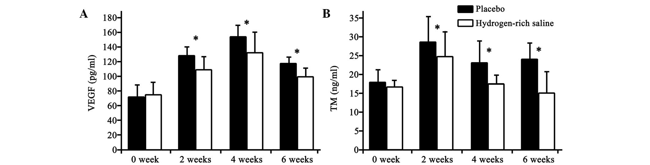
The concentrations of VEGF and TM in the plasma
decreased significantly following the hydrogen-rich saline
treatment (Fig. 1). The differences
between the hydrogen-rich saline and placebo groups were
significant (P<0.05).
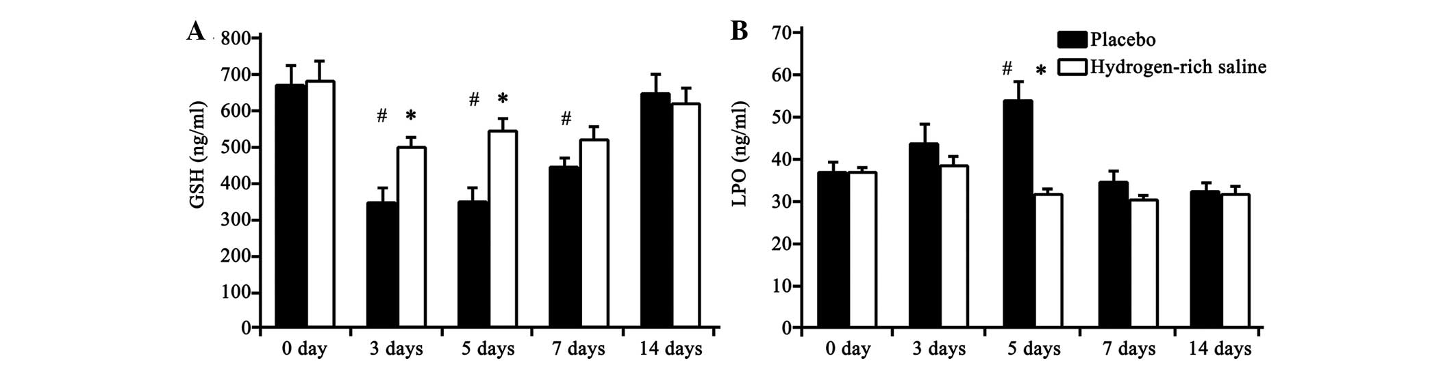
The activity of GSH and LPO was determined as an
indicator of oxidative stress. As shown in Fig. 2, the GSH concentration in the plasma
was significantly increased in the hydrogen-rich saline group at
days 3 and 5 compared with that in the placebo group, and the LPO
concentration was significantly decreased at day 5. No significant
differences between the groups were observed on days 7 and 14.
Compared with baseline levels (immediately prior to
the LPS injection), significant differences were observed in the
GSH level in both the placebo and hydrogen-rich saline groups
between days 3 and 7. With regard to LPO, a significant increase
was only identified on day 5 in the placebo group. These
differences gradually diminished with the passage of time.
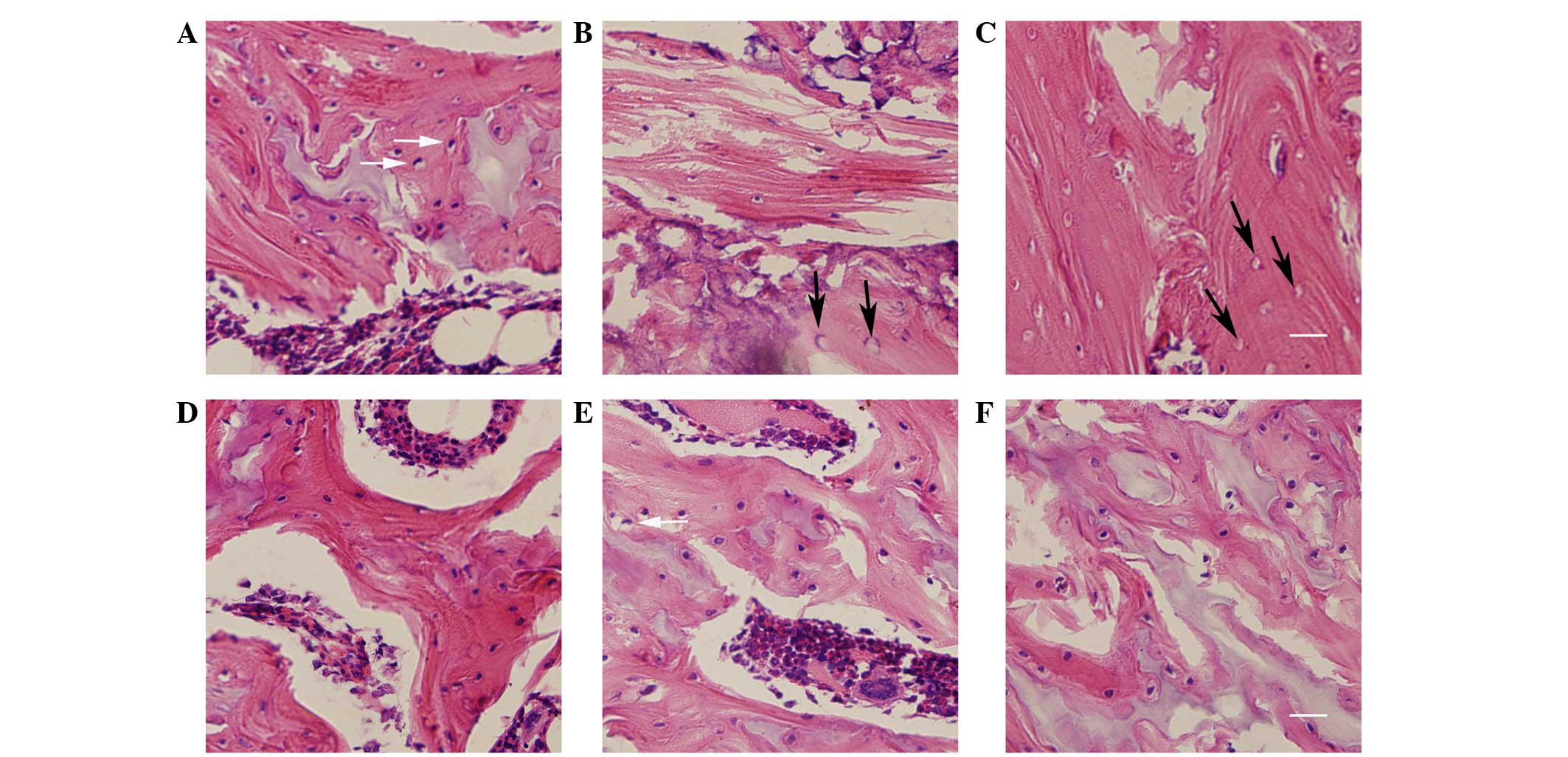
Histopathological observations
The degree of osteonecrosis in the femoral head was
assessed using H&E staining. The trabecular bone, lacunae, bone
marrow and fat tissue were visualized using light microscopy
(Fig. 3). Histological examination
demonstrated that the condition of the femoral head in the placebo
group became gradually aggravated as the time passed. A diffuse
presence of empty lacunae or pyknotic nuclei of osteocytes was
observed in the trabeculae at week 2, and thin trabeculae with a
disordered texture and hypertrophic fat cells were noted. At week
4, thinner bone trabeculae were observed, with breakage of a part
of the bone trabeculae. A number of lacunae were empty, and
abundant adipose cells were found in the marrow cavity. At week 6,
there was sparser trabecular bone with empty lacunae compared with
week 4; however, the empty bone lacunae had clearly increased. The
bone marrow had been partly replaced by fat tissue. There was no
clear indication of new bone formation.
Histological examination revealed improvement in the
trabecular bone, lacunae, bone marrow and fat tissue following
treatment with hydrogen-rich saline. In the hydrogen-rich saline
group, the bone trabeculae were thinner and no diffuse, empty
lacunae were observed. The number of adipose cells had almost
returned to normal at week 2. Some cells exhibited vacuolization or
pyknosis at week 4, and empty bone lacunae were occasionally found.
At the end of the experiment (week 6) a number of empty lacunae and
osteocytes with pyknotic nuclei were found within the trabeculae.
There were no signs of an inflammatory response (inflammatory
cells).
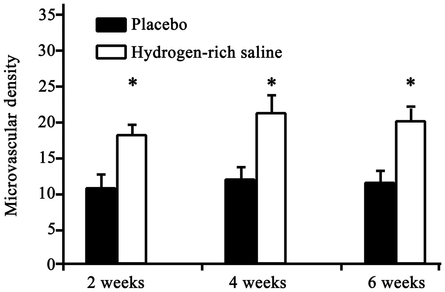
Microvascular characteristics
Microvascular density was evaluated in order to
ascertain vascular changes. All microvessels were highlighted using
immunohistochemical staining. The results revealed a significant
increase in the microvascular density in the hydrogen-rich saline
group compared with that in the placebo group (Fig. 4) (P<0.05).
Discussion
Accumulating evidence suggests that hydrogen has
medicinal properties (4–11,19);
however, the molecular mechanism has yet to be elucidated. Little
is known about the potential role of hydrogen in femoral head
necrosis. In the present study, the effect of hydrogen-rich saline
on femoral head necrosis was evaluated using histopathology,
immunohistochemistry and the quantitative determination of the
plasma levels of VEGF, TM, GSH and LPO in an animal model, which
was established using prednisolone. The levels of the biochemical
indicators VEGF and TM were decreased in the plasma samples treated
with hydrogen-rich saline, suggesting that the treatment relieved
the microvascular injury. The microvascular and histopathological
results were similar, indicating a therapeutic effect of
hydrogen-rich saline on femoral head necrosis. To the best of our
knowledge, the present study is the first to demonstrate a
protective role of hydrogen-rich saline in femoral head necrosis.
Furthermore, it was found that hydrogen-rich saline could markedly
decrease oxidative stress.
Femoral head necrosis is known as Chandler's disease
in adults and Legg-Calve-Perthes disease in children (20). Osteonecrosis indicates cellular death
in both bone and marrow tissue. The pathogenesis of this condition
is extremely complex and still not completely understood, and there
is currently no satisfactory clinical treatment available. Animal
models of corticosteroid-induced bone necrosis have been widely
used in the investigation of the etiology and therapeutics of
femoral head necrosis (21–23). In the present study, the animal
models were established in adult male New Zealand white rabbits
using prednisolone. Only male rabbits were selected in order to
avoid gender variation in the incidence of osteonecrosis. The high
degree of osteonecrosis revealed by the histological examination in
the placebo group indicated successful modeling.
VEGF and TM levels in the plasma are indexes of
endothelial injury (24,25). In the present experiment, the plasma
level of VEGF increased in the first 4 weeks and then decreased
thereafter. This indicates that early necrosis of the femoral head
promotes VEGF secretion. The TM level peaked at week 2 and began to
fall at week 4. The changes in VEGF lagged behind TM. The treatment
with hydrogen-rich saline significantly decreased the levels of
both VEGF and TM. These results indicate that hydrogen is able to
prevent endothelial injury, as shown by the fact that the
significantly increased VEGF and TM levels in the placebo group
were attenuated in the hydrogen-rich saline group. We therefore
suggest that hydrogen-rich saline ameliorates femoral head necrosis
by inhibiting the VEGF and TM expression. The determination of
microvessel density in the present study provided additional
information and showed that the hydrogen-rich saline treatment
resulted in the significant improvement in the microvascular
condition. Hydrogen-rich saline therefore prevents the decrease in
microvascular density. The protective effect of hydrogen-rich
saline against femoral head necrosis was further confirmed
histologically. The significant decrease in VEGF and TM
concentrations and the marked increase in microvascular density
following hydrogen-rich saline treatment indicate that
hydrogen-rich saline could prevent the development of
osteonecrosis.
Since the hydrogen molecule is electrically neutral
and considerably smaller than other antioxidants, it can easily
penetrate membranes and enter organelles (12,26);
however, it remains unclear exactly what type of oxidative stress
is involved in femoral head necrosis. GSH stabilizes the lysosomal
membranes to suppress injury to the vascular endothelium. LPO is a
biochemical indicator of tissue injury and is closely associated
with vascular injury (27). GSH and
LPO in the plasma are biochemical markers indicating the level of
oxidative stress, and GSH has often been used in the study of
osteonecrosis, due to the fact that GSH levels and oxidative stress
are associated with osteonecrosis (28–30). In
the present experiment, hydrogen-rich saline treatment
significantly increased the GSH levels in the plasma, while it
decreased the LPO levels, indicating that hydrogen-rich saline has
a protective effect against oxidative stress in femoral head
necrosis. The results therefore showed that hydrogen-rich saline
could suppress activated oxidative stress in endothelial injury at
an early stage during the development of femoral head necrosis.
In conclusion, the results of this study validated
the therapeutic potential of hydrogen-rich saline by demonstrating
that an injection of this agent can alleviate steroid-associated
osteonecrosis in a rabbit model. The present results also indicate
that hydrogen can protect the endothelium from oxidative stress.
The suppression of the activated oxidative stress leading to
intravascular endothelium injury could be involved in the
underlying mechanism; however, the exact mechanism of the
therapeutic role of hydrogen-rich saline in femoral head necrosis
requires further research.
References
|
1
|
Pietrogrande V and Mastomarino R:
Osteopathy from prolonged cortisone treatment. Ortop Traumatol
Apparate Motore. 25:791–810. 1957.(In Italian).
|
|
2
|
Zhang YG, Wang X, Yang Z, Zhang H, Liu M,
Qiu Y and Guo X: The therapeutic effect of negative pressure in
treating femoral head necrosis in rabbits. PLoS One. 8:e557452013.
View Article : Google Scholar : PubMed/NCBI
|
|
3
|
Wang C, Peng J and Lu S: Summary of the
various treatments for osteonecrosis of the femoral head by
mechanism: A review. Exp Ther Med. 8:700–706. 2014.PubMed/NCBI
|
|
4
|
Ji Q, Hui K, Zhang L, Sun X, Li W and Duan
M: The effect of hydrogen-rich saline on the brain of rats with
transient ischemia. J Surg Res. 168:e95–e101. 2011. View Article : Google Scholar : PubMed/NCBI
|
|
5
|
Ohsawa I, Ishikawa M, Takahashi K,
Watanabe M, Nishimaki K, Yamagata K, Katsura K, Katayama Y, Asoh S
and Ohta S: Hydrogen acts as a therapeutic antioxidant by
selectively reducing cytotoxic oxygen radicals. Nat Med.
13:688–694. 2007. View
Article : Google Scholar : PubMed/NCBI
|
|
6
|
Hayashida K, Sano M, Ohsawa I, Shinmura K,
Tamaki K, Kimura K, Endo J, Katayama T, Kawamura A, Kohsaka S, et
al: Inhalation of hydrogen gas reduces infarct size in the rat
model of myocardial ischemia-reperfusion injury. Biochem Biophys
Res Commun. 373:30–35. 2008. View Article : Google Scholar : PubMed/NCBI
|
|
7
|
Fukuda K, Asoh S, Ishikawa M, Yamamoto Y,
Ohsawa I and Ohta S: Inhalation of hydrogen gas suppresses hepatic
injury caused by ischemia/reperfusion through reducing oxidative
stress. Biochem Biophys Res Commun. 361:670–674. 2007. View Article : Google Scholar : PubMed/NCBI
|
|
8
|
Wang F, Yu G, Liu SY, Li JB, Wang JF, Bo
LL, Qian LR, Sun XJ and Deng XM: Hydrogen-rich saline protects
against renal ischemia/reperfusion injury in rats. J Surg Res.
167:e339–e344. 2011. View Article : Google Scholar : PubMed/NCBI
|
|
9
|
Buchholz BM, Kaczorowski DJ, Sugimoto R,
Yang R, Wang Y, Billiar TR, McCurry KR, Bauer AJ and Nakao A:
Hydrogen inhalation ameliorates oxidative stress in transplantation
induced intestinal graft injury. Am J Transplant. 8:2015–2024.
2008. View Article : Google Scholar : PubMed/NCBI
|
|
10
|
Kawamura T, Huang CS, Tochigi N, Lee S,
Shigemura N, Billiar TR, Okumura M, Nakao A and Toyoda Y: Inhaled
hydrogen gas therapy for prevention of lung transplant-induced
ischemia/reperfusion injury in rats. Transplantation. 90:1344–1351.
2010. View Article : Google Scholar : PubMed/NCBI
|
|
11
|
Huang Y, Xie K, Li J, Xu N, Gong G, Wang
G, Yu Y, Dong H and Xiong L: Beneficial effects of hydrogen gas
against spinal cord ischemia-reperfusion injury in rabbits. Brain
Res. 1378:125–136. 2011. View Article : Google Scholar : PubMed/NCBI
|
|
12
|
Li S, Lu D and Zhang Y and Zhang Y:
Long-term treatment of hydrogen-rich saline abates testicular
oxidative stress induced by nicotine in mice. J Assist Reprod
Genet. 31:109–114. 2014. View Article : Google Scholar : PubMed/NCBI
|
|
13
|
Du Z, Jia H, Liu J, Zhao X, Wang Y and Sun
X: Protective effects of hydrogen-rich saline in uncontrolled
hemorrhagic shock. Exp Ther Med. 7:1253–1258. 2014.PubMed/NCBI
|
|
14
|
Zheng H and Yu YS: Chronic hydrogen-rich
saline treatment attenuates vascular dysfunction in spontaneous
hypertensive rats. Biochem Pharmacol. 83:1269–1277. 2012.
View Article : Google Scholar : PubMed/NCBI
|
|
15
|
Zhang G, Qin L, Sheng H, Wang XL, Wang YX,
Yeung DK, Griffith JF, Yao XS, Xie XH, Li ZR, et al: A novel
semisynthesized small molecule icaritin reduces incidence of
steroid-associated osteonecrosis with inhibition of both thrombosis
and lipid-deposition in a dose-dependent manner. Bone. 44:345–356.
2009. View Article : Google Scholar : PubMed/NCBI
|
|
16
|
Qin L, Zhang G, Sheng H, Yeung KW, Yeung
HY, Chan CW, Cheung WH, Griffith J, Chiu KH and Leung KS: Multiple
bioimaging modalities in evaluation of an experimental
osteonecrosis induced by a combination of lipopolysaccharide and
methylprednisolone. Bone. 39:863–871. 2006. View Article : Google Scholar : PubMed/NCBI
|
|
17
|
Weidner N, Folkman J, Pozza F, Bevilacqua
P, Allred EN, Moore DH, Meli S and Gasparini G: Tumor angiogenesis:
A new significant and independent prognostic indicator in
early-stage breast carcinoma. J Natl Cancer Inst. 84:1875–1887.
1992. View Article : Google Scholar : PubMed/NCBI
|
|
18
|
Weidner N, Semple JP, Welch WR and Folkman
J: Tumor angiogenesis and metastasis-correlation in invasive breast
carcinoma. N Engl J Med. 324:1–8. 1991. View Article : Google Scholar : PubMed/NCBI
|
|
19
|
Hanaoka T, Kamimura N, Yokota T, Takai S
and Ohta S: Molecular hydrogen protects chondrocytes from oxidative
stress and indirectly alters gene expressions through reducing
peroxynitrite derived from nitric oxide. Med Gas Res. 1:182011.
View Article : Google Scholar : PubMed/NCBI
|
|
20
|
McLennan MK and Margolis M: Radiology
rounds. Inflammatory bowel disease (ulcerative colitis) with
steroid-induced avascular necrosis of the femoral heads. Can Fam
Physician. 40:1272–1274. 1994.PubMed/NCBI
|
|
21
|
Guan XY and Han D: Role of
hypercoagulability in steroid-induced femoral head necrosis in
rabbits. J Orthop Sci. 15:365–370. 2010. View Article : Google Scholar : PubMed/NCBI
|
|
22
|
He W, Xu C, Fan Y, Fang B, Li X, Wang H,
Liu S and Yuan H: Effects of the Chinese drugs for activating blood
circulation on plasma TXB2 and 6-keto-PGF1alpha contents in rabbits
with glucocorticoid-induced femoral head necrosis. J Tradit Chin
Med. 24:233–237. 2004.PubMed/NCBI
|
|
23
|
Zhu H, Cai X, Lin T, Shi Z and Yan S:
Low-intensity pulsed ultrasound enhances bone repair in a rabbit
model of steroid-associated osteonecrosis. Clin Orthop Relat Res.
473:1830–1839. 2015. View Article : Google Scholar : PubMed/NCBI
|
|
24
|
Zhang C, Li Y, Cornelia R, Swisher S and
Kim H: Regulation of VEGF expression by HIF-1α in the femoral head
cartilage following ischemia osteonecrosis. Sci Rep. 2:6502012.
View Article : Google Scholar : PubMed/NCBI
|
|
25
|
Aksoy MCI, Aksoy DY, Haznedaroglu IC,
Sayinalp N, Kirazli S and Alpaslan M: Thrombomodulin and GFC levels
in Legg-Calve-Perthes disease. Hematology. 13:324–328. 2008.
View Article : Google Scholar : PubMed/NCBI
|
|
26
|
Zhang CB, Tang YC, Xu XJ, Guo SX and Wang
HZ: Hydrogen gas inhalation protects against liver
ischemia/reperfusion injury by activating the NF-κB signaling
pathway. Exp Ther Med. 9:2114–2120. 2015.PubMed/NCBI
|
|
27
|
Uzel N, Sivas A, Uysal M and Oz H:
Erythrocyte lipid peroxidation and glutathione peroxidase
activities in patients with diabetes mellitus. Horm Metab Res.
19:89–90. 1987. View Article : Google Scholar : PubMed/NCBI
|
|
28
|
Ichiseki T, Kaneuji A, Ueda Y, Nakagawa S,
Mikami T, Fukui K and Matsumoto T: Osteonecrosis development in a
novel rat model characterized by a single application of oxidative
stress. Arthritis Rheum. 63:2138–2141. 2011. View Article : Google Scholar : PubMed/NCBI
|
|
29
|
Ichiseki T, Kaneuji A, Katsuda S, Ueda Y,
Sugimori T and Matsumoto T: DNA oxidation injury in bone early
after steroid administration is involved in the pathogenesis of
steroid-induced osteonecrosis. Rheumatology (Oxford). 44:456–460.
2005. View Article : Google Scholar : PubMed/NCBI
|
|
30
|
Ichiseki T, Matsumoto T, Nishino M,
Kaneuji A and Katsuda S: Oxidative stress and vascular permeability
in steroid-induced osteonecrosis model. J Orthop Sci. 9:509–515.
2004. View Article : Google Scholar : PubMed/NCBI
|