Introduction
An important characteristic of aging is cognitive
decline. Previous findings have shown that the decline of cognitive
function was associated with reduced plasticity in the hippocampus
(1–2). Decreased androgen levels associated
with aging have always been considered to be associated with an
increased risk of mild cognitive impairment (MCI) and Alzheimer's
disease (AD). In 2011, the National Institute on Aging and the
Alzheimer's Association proposed new guidelines for AD (1–4). These
guidelines recommend that MCI be considered a transitional
condition with cognitive changes between normal aging and AD
(5,6). Through evaluation and control of risk
factors for conversion from MCI to AD, onset of the disease can be
prevented or delayed. The pathophysiology of MCI includes
significant neuronal loss in the hippocampus and decreased blood
flow in the subiculum (7).
Neurofibrillary tangles and β-amyloid (Aβ) were observed in
individuals with MCI (8,9). Cerebrospinal fluid also shows a
disproportionate increase in τ protein levels relative to
individuals from a similar age group (10). Patients with MCI usually have
decreased gonadal steroids at the same time.
Depletion of sex steroid hormones with increasing
age is the most significant risk factor for the development of AD.
Previous findings indicated that estrogen and progesterone (P4) at
menopause in women and testosterone in aging men are AD risk
factors (11–18). Women have greater vulnerability to AD
than men, as observed by relatively sudden and extensive loss of
17β-estradiol (E2) and P4 at menopause. A decrease of total
testosterone (12,13) or free testosterone levels in plasma
have been identified in men with AD when compared to patients
(15) of vascular dementia and
age-matched controls (11,14,19).
Estrogens and androgens can play a protective role
against AD-related neurodegeneration (14,20). Men
who become hypogonadal in later life often report problems with
their memory. Lower than normal testosterone levels have also been
detected in patients prior to the onset of AD and in younger,
late-onset, male AD patients when compared to the appropriate
controls. Age-related depletion of testosterone is a risk factor
for AD in men (20). Since
testosterone is metabolized in the brain into the androgen
dihydrotestosterone (DHT) and estrogen E2, testosterone can mediate
its effects through androgen and/or estrogen pathways. The
reduction of DHT in elderly males was negatively associated with
the incidence of AD, and it may be able to decrease the activity of
the androgen receptor. Previous findings showed that androgen is
involved in the development of the central nervous system as well
as the maintenance of normal function during maturation (21–23).
In recent decades, the progressive reduction of
structural and functional plasticity in hippocampus and the
prefrontal cortex were thought to be associated with the gradual
decline in cognitive function (24).
There was a close associated between synaptic plasticity and sex
hormones. It was previously reported that the density of synapses
in the hippocampus of adult female rat fluctuates naturally with
the levels of ovarian steroids. In adult female rats, the estradiol
level in vivo was associated with hippocampal pyramidal cell
synapse density and quantity, as well as dendritic spine density
(25). The density of the dendritic
spines of the pyramidal neurons in the brain regions related to
learning and memory, such as the hippocampus, is associated with
the estrogen level (26). Thus, the
regulatory mechanism of androgens on synaptic plasticity has been
considered. Similarly, the density of the dendritic spines of the
pyramidal neurons in the hippocampus is also modulated in
vivo by the depletion and replacement of androgens (27).
The senescence-accelerated mouse prone 8 (SAMP8) is
characterized by an age-related spontaneous deterioration in
learning and memory that are similar to AD (28). Previous findings have shown that
SAMP8 is a good model of cognitive decline with aging (29–30). Our
previous studies (31) identified a
fluctuant trend of the density of dendritic spines and, Aβ
deposits, leading to cognitive and neurobiological changes in
5-month-old SAMP8 mice. Thus, ‘middle-aged’ SAMP8 mice are a
suitable model for basic MCI research (31). In the MCI stage of SAMP8 mice, the
synaptic injuries have already appeared. As such, the pathological
changes of the synapses are a key problem in cognitive dysfunction
(32).
Gonadectomy (GDNX) in castrated male rats reduced
CA1 spine synapse density compared with the sham-operated controls
(32). Treatment of GDNX rats with
DHT or testosterone propionate increased spine synapse density to
approximately the same levels when compared with intact males.
However, an increase in synapse density was not observed in the
GDNX animals after treatment with estradiol. These data indicated
that androgen is essential for the normal spine synapse density in
the CA1 region of the male rat hippocampus. The lack of response to
estradiol suggests that testosterone acts directly on hippocampal
androgen receptors rather than indirectly via local estrogen
biosynthesis (27).
Previous studies conducted on SAMP8 mice identified
that the serum testosterone levels quickly decreased in the aging
process, with a similar gradual reduction in dendritic spine
density and quantity (30–32). The aging core and behavioral
experiments were also improved after providing a physiological dose
of DHT (1 mg/kg) compared with the castration group (33). Therefore, it is essential to
determine the mechanisms of DHT in hippocampal synaptic
plasticity.
Postsynaptic density protein 95 (PSD95) in
hippocampus exhibited a significant reduction in MCI compared to
subjects without dementia. The expression of synaptophysin (SYN), a
key player in membrane trafficking events preceding exocytosis that
modulates activity-dependent exocytosis, was relatively decreased
in castrated aging mice (34).
Drebrin is an f-actin postsynaptic binding protein that is
associated with synaptic plasticity (35). Previous studies have reported that
the levels of Drebrin decreased in the hippocampus of MCI cases,
and were associated with the cognitive decline (12). Synaptic plasticity-associated
proteins [e.g., SYN, PSD95, developmentally regulated brain protein
(Drebrin)] and the memory formation-related protein cAMP-response
element binding protein (CREB), were correlated with animal
behavior and synaptic morphology (36–39).
The present study focused on whether DHT altered the
expression of CREB, PSD95, SYN and Drebrin and examined the
protective mechanisms and neurological basis of DHT. In our
experiment, immunohistochemistry, western blot analysis and
quantitative polymerase chain reaction (qPCR) analysis were used to
detect the expression of the aforementioned proteins and to observe
the regulation of DHT on synaptic plasticity in the hippocampus of
MCI SAMP8 mice. The results confirmed those of our previous study
regarding the effects of DHT on the behavior and synaptic
plasticity in MCI SAMP8 mice, and provided a valuable theoretical
basis for the protective effects of DHT in MCI.
Materials and methods
Animals and study groups
Five-month-old SAMP8 mice were used as experimental
animals (n=45, 28–32 g), and randomly divided into the
sham-operated control group (P8 group), castration group, and
castration plus dihydrotestosterone group (DHT group). Animals in
this group received the same surgical castration operation as the
castrated group, but without removing the testicles. Mice in the
castrated group underwent bilateral orchiectomy. Mice in the DHT
group received bilateral orchiectomy and DHT injection. Medicine
was administered three days after castration (product code:
2500981; International Laboratory, South San Francisco, CA, USA),
for 60 days by subcutaneous injection (1 mg/kg per day administered
between 5:00 and 6:00 p.m.). Other groups were injected with the
same dose of sterile medical corn oil (0.1 ml). SAMP8 male mice
were given as gifts by David Yew, Professor of the Chinese
University of Hong Kong and bred by our laboratory. For breeding of
mice standard rodent diet and water method were used, with a 12 h
light-dark cycle (lights at 6:00 a.m.) and room temperature
maintained at 21±2°C.
The experimental procedures were implemented in
accordance with the use of mammal in neuroscience health
guidelines, recognized by the Association of Animal Experimentation
Ethics, Hebei Medical University.
Immunohistochemical staining
Each group of mice (n=5) were anesthetized by 1%
sodium pentobarbital [80 mg/kg, intraperitoneal (i.p.) injection]
and perfused with 0.9% saline through the left ventricle, and fixed
with 4% paraformaldehyde, pH 7.4. The brain was carefully removed,
specimens were separated between the superior colliculus and
optical chiasm, and fixed overnight at 4°C. The tissue was divided
into two halves along the median plane, and the gradient ethanol
was dehydrated. The tissue was cleared in xylene and embedded in
paraffin. Paraffin blocks were cut into 5 µm serial sections along
the sliding microtome (Leica-RM2145; Leica, Mannheim, Germany) in
the coronal position. Two consecutive 50 µm tissues were collected
and placed on polylysine-coated slides to detect the protein
expression of CREB, SYN, PSD95 and Drebrin. Following
deparaffinization and hydration, the sections were subjected to
antigen retrieval using a microwave for 30 min and immersed in 3%
hydrogen peroxide in methanol for 30 min to eliminate endogenous
peroxidase activity. The sections were incubated with 5% goat serum
to block non-specific binding, followed by overnight incubation
with rabbit anti-SYN antibody (1:200, product code: ab14692; Abcam,
Cambridge, MA, USA), rabbit anti-PSD95 antibody (1:200, product
code: ab12093; Abcam), rabbit anti-CREB antibody (1:200, code:
ab32515; Abcam), and rabbit anti-Drebrin antibody (1:100, code:
ab60933; Abcam) at 4°C. After washing with PBS, the sections were
incubated with biotinylated goat anti-rabbit IgG for 2 h. A
computer image analysis system Image-Pro Plus 6.0 (Media
Cybernetics, Inc., Rockville, MD, USA) was used to determine the
average values of the optical density (OD) of SYN, PSD95, Drebrin
and CREB in the hippocampal CA1 region. Ten sections were analyzed
for each mouse, and the average OD value was determined for each
mouse.
Western blot analysis
After sample collection, lysis buffer was added, and
the samples were homogenized under ice-cooling, and incubated for
30 min. The samples were then centrifuged for 15 min at 4°C at
10,000 × g and the supernatant was collected. A BAC microplate
reader (VersaMax enzyme standard instrument, Molecular Devices,
LLC. Sunnyvale CA, USA) was used to determine protein content.
Subsequently, 100 µl protein was added to an equal volume of 2X
upper sample buffer, and mixed at 95°C for 5 min, for denaturation
of the protein. The samples were then centrifuged and cooled to
room temperature 21±2°C. A constant voltage of electrophoresis of
upper sample was 80 V. When the sample reached the junction between
stacking and separating gel, the constant voltage was adjusted to
120 V, stopping electrophoresis. The constant voltage of the
cutting gel and transferring membrane was 300 V, which was stopped
after a 60-min wet transfer. Non-fat dry milk (5%) was used for
blocking for 1 h at room temperature. Then, 1X TBS was used to
dilute SYN, PSD95, Drebrin, CREB antibody at 1:5,000, followed by
overnight incubation at 4°C. Fluorescent secondary antibody was
added, and kept in the dark at 37°C prior to agitation for 1 h. The
Li-Cor Odyssey fluorescence scanning imaging system (Gene Company
Limited, Pleasanton, California, USA) was employed to perform
semi-quantitative analysis to yield relevant bands. Arbitrary units
(AU) (darea • density) were used to represent the area × gray value
of protein bands. GAPDH expression was viewed as an internal
reference. The ratio between the target protein and internally
referred AU represented the relative protein expression levels of
each group.
qPCR analysis
Each group of mice (n=5) was anesthetized with 1%
sodium pentobarbital (80 mg/kg, i.p. injection). RNA samples were
extracted using TRIzol reagent from the hippocampus. Ultraviolet
NanoDrop lite spectrophotometer (Thermo Fisher Scientific, Waltham,
MA, USA) (the A260) was used to measure the amount of RNA. The
superscript first-strand synthesis system (Invitrogen Life
Technologies, Carlsbad, CA, USA) was used to reverse transcribe 500
ng RNA into complementary DNA. Following transcription, 1 µl RT
product combined 2.5 units was used to start the volume of Taq DNA
polymerase (Sigma-Aldrich, St. Louis, MO, USA) (50 µl buffer
contains 20 pmol antisense primer, 10 mM Tris-HCl, 50 mM KCl, 2.5
mM MgCl2 and 0.2 mM dNTP). Chemically produced primers
(Sangon Biotech Co., Ltd., Shanghai, China) are listed in Table I.
 | Table I.Primers. |
Table I.
Primers.
| Gene | NCBI GeneID | Forward/reverse
primer | Amplicon size
(bp) |
|---|
| drebrin | 56320 |
5′-AGGAGCAGTCTATCTTTGGTGA-3′ | 246 |
|
|
|
5′-CACTGTCCGATGCCTTTATGAA-3′ |
|
| syn | 20977 |
5′-CAGTTCCGGGTGGTCAAGG-3′ | 138 |
|
|
|
5′-ACTCTCCGTCTTGTTGGCAC-3′ |
|
| creb | 12912 |
5′-CAGGGGTCGCAAGGATTGAAG-3′ | 101 |
|
|
|
5′-ATCGCCTGAGGCAGTGTACT-3′ |
|
| psd95 | 13385 |
5′-TGAGATCAGTCATAGCAGCTACT-3′ | 107 |
|
|
|
5′-CTTCCTCCCCTAGCAGGTCC-3′ |
|
| GAPDH | 14433 |
5′-AGGTCGGTGTGAACGGATTTG-3′ | 95 |
|
|
|
5′-GGGGTCGTTGATGGCAACA-3′ |
|
The Eco™ Real-Time PCR system (Illumina Inc., San
Diego, CA, USA) was used to quantitatively determine the relative
levels of mRNAs. GAPDH expression was viewed as an internal
reference, and the ratio between the data and internal reference
indicated the relative level of the target gene.
Results
Immunohistochemical staining
Immunohistochemical staining showed that the
positive products of CREB protein were mainly distributed in the
neuronal membrane, cytoplasm and projections. Compared with the
castration group, the staining in projections of the P8 and DHT
groups was dense and thick, and the cytoplasmic membrane
immunoreactive substance increased significantly (Fig. 1A-C). The OD value of the castration
group was 0.135±0.025, which was significantly lower than that of
the P8 group value of 0.270±0.046 (P<0.01). The OD value of DHT
group was 0.216±0.034 and was significantly increased compared to
the Castration group (P<0.01).
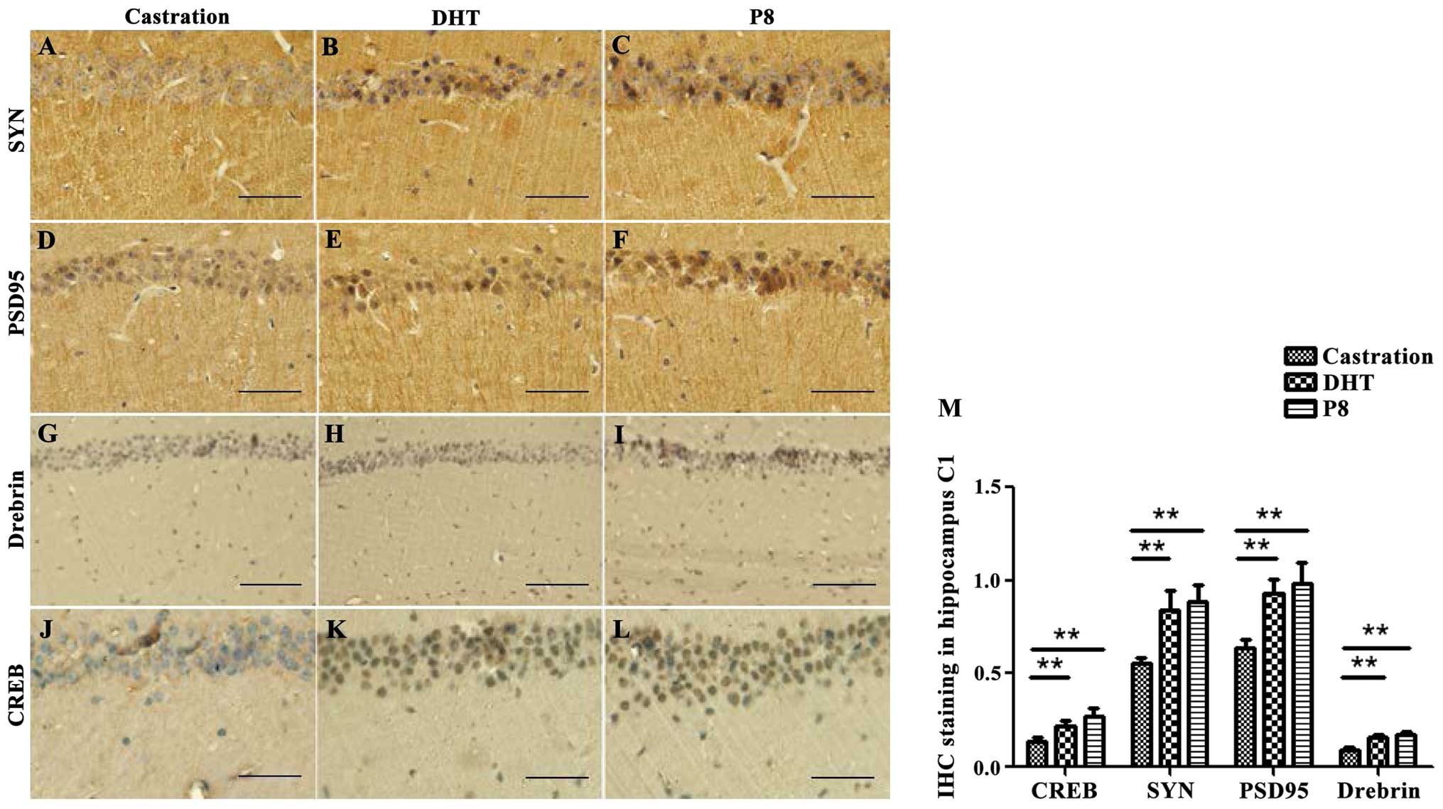 | Figure 1.Immunohistochemical staining shows
the effects of castration and DHT intervention on the conversion
from MCI to Alzheimer's dementia in SAMP8 mice. Immunohistochemical
staining detected (A-C) SYN, (D-F) PSD95, (G-I) Drebrin and (J-K)
CREB protein expression in the (A, D, G and J) castration, (B, E, H
and K) DHT and (C, F, I and L) P8 groups. A statistical graph
showing the difference of protein expression in the (M) four
groups. The results are presented as the mean ± standard deviation
(n=5). Statistical analysis was performed using one-way ANOVA with
LSD post-hoc test. **P<0.01. DHT, dihydrotestosterone; MCI, mild
cognitive impairment; SAMP8, senescence-accelerated mouse prone 8;
SYN, synaptophysin; PSD95, postsynaptic density protein 95; CREB,
cAMP-response element binding protein; Drebrin, developmentally
regulated brain protein. |
Immunohistochemical staining for PSD95, SYN and
Drebrin showed that brownish yellow granules were located in the
neuropil of pyramidal cells (Fig.
1D-L). The expression of the PSD95, SYN and Drebrin proteins
were consistent with CREB protein expression among the different
groups. The OD values were: PSD95, 0.636±0.046, 0.935±0.073 and
0.983±0.116; SYN, 0.553±0.034, 0.843±0.107 and 0.887±0.089; and
Drebrin, 0.089±0.014, 0.159±0.015 and 0.173±0.018 in the
castration, DHT and P8 groups, respectively (Table II, Fig.
1D-L).
| Table II.Results of immunohistochemical
staining shows the effects of castration and DHT intervention on
the conversion from MCI to Alzheimer's dementia in SAMP8
mice.a |
Table II.
Results of immunohistochemical
staining shows the effects of castration and DHT intervention on
the conversion from MCI to Alzheimer's dementia in SAMP8
mice.a
| Group | CREB | SYN | PSD95 | Drebrin |
|---|
| Castration | 0.136±0.025 | 0.553±0.034 | 0.636±0.046 | 0.089±0.014 |
| DHT |
0.216±0.035b |
0.843±0.107b |
0.935±0.073b |
0.159±0.015b |
| P8 |
0.270±0.046b |
0.887±0.089b |
0.983±0.116b |
0.174±0.018b |
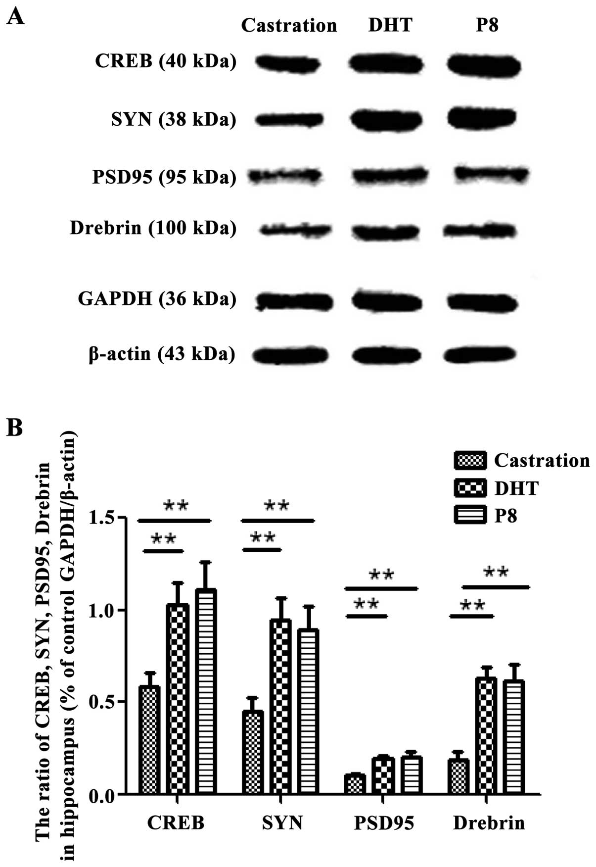
Western blotting
The protein bands for CREB in hippocampus were
clearly visible in the different groups (Fig. 2). The integrated OD (IOD) value of
CREB in the castration group was 0.589±0.075, which was
significantly lower compared to the P8 group at 1.113±0.152
(P<0.01). However, the IOD value for CREB in the DHT group was
1.032±0.118, which was significantly different compared to the
castration group (P<0.01). The expression levels of SYN
(0.453±0.074, 0.947±0.117, 0.891±0.126), PSD95 (0.109±0.007,
0.193±0.018, 0.205±0.025), and Drebrin (0.189±0.045, 0.632±0.058,
0.613±0.092) was consistent with that of CREB protein. There was a
statistically significant difference (P<0.01) between the
castration group and P8 groups (Table
III).
| Table III.Results of western blotting shows the
effects of castration and DHT intervention on the conversion from
MCI to Alzheimer's dementia in SAMP8 mice.a |
Table III.
Results of western blotting shows the
effects of castration and DHT intervention on the conversion from
MCI to Alzheimer's dementia in SAMP8 mice.a
| Group | CREB | SYN | PSD95 | Drebrin |
|---|
| Castration | 0.589±0.075 | 0.453±0.074 | 0.109±0.007 | 0.189±0.045 |
| DHT |
1.032±0.118b |
0.947±0.117b |
0.193±0.018b |
0.632±0.058b |
| P8 |
1.113±0.152b |
0.891±0.126b |
0.205±0.025b |
0.613±0.092b |
qPCR
The mRNA expression of creb, psd95,
syn and drebrin in hippocampal neurons in different
groups was similar to that of their protein expression levels
(Fig. 3). The creb,
psd95, syn and drebrin mRNAs levels in the
castration group were the lowest (0.597±0.075, 0.853±0.079,
0.420±0.057 and 0.328±0.019, respectively) and were significantly
decreased compared with the P8 group (1.245±0.059, P<0.01;
1.231±0.116, P<0.01; 0.894±0.052, P<0.01; 0.452±0.041,
P<0.01, respectively) and the DHT group (1.177±0.101, P<0.01;
1.194±0.064, P<0.01; 0.861±0.026, P<0.01; 0.422±0.018,
P<0.01, respectively) (Table IV,
Fig. 3).
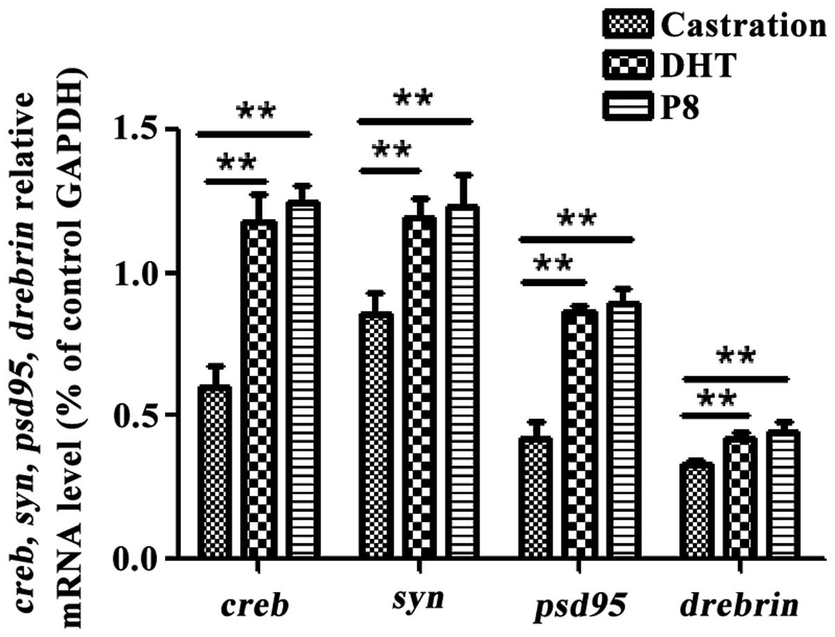 | Figure 3.Quantitative polymerase chain
reaction showing effects of castration and DHT intervention on the
conversion from MCI to Alzheimer's dementia in SAMP8 mice, showing
gene expression of SYN, PSD95, Drebrin, and CREB in the Castration,
DHT and P8 groups. **P<0.01. DHT, dihydrotestosterone; SAMP8,
senescence-accelerated mouse prone 8; SYN, synaptophysin; PSD95,
post synaptic density protein 95; CREB, cAMP-response element
binding protein; Drebrin, developmentally regulated brain
protein. |
| Table IV.Results of quantitative polymerase
chain reaction (qPCR) display the effects of castration and DHT
intervention on the conversion from MCI to Alzheimer's dementia in
SAMP8 mice.a |
Table IV.
Results of quantitative polymerase
chain reaction (qPCR) display the effects of castration and DHT
intervention on the conversion from MCI to Alzheimer's dementia in
SAMP8 mice.a
| Group | creb | syn | psd95 | drebrin |
|---|
| Castration | 0.597±0.075 | 0.853±0.079 | 0.420±0.057 | 0.328±0.019 |
| DHT |
1.177±0.101b |
1.194±0.064b |
0.861±0.026b |
0.422±0.018b |
| P8 |
1.245±0.059b |
1.231±0.116b |
0.894±0.052b |
0.452±0.041b |
Discussion
The brain is an androgen-sensitive tissue and is
influenced by age-related androgen deficiency (12–14).
Research on testosterone and its metabolite DHT in rodent and human
brains has indicated that reduced brain function is closely related
to androgen loss. In particular, some research suggests that sex
hormone loss occurs prior to the occurrence of AD, and can be
regarded as a cause of AD, rather than the result of AD. This fact
suggests that androgen depletion is a precursor event that changes
AD clinical manifestation and pathology, which may contribute to
the understanding of the progression of the disease (40). Despite intensive laboratory and
clinical research over the past three decades, effective treatment
to delay the onset and progression of AD is not available. The
repetitive failures of clinical trials targeting mild to moderate
AD suggest that MCI is a potential way to treat AD (41).
Compared to other pathological lesions, in the
pathological change of AD, synaptic density changes and cognitive
impairment are closely related, indicating the extreme importance
of mechanisms of contact obstacles of synapses (42). Previous findings suggest that the
hippocampus shows decreases of synaptic plasticity in MCI (31,33).
However, whether the protein change in the hippocampus reflects the
correlation between changes in the number of synapses and low MCI
cognitive level is worthy of further investigation.
In young and middle-aged castrated rats, the levels
of endogenous androgens were reduced, aggravating pathological
changes and hippocampus-related behavior in the animals. Treatment
with testosterone or DHT in these castrated rats may significantly
reduce pathological damage (43). It
seems possible to develop an effective hormone or hormone mimic
therapy for prevention of the conversion from MCI to AD (44). Previous findings have indicated that
there is close relationship between androgens and synapses
(31,32). Experimentally, androgens can reverse
the reduced density of hippocampus dendritic spines in castrated
male rats, and at the same time an androgen antagonist may
significantly weaken this effect (45–47).
Those reports suggested that androgens play an important role in
maintaining the density of dendritic spine synapses in the
hippocampus of normal male animals. Therefore, we hypothesize that
the synaptic plasticity of hippocampal structures is correlated
with the cognitive function. Studies have reported the existence of
a pathway for androgen regulation of synaptic function that is
distinct from estrogens; however, the mechanism of this independent
androgen pathway was not previously evident (48).
Our previous studies (31,33) have
shown that, compared with the control group, the cognitive function
of castrated MCI and SAMP8 mice during AD period was impaired.
Additionally, DHT improves the aging mice hippocampus-dependent
spatial cognitive ability, and castration increases learning and
memory disorders. These functions may be associated with changes in
the hippocampal formation and morphology of MCI SAMP8 mice. From
this perspective, hippocampal neurons and synaptic morphology and
structure are worthy of exploration in further experiments.
Castration-induced cognitive dysfunction may cause morphological
change (32). Castration reduces the
thickness of postsynaptic density and postsynaptic PSD, and
significantly increases Aβ accumulation and degeneration of
organelles in cytoplasm. It is thought that synaptic loss and
cognitive decline in AD are closely related, and senile plaque
pathology and partial loss of synapses and deduction of dendritic
spines are related (49).
Conversely, cognitive disorders, especially in executive function
and negative symptoms are considered to be associated with defects
of neural plasticity (50). These
views explain the correlation between castration-induced cognitive
decline and morphological changes.
Our previous results showed that castration
aggravates cognitive decline and the loss of synapses and dendritic
spines in MCI SAMP8 (31,33). In the process of natural aging, a
decrease in testosterone induces the AD pathological change process
and decreases synaptic plasticity. Modulation of hippocampal
synaptic plasticity by androgens has attracted substantial
scientific attention. However, more studies are needed to elucidate
the effects of DHT on synaptic plasticity in MCI research.
Therefore, we investigated the effect of DHT on the modulation of
spine structure and proteins related to synaptic plasticity in the
MCI SAMP8 mouse hippocampus. In the present study, castration
reduced the levels of synaptic protein markers (PSD95, SYN and
Drebrin), which coincided with smaller numbers of synapses and
thinner PSDs, as observed in our previous results (31,32).
This finding demonstrates that cell viability is low and neural
remodeling is suppressed during MCI, and worse structural and
morphological changes in castration lead to further decline in
spatial learning and memory in the P8 mice.
The above data show that DHT is involved in the
regulation of learning and memory, suggesting that physiological
levels of DHT can regulate hippocampal function and maintain the
hippocampal formation of normal male mice. However, the role of DHT
in some proteins, receptors, neurotransmitters and messenger
molecules associated with structural basis and physiological
functions of synaptic plasticity (51), as well as transcription and protein
synthesis needed in long-term memory potentiation remain to be
investigated (52).
DHT is thought to increase the thickness of
postsynaptic density and postsynaptic density of neurons in
castrated animals. These studies indicate that androgen therapy may
be effective in delaying the deduction of synapses of hippocampal
neurons and dendritic spines caused by castration. Thus, DHT can
also enhance the synaptic plasticity of hippocampal neurons of AD
mice, to maintain the structure and function of synapses. In this
experiment, with P8 sham operation mice as the control group, we
studied whether DHT was conducive to maintaining hippocampal
neurons and hippocampal synaptic plasticity of castrated mice
during MCI period. Synaptic plasticity is the neurobiological basis
of learning and memory, and can affect cognitive ability by
adjusting the hippocampal synaptic structure and function. Based on
this, it is recognized that DHT-induced structural changes of
neural plasticity improve the morphological basis of cognitive
function (53).
We hypothesized that expression changes of
differentiated genes among individuals may be associated with the
memory-related behavior of aging rodents. Furthermore, mechanisms
of DHT regulating hippocampal synaptic plasticity of MCI SAMP8 mice
is unclear. Therefore, we focused on the expression of synaptic
plasticity markers (SYN, PSD95, Drebrin) in pyramidal cells of
hippocampus, and examined the regulatory mechanism and neural basis
effect of DHT.
SYN, PSD95 and Drebrin are involved in synaptic
formation and reconstruction (54–56). The
immunohistochemical results revealed that DHT decreased SYN, PSD95,
Drebrin protein levels of castrated SAMP8 mice. SYN involves the
release of presynaptic vesicle protein neurotransmitter and a sign
of density of synapses. PSD95 is glutamatergic excitatory
postsynaptic density, interacting with NMDA receptor subunits, and
participating in the information storage procedure (57). Increased PSD95 expression is capable
of adjusting the increase of synaptic activity. Drebrin is an
F-actin binding protein expressed in excitatory postsynaptic
dendritic spines. It affects synaptic plasticity by regulating the
synaptic morphogenesis, and reorganizing postsynaptic density and
target receptors (39,58,59).
Drebrin, a synaptic marker, is used as a marker of synaptic
structural changes. Experimental results show that the effect of
DHT on synaptic structure is associated with the expression of
protein markers SYN, PSD95 and Drebrin (60,61).
Expression of synaptic proteins SYN, PSD95 and
Drebrin reflects changes in cognitive behavior, and shows a
positive correlation with synaptic plasticity, a result that is
consistent with the results of the present study. This also
corroborates our previous studies (31,33)
where, after DHT treatment, learning and memory of castrated mice
were significantly improved, Aβ levels were reduced, and the number
of dendritic spines density increased (27,62).
Expression of SYN and Drebrin in the hippocampus of patients with
advanced AD decreased. However, in other studies, after comparing
patients with no cognitive impairment (NCI), mild cognitive
impairment (MCI) and AD, it was identified that, SYN and Drebrin
expression in male hippocampus is different. For SYN expression,
MCI and AD were reduced compared to NCI, although the differences
were not statistically significant, whereas Drebrin in MCI and AD
were reduced by approximately 40% compared to NCI. In the MCI and
AD patients, the difference of synaptic marker protein expression
indicates that the plasticity of postsynaptic membrane dendritic
spines may be damaged selectively. Furthermore, Drebrin may serve
as a protein marker predictor for the onset of memory impairment.
(63).
The role of androgen in hippocampal pyramidal
neurons in male rats has been investigated and with regard to the
expression of SYN, it was found that castration does not
significantly affect the number of pyramidal cells in the
hippocampal CA1 area, although the SYN level is reduced (27). The SYN level of castrated rats is
increased after DHT supplementation. Similarly, DHT and estradiol
SYN expression levels were increased for castrated male and female
rodent animals. The data suggested that sex hormones were closely
associated with maintaining a normal SYN expression level in the
CA1 area of the rodent hippocampus, indicatign AD-like pathological
change caused by lack of sex hormone and pathogenesis that is
different to humans. A comparison of testosterone and non-aromatic
DHT has shown that, testosterone may, without the aid of a local
estrogen pathway, directly affect the hippocampus of the androgen
receptor.
Previous studies showed that, CREB has a potential
therapeutic effect for AD patients (64). In the early stage of AD, CREB was the
most important transcription control factor in the transport
network, suggesting that, in the early stage of the disease,
dysfunction caused by CREB emerges (65). CREB plays an important role in the
neuron plasticity of the brain and long-term memory, that is,
synaptic transmission and restructuring. In the pathogenesis of the
Aβ-induced pathological change of AD, mediated by the CREB pathway,
Aβ changes hippocampus-dependent synaptic plasticity alters and
reduces memory loss caused by a number of synapses (66). Thus, CREB protein expression is
detected to analyze the impact of DHT in AD pathogenesis.
Our results indicate that CREB protein expression,
and the mRNA transcription, protein and expression of PSD95, SYN
and Drebrin of castrated SAMP8 mice was decreases. In the present
study, CREB protein and mRNA expression of MCI SAMP8 mice was
increased after DHT supplementation, as were the synaptic marker
protein and mRNA expression. Thus, these data and our results
indicate that enhanced synaptic plasticity and cognitive function
are closely related to the improved expression of CREB following
DHT increase. Cognitive decline and a decrease of DHT levels are
consistent, thus, DHT may be involved in the CREB-induced
regulation of synaptic plasticity and cognitive function. DHT, a
non-aromatized androgen, cannot be converted to estradiol, and thus
plays a role through the estrogen pathway, indicating that the
androgen pathway plays a role in synaptic plasticity and the
cognitive function of SAMP8 male mice (31,33).
These results suggest that, DHT promotes the expression of
dendritic spine density and constitutive proteins, rather than
converse androgens into estrogens and play a role through the
estrogen pathway. Thus, our results are useful for understanding
the differences of signaling pathways across the brain between
different genders. The results are also consistent with our
previous results (31,33), and directly or indirectly support
that testosterone and DHT can improve the cognitive behavior of
castrated rats and mice, eliminating biological markers (Aβ) and
increasing the number and density of Nissl staining dendritic
spines and other experimental results.
The present results indicate that, DHT can affect
cognitive and synaptic plasticity of SAMP8 mice during MCI period.
Previous findings have shown that cognitive competence and neural
plasticity of SAMP8 mice during MCI period decline (31,33).
Synaptic plasticity in the hippocampus structure may be the cause
of reduced spatial memory impairment and this function is related
to androgen decline in male mice. In turn, we believe that
androgens play an important role in maintaining and regulating
hippocampal synaptic plasticity structure in aging mice. Androgen
maintains synaptic marker protein (SYN, PSD95 and Drebrin)
expression and activates CREB gene expression, thereby regulating
synaptic plasticity changes in the basic structure of MCI
hippocampus and improving cognitive function. It may be the neural
basis mechanism of the wholesome effects which androgen applied to
synaptic plasticity in hippocampus. Androgen increases synaptic
activity, thereby promoting neural proliferation and maintenance of
cells in the MCI SAMP8 mouse hippocampus area, and plays a
neuro-protective role. However, the protective effect of androgens
in neuroplasticity is worthy of further investigation.
In summary, this study was performed in SAMP8 male
mice, and it was determined that the role of mechanism of action of
androgens in humans requires additional investigation.
Acknowledgements
The present study was supported by the National
Natural Science Foundation of China Grants (nos. 31271191 and
31471145) and the Natural Science Foundation of Hebei Province
Grants (no. C2012206128).
References
|
1
|
Albert MS, DeKosky ST, Dickson D, Dubois
B, Feldman HH, Fox NC, Gamst A, Holtzman DM, Jagust WJ, Petersen
RC, et al: The diagnosis of mild cognitive impairment due to
Alzheimer's disease: Recommendations from the National Institute on
Aging-Alzheimer's Association workgroups on diagnostic guidelines
for Alzheimer's disease. Alzheimers Dement. 7:270–279. 2011.
View Article : Google Scholar : PubMed/NCBI
|
|
2
|
Alzheimer's Association: 2013 Alzheimer's
disease facts and figures. Alzheimers Dement. 9:208–245. 2013.
View Article : Google Scholar : PubMed/NCBI
|
|
3
|
Jack CR Jr, Albert MS, Knopman DS, McKhann
GM, Sperling RA, Carrillo MC, Thies B and Phelps CH: Introduction
to the recommendations from the National Institute on
Aging-Alzheimer's Association workgroups on diagnostic guidelines
for Alzheimer's disease. Alzheimers Dement. 7:257–262. 2011.
View Article : Google Scholar : PubMed/NCBI
|
|
4
|
Sperling RA, Aisen PS, Beckett LA, Bennett
DA, Craft S, Fagan AM, Iwatsubo T, Jack CR Jr, Kaye J, Montine TJ,
et al: Toward defining the preclinical stages of Alzheimer's
disease: Recommendations from the National Institute on
Aging-Alzheimer's Association workgroups on diagnostic guidelines
for Alzheimer's disease. Alzheimers Dement. 7:280–292. 2011.
View Article : Google Scholar : PubMed/NCBI
|
|
5
|
DeCarli C: Mild cognitive impairment:
Prevalence, prognosis, aetiology, and treatment. Lancet Neurol.
2:15–21. 2003. View Article : Google Scholar : PubMed/NCBI
|
|
6
|
Petersen RC: Mild cognitive impairment:
Current research and clinical implications. Semin Neurol. 27:22–31.
2007. View Article : Google Scholar : PubMed/NCBI
|
|
7
|
Jack CR Jr, Petersen RC, Xu YC, O'Brien
PC, Smith GE, Ivnik RJ, Boeve BF, Waring SC, Tangalos EG and Kokmen
E: Prediction of AD with MRI-based hippocampal volume in mild
cognitive impairment. Neurology. 52:1397–1403. 1999. View Article : Google Scholar : PubMed/NCBI
|
|
8
|
Mufson EJ, Chen EY, Cochran EJ, Beckett
LA, Bennett DA and Kordower JH: Entorhinal cortex beta-amyloid load
in individuals with mild cognitive impairment. Exp Neurol.
158:469–490. 1999. View Article : Google Scholar : PubMed/NCBI
|
|
9
|
Price JL and Morris JC: Tangles and
plaques in nondemented aging and ‘preclinical’ Alzheimer's disease.
Ann Neurol. 45:358–368. 1999. View Article : Google Scholar : PubMed/NCBI
|
|
10
|
Lautenschlager NT, Riemenschneider M,
Drzezga A and Kurz AF: Primary degenerative mild cognitive
impairment: Study population, clinical, brain imaging and
biochemical findings. Dement Geriatr Cogn Disord. 12:379–386. 2001.
View Article : Google Scholar : PubMed/NCBI
|
|
11
|
Hogervorst E, Bandelow S, Combrinck M and
Smith AD: Low free testosterone is an independent risk factor for
Alzheimer's disease. Exp Gerontol. 39:1633–1639. 2004. View Article : Google Scholar : PubMed/NCBI
|
|
12
|
Hogervorst E, Combrinck M and Smith AD:
Testosterone and gonadotropin levels in men with dementia. Neuro
Endocrinol Lett. 24:203–208. 2003.PubMed/NCBI
|
|
13
|
Hogervorst E, Williams J, Budge M,
Barnetson L, Combrinck M and Smith AD: Serum total testosterone is
lower in men with Alzheimer's disease. Neuro Endocrinol Lett.
22:163–168. 2001.PubMed/NCBI
|
|
14
|
Paoletti AM, Congia S, Lello S, Tedde D,
Orrù M, Pistis M, Pilloni M, Zedda P, Loddo A and Melis GB: Low
androgenization index in elderly women and elderly men with
Alzheimer's disease. Neurology. 62:301–303. 2004. View Article : Google Scholar : PubMed/NCBI
|
|
15
|
Watanabe T, Koba S, Kawamura M, Itokawa M,
Idei T, Nakagawa Y, Iguchi T and Katagiri T: Small dense
low-density lipoprotein and carotid atherosclerosis in relation to
vascular dementia. Metabolism. 53:476–482. 2004. View Article : Google Scholar : PubMed/NCBI
|
|
16
|
Manly JJ, Merchant CA, Jacobs DM, Small
SA, Bell K, Ferin M and Mayeux R: Endogenous estrogen levels and
Alzheimer's disease among postmenopausal women. Neurology.
54:833–837. 2000. View Article : Google Scholar : PubMed/NCBI
|
|
17
|
Moffat SD, Zonderman AB, Metter EJ,
Blackman MR, Harman SM and Resnick SM: Longitudinal assessment of
serum free testosterone concentration predicts memory performance
and cognitive status in elderly men. J Clin Endocrinol Metab.
87:5001–5007. 2002. View Article : Google Scholar : PubMed/NCBI
|
|
18
|
Tsolaki M, Grammaticos P, Karanasou C,
Balaris V, Kapoukranidou D, Kalpidis I, Petsanis K and Dedousi E:
Serum estradiol, progesterone, testosterone, FSH and LH levels in
postmenopausal women with Alzheimer's dementia. Hell J Nucl Med.
8:39–42. 2005.PubMed/NCBI
|
|
19
|
Chu LW, Tam S, Wong RL, Yik PY, Song Y,
Cheung BM, Morley JE and Lam KS: Bioavailable testosterone predicts
a lower risk of Alzheimer's disease in older men. J Alzheimers Dis.
21:1335–1345. 2010.PubMed/NCBI
|
|
20
|
Pike CJ, Rosario ER and Nguyen TV:
Androgens, aging, and Alzheimer's disease. Endocrine. 29:233–241.
2006. View Article : Google Scholar : PubMed/NCBI
|
|
21
|
Liu L, Orozco IJ, Planel E, Wen Y,
Bretteville A, Krishnamurthy P, Wang L, Herman M, Figueroa H, Yu
WH, et al: A transgenic rat that develops Alzheimer's disease-like
amyloid pathology, deficits in synaptic plasticity and cognitive
impairment. Neurobiol Dis. 31:46–57. 2008. View Article : Google Scholar : PubMed/NCBI
|
|
22
|
Corbyn Z: New set of Alzheimer's trials
focus on prevention. Lancet. 381:614–615. 2013. View Article : Google Scholar : PubMed/NCBI
|
|
23
|
Ambati J and Fowler BJ: Mechanisms of
age-related macular degeneration. Neuron. 75:26–39. 2012.
View Article : Google Scholar : PubMed/NCBI
|
|
24
|
Bisaz R, Boadas-Vaello P, Genoux D and
Sandi C: Age-related cognitive impairments in mice with a
conditional ablation of the neural cell adhesion molecule. Learn
Mem. 20:183–193. 2013. View Article : Google Scholar : PubMed/NCBI
|
|
25
|
Woolley CS and McEwen BS: WoolleyCS:
Estradiol mediates fluctuation in hippocampal synapse density
during the estrous cycle in the adult rat. J Neurosci.
12:2549–2554. 1992.PubMed/NCBI
|
|
26
|
Scheff SW and Price DA: Synaptic density
in the inner molecular layer of the hippocampal dentate gyrus in
Alzheimer disease. J Neuropathol Exp Neurol. 57:1146–1153. 1998.
View Article : Google Scholar : PubMed/NCBI
|
|
27
|
Leranth C, Petnehazy O and MacLusky NJ:
Gonadal hormones affect spine synaptic density in the CA1
hippocampal subfield of male rats. J Neurosci. 23:1588–1592.
2003.PubMed/NCBI
|
|
28
|
Takeda T, Hosokawa M and Higuchi K:
Senescence-accelerated mouse (SAM): A novel murine model of
senescence. Exp Gerontol. 32:105–109. 1997. View Article : Google Scholar : PubMed/NCBI
|
|
29
|
Tresguerres JA, Kireev R, Forman K, Cuesta
S, Tresguerres AF and Vara E: Effect of chronic melatonin
administration on several physiological parameters from old Wistar
rats and SAMP8 mice. Curr Aging Sci. 5:242–253. 2012. View Article : Google Scholar : PubMed/NCBI
|
|
30
|
Morley JE, Farr SA, Kumar VB and Armbrecht
HJ: The SAMP8 mouse: A model to develop therapeutic interventions
for Alzheimer's disease. Curr Pharm Des. 18:1123–1130. 2012.
View Article : Google Scholar : PubMed/NCBI
|
|
31
|
Kang L, Li S, Xing Z, Li J, Su Y, Fan P,
Wang L and Cui H: Dihydrotestosterone treatment delays the
conversion from mild cognitive impairment to Alzheimer's disease in
SAMP8 mice. Horm Behav. 65:505–515. 2014. View Article : Google Scholar : PubMed/NCBI
|
|
32
|
Jia J, Kang L, Li S, Geng D, Fan P, Wang L
and Cui H: Amelioratory effects of testosterone treatment on
cognitive performance deficits induced by soluble Aβ1-42 oligomers
injected into the hippocampus. Horm Behav. 64:477–486. 2013.
View Article : Google Scholar : PubMed/NCBI
|
|
33
|
Li S, Kang L, Zhang C, Xie G, Li N, Zhang
Y, Du J and Cui H: Effects of dihydrotestosterone on synaptic
plasticity of hippocampus in male SAMP8 mice. Exp Gerontol.
48:778–785. 2013. View Article : Google Scholar : PubMed/NCBI
|
|
34
|
Ma J, Zhang Z, Su Y, Kang L, Geng D, Wang
Y, Luan F, Wang M and Cui H: Magnetic stimulation modulates
structural synaptic plasticity and regulates BDNF-TrkB signal
pathway in cultured hippocampal neurons. Neurochem Int. 62:84–91.
2013. View Article : Google Scholar : PubMed/NCBI
|
|
35
|
Mufson EJ, Binder L, Counts SE, DeKosky
ST, de Toledo-Morrell L, Ginsberg SD, Ikonomovic MD, Perez SE and
Scheff SW: Mild cognitive impairment: Pathology and mechanisms.
Acta Neuropathol. 123:13–30. 2012. View Article : Google Scholar : PubMed/NCBI
|
|
36
|
Terakawa Y, Agnihotri S, Golbourn B, Nadi
M, Sabha N, Smith CA, Croul SE and Rutka JT: The role of drebrin in
glioma migration and invasion. Exp Cell Res. 319:517–528. 2013.
View Article : Google Scholar : PubMed/NCBI
|
|
37
|
Tu JC, Xiao B, Naisbitt S, et al: Coupling
of mGluR/Homer and PSD-95 complexes by the Shank family of
postsynaptic density proteins. Neuron. 23:583–592. 1999. View Article : Google Scholar : PubMed/NCBI
|
|
38
|
Du H, Guo L, Yan S, Sosunov AA, McKhann GM
and Yan SS: Early deficits in synaptic mitochondria in an
Alzheimer's disease mouse model. Proc Natl Acad Sci USA.
107:18670–18675. 2010. View Article : Google Scholar : PubMed/NCBI
|
|
39
|
Hatanaka Y, Mukai H, Mitsuhashi K, Hojo Y,
Murakami G, Komatsuzaki Y, Sato R and Kawato S: Androgen rapidly
increases dendritic thorns of CA3 neurons in male rat hippocampus.
Biochem Biophys Res Commun. 381:728–732. 2009. View Article : Google Scholar : PubMed/NCBI
|
|
40
|
Moffat SD, Zonderman AB, Metter EJ, Kawas
C, Blackman MR, Harman SM and Resnick SM: Free testosterone and
risk for Alzheimer disease in older men. Neurology. 62:188–193.
2004. View Article : Google Scholar : PubMed/NCBI
|
|
41
|
Selkoe DJ: Preventing Alzheimer's disease.
Science. 337:1488–1492. 2012. View Article : Google Scholar : PubMed/NCBI
|
|
42
|
Bell KF and Claudio Cuello A: Altered
synaptic function in Alzheimer's disease. Eur J Pharmacol.
545:11–21. 2006. View Article : Google Scholar : PubMed/NCBI
|
|
43
|
Bilkei-Gorzo A: Genetic mouse models of
brain ageing and Alzheimer's disease. Pharmacol Ther. 142:244–257.
2014. View Article : Google Scholar : PubMed/NCBI
|
|
44
|
Demartini DR, Schilling LP, da Costa JC
and Carlini CR: Alzheimer's and Parkinson's diseases: An
environmental proteomic point of view. J Proteomics. 104:24–36.
2014. View Article : Google Scholar : PubMed/NCBI
|
|
45
|
Balthazart J, Baillien M, Charlier TD and
Ball GF: Calcium-dependent phosphorylation processes control brain
aromatase in quail. Eur J Neurosci. 17:1591–1606. 2003. View Article : Google Scholar : PubMed/NCBI
|
|
46
|
Takeuchi Y, Miyamoto E and Fukunaga K:
Activation of the rat dopamine D2 receptor promoter by
mitogen-activated protein kinase and Ca2+/calmodulin-dependent
protein kinase II pathways. J Neurochem. 83:784–796. 2002.
View Article : Google Scholar : PubMed/NCBI
|
|
47
|
Ishida A, Shigeri Y, Taniguchi T and
Kameshita I: Protein phosphatases that regulate multifunctional
Ca2+/calmodulin-dependent protein kinases: From biochemistry to
pharmacology. Pharmacol Ther. 100:291–305. 2003. View Article : Google Scholar : PubMed/NCBI
|
|
48
|
Ooishi Y, Kawato S, Hojo Y, Hatanaka Y,
Higo S, Murakami G, Komatsuzaki Y, Ogiue-Ikeda M, Kimoto T and
Mukai H: Modulation of synaptic plasticity in the hippocampus by
hippocampus-derived estrogen and androgen. J Steroid Biochem Mol
Biol. 131:37–51. 2012. View Article : Google Scholar : PubMed/NCBI
|
|
49
|
Spires TL, Meyer-Luehmann M, Stern EA,
McLean PJ, Skoch J, Nguyen PT, Bacskai BJ and Hyman BT: Dendritic
spine abnormalities in amyloid precursor protein transgenic mice
demonstrated by gene transfer and intravital multiphoton
microscopy. J Neurosci. 25:7278–7287. 2005. View Article : Google Scholar : PubMed/NCBI
|
|
50
|
Voineskos D, Rogasch NC, Rajji TK,
Fitzgerald PB and Daskalakis ZJ: A review of evidence linking
disrupted neural plasticity to schizophrenia. Can J Psychiatry.
58:86–92. 2013.PubMed/NCBI
|
|
51
|
Sheng M and Kim MJ: Postsynaptic signaling
and plasticity mechanisms. 298:776–780. 2002.
|
|
52
|
Hernandez PJ and Abel T: The role of
protein synthesis in memory consolidation: Progress amid decades of
debate. Neurobiol Learn Mem. 89:293–311. 2008. View Article : Google Scholar : PubMed/NCBI
|
|
53
|
Hasan A, Nitsche MA, Rein B,
Schneider-Axmann T, Guse B, Gruber O, Falkai P and Wobrock T:
Dysfunctional long-term potentiation-like plasticity in
schizophrenia revealed by transcranial direct current stimulation.
Behav Brain Res. 224:15–22. 2011. View Article : Google Scholar : PubMed/NCBI
|
|
54
|
Honer WG, Dickson DW, Gleeson J and Davies
P: Regional synaptic pathology in Alzheimer's disease. Neurobiol
Aging. 13:375–382. 1992. View Article : Google Scholar : PubMed/NCBI
|
|
55
|
Davidsson P and Blennow K: Neurochemical
dissection of synaptic pathology in Alzheimer's disease. Int
Psychogeriatr. 10:11–23. 1998. View Article : Google Scholar : PubMed/NCBI
|
|
56
|
Harigaya Y, Shoji M, Shirao T and Hirai S:
Disappearance of actin-binding protein, drebrin, from hippocampal
synapses in Alzheimer's disease. J Neurosci Res. 43:87–92. 1996.
View Article : Google Scholar : PubMed/NCBI
|
|
57
|
Gardoni F, Marcello E and Di Luca M:
Postsynaptic density-membrane associated guanylate kinase proteins
(PSD- MAGUKs) and their role in CNS disorders. Neuroscience.
158:324–333. 2009. View Article : Google Scholar : PubMed/NCBI
|
|
58
|
Kirchberg BC, Cohen JR, Adelsky MB,
Buthorn JJ, Gomar JJ, Gordon M, Koppel J, Christen E,
Conejero-Goldberg C, Davies P, et al: Semantic distance
abnormalities in mild cognitive impairment: Their nature and
relationship to function. Am J Psychiatry. 169:1275–1283. 2012.
View Article : Google Scholar : PubMed/NCBI
|
|
59
|
Verma M and Howard RJ: Semantic memory and
language dysfunction in early Alzheimer's disease: A review. Int J
Geriatr Psychiatry. 27:1209–1217. 2012. View Article : Google Scholar : PubMed/NCBI
|
|
60
|
Kwon SE and Chapman ER: Synaptophysin
regulates the kinetics of synaptic vesicle endocytosis in central
neurons. Neuron. 70:847–854. 2011. View Article : Google Scholar : PubMed/NCBI
|
|
61
|
Sultana R, Banks WA and Butterfield DA:
Decreased levels of PSD95 and two associated proteins and increased
levels of BCl2 and caspase 3 in hippocampus from subjects with
amnestic mild cognitive impairment: Insights into their potential
roles for loss of synapses and memory, accumulation of Abeta, and
neurodegeneration in a prodromal stage of Alzheimer's disease. J
Neurosci Res. 88:469–477. 2010.PubMed/NCBI
|
|
62
|
Leranth C, Hajszan T and MacLusky NJ:
Androgens increase spine synapse density in the CA1 hippocampal
subfield of ovariectomized female rats. J Neurosci. 24:495–499.
2004. View Article : Google Scholar : PubMed/NCBI
|
|
63
|
Counts SE, He B, Nadeem M, Wuu J, Scheff
SW and Mufson EJ: Hippocampal drebrin loss in mild cognitive
impairment. Neurodegener Dis. 10:216–219. 2012. View Article : Google Scholar : PubMed/NCBI
|
|
64
|
Pugazhenthi S, Wang M, Pham S, Sze CI and
Eckman CB: Downregulation of CREB expression in Alzheimer's brain
and in Aβ-treated rat hippocampal neurons. Mol Neurodegener.
6:602011. View Article : Google Scholar : PubMed/NCBI
|
|
65
|
Satoh J, Tabunoki H and Arima K: Molecular
network analysis suggests aberrant CREB-mediated gene regulation in
the Alzheimer disease hippocampus. Dis Markers. 27:239–252. 2009.
View Article : Google Scholar : PubMed/NCBI
|
|
66
|
Saura CA and Valero J: The role of CREB
signaling in Alzheimer's disease and other cognitive disorders. Rev
Neurosci. 22:153–169. 2011. View Article : Google Scholar : PubMed/NCBI
|