Introduction
Cardiomyopathy is a heterogeneous heart disease. The
European Society of Cardiology has defined cardiomyopathy in
children as ‘a heart disease generated by an abnormal cardiac
structure and function caused by non-coronary heart disease,
hypertension, valvular heart disease or congenital heart disease’
(1,2). Statistics show the morbidity of
pediatric cardiomyopathy has been on the increase annually probably
due to the increasing number of immunocompromised infants and the
aggravation of environmental pollution and other environmental
problems (3). Relevant research
results show that the current incidence of cardiomyopathy in
children is ~0.001% (4) while
postoperative rehabilitation is extremely poor for children with
cardiomyopathy (5), the mortality
rate after surgical procedures is approximately 30% and there is a
high need for cardiac transplantation (6). According to the published results from
the National Child Health Survey Statistics released in 2104, the
current incidence of cardiomyopathy of children in China is
approximately 30% higher than the incidence in the United States
(7). In spite of the bleak prognosis
for pediatric cardiomyopathy, investigations have not produced
significant advances (7), and the
etiology and pathogenesis of the disease remain uncertain providing
a weak basis for educated treatment approaches (8).
Previous findings show the ACR1 gene is
important for the growth and repair of myocardial cells in mice,
where it is involved in the promotion of the rehabilitation of
dysfunctional cardiomyocytes (9).
The aim of the present study was to examine the
association between the expression of the ACR1 gene and
cardiomyopathy in children, providing theoretical and practical
preliminary references that may be useful in the elucidation of the
pathogenesis of cardiomyopathy in children.
Materials and methods
General data
In total, 73 children with cardiomyopathy treated at
the Xuzhou Children's Hospital (Jiangsu, China) from April, 2013 to
April, 2015 were enrolled in the study as the observational group.
The patients comprised 38 male and 35 female children, with an
average age of 6.2±3 years. During the same period, 76 healthy
children were selected for the control group. The healthy children
included 34 males and 32 females, with an average age of 5.8±3.2
years. The study subjects were evaluated in accordance with the
relevant standards for children with cardiomyopathy and were found
to suffer no other additional diseases.
Approval for the study was provided by the ethics
committee of Xuzhou Children's Hospital. Written informed consent
was provided by the children's guardians.
Methods
Venous blood samples (6 ml each) were drawn from
each study subject. Samples were centriguged at 2,000 × g for 10
min, and the supernatant serum was stored at −80°C. The cell
pellets were resuspended in frozen stock solution (Biosharp Co.,
Hefei, China), and then cryopreserved at −80°C. ACR1 antibody used
in the present study was purchased from Roche (Mannheim, Germany).
RNA extraction kits were purchased from Axygen (Union City, CA,
USA) and their associated molecular reagents were purchased from
Takara Bio (Dalian, China). RT-qPCR primers were produced by
Shanghai Biological Engineering Co., Ltd. (Shanghai, China).
RT-PCR
RNA extraction
RNA was extracted as per the instructions of the
Axygen kit (10). Briefly, frozen
pelleted cells were mixed with RNA Plus (KeyGen BioTech, Nanjing,
China) to separate the cells and extract the RNA by sequential
centrifugation steps. The purified RNA was resuspended in
H2O prior to verifying the quality of the RNA via
spectroscopy. An aliquot from each sample was used for a reverse
transcription reaction.
RT-qPCR
The RT-qPCR kit was purchased from Takara Bio. A
three-step method was used in the experimental protocol, and the
specific scheme was carried out as per the manufacturer's
instructions. GAPDH was used as an internal control. The primers
used are shown in Table I.
 | Table I.RT-qPCR primers. |
Table I.
RT-qPCR primers.
| Gene | Primer sequence | Fragment length |
|---|
| AVR1 | F:
5′-ACGGTCGATGCAGGTCAGC-3′ | 138 bp |
|
| R:
5′-TGCTCGGACCTAGCATGCAG-3′ |
|
| GAPDH | F:
5′-GAAGGTGAAGGTCGGAGTC-3′ | 226 bp |
|
| R:
5′-GAAGATGGTGATGGGATTTC-3′ |
|
Enzyme-linked immunosorbent assay (ELISA)
An antibody-sandwich method was used to detect the
protein expression level of ACR1 (11), and the specific methods used were: i)
Coating: The protein was diluted with phosphate-buffered saline
buffer solution (pH 9.0) to a concentration of ~5 µg/ml. Then, 0.1
ml of the sample were added to a well in 96-well plates, and stored
at 4°C overnight. Once the protein was sufficiently incubated to
coat the plate, the solution in the 96-well plates was discarded,
and the plate was washed with scrubbing solution five times for 2
min at a time. ii) Sample addition: Each 0.1 ml serum sample was
added to a well in the abovementioned 96-well plates, and then
incubated at 37°C for 1 h. Five washes with scrubbing solution for
2 min at a time were then performed. Blank, negative control and
positive control plates were set up. ii) Secondary antibody
addition: After washing, 0.1 ml secondary antibody solution
[HRP-labeled goat anti-rabbit IgG (H+L) monoclonal antibody,
dilution: 1:5000; Suzhou Alpha BioTech Co., Ltd., Suzhou, China,
cat no.: 456213] were added to the 96-well plates, and the samples
were incubated at 37°C for 0.5–1.2 h. The plates were then washed
with buffer solution 5 times for 2 min at a time. iv) Chromogenic
substrate addition: 0.1 ml of the freshly prepared chromogenic
substrate solution were added to each well, and 96-well plates were
incubated at 37°C for 30 min. v) Stop buffer addition: 0.005 ml of
0.2 M sulfuric acid were added to each well to stop the reaction.
vi) Reading: The 96-well plates were placed on top of a blank paper
to conduct qualitative observation by comparing the color depth.
The darker the color, the higher the protein content of ACR1. The
negative plate control was colorless. The 96-well plates were
placed on a microplate reader (Bio-Tek Instruments Inc., Winooski,
VT, USA) for quantitative detection and the wavelength was set to
450 nm. A blank plate was used to zero the reader and the OD values
for each well were determined. A sample optical density (OD) value
greater than that of the control by >1.2-fold was considered as
positive.
Western blotting
The Roche animal cell protein extraction kit was
used to extract the total protein of samples according to the
manufacturer's intructions (12).
Antibody dilution was in accordance with the manual and the final
dilution ratio was 5,000:1. The remaining related operations were
operated according to the manufacturer's instructions.
Immunohistochemical detection of AVR1 in
myocardial tissue
Conventional antibody incubation and staining with
cardiomyopathy tissue samples from children using streptomycin and
enzymes (S-P) was performed. A published immunohistochemical
standard was used (9): Briefly,
membrane staining of <10% was considered negative, and membrane
staining >10% was considered positive (+, for weak; ++, for
moderate; and +++, for strong staining).
Data processing
SPSS 20.2 software (IBM SPSS, Armonk, NY, USA) was
used for the statistical analysis on experimental data. Measurement
results were presented as mean ± standard deviation and tested
using the χ2 test. P<0.05 was considered
statistically significant.
Results
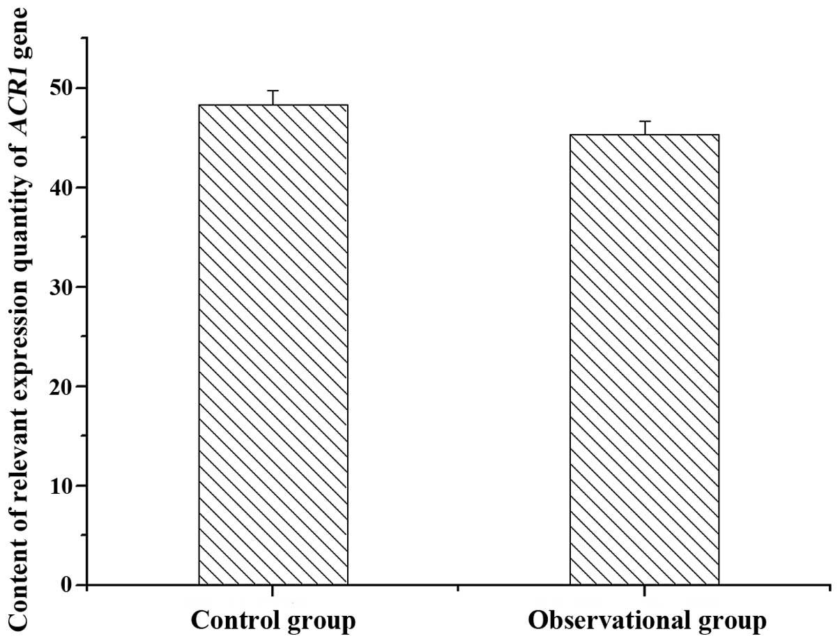
Relative ACR1 mRNA expression
measurements
The ACR1 mRNA expression in the samples of the
control and observation groups was quantified using fluorescence
methods. The differences between the two groups were not
significant (P>0.05; Fig. 1).
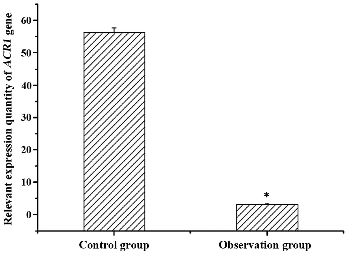
ELISA relative ACR1 protein
expression
The ACR1 protein expression in the samples of the
control and observation groups was quantified using ELISA. The
observational group showed a lower protein level than that in the
control group, and the difference was significant (P<0.05;
Fig. 2).
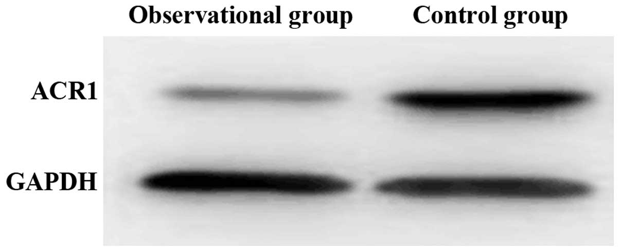
Western blotting of ACR1 proteins
Western blotting was used to verify the results
obtained by ELISA in the serum protein samples of the two groups.
The results confirmed the former findings, and the ACR1 protein
content in the sera of children with cardiomyopathy was lower than
that in the control group (Fig.
3).
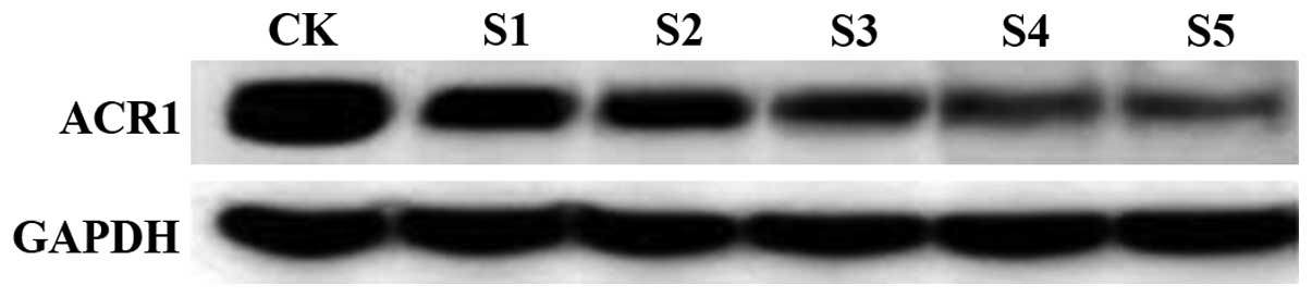
Relative ACR1 protein expression in
patients at different disease duration times
Western blotting was used to assess the expression
of ACR1 protein in the sera of children with cardiomyopathy, at
different time points of the disease course. The levels of ACR1
protein decreased with a longer disease course (Fig. 4).
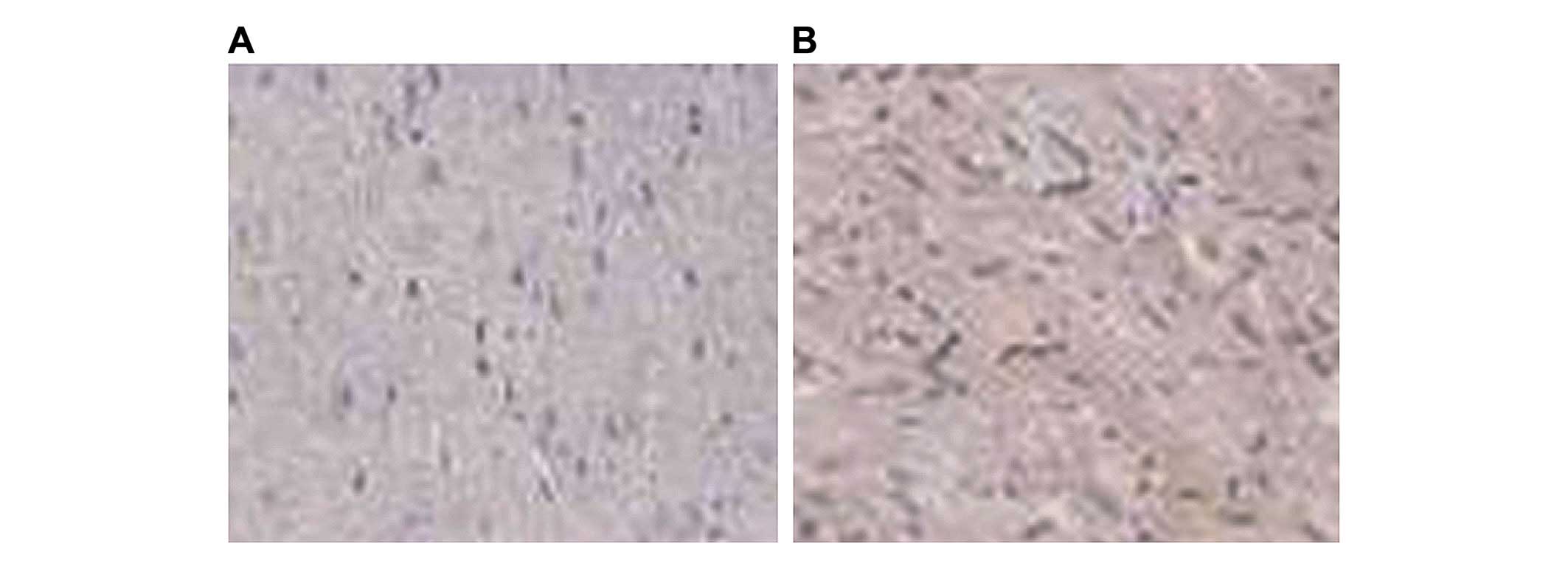
ACR1 in myocardial tissue tested by
immunohistochemistry
ACR1 staining in cardiomyopathy tissues was mainly
concentrated on the membranes (brown-yellow small particles of
heterogeneous size). The normal staining observed in healthy
cardiomyocytes was not present in the cardiocytes of patients
(Fig. 5).
Discussion
Previous findings have shown that cardiomyopathy is
often associated with disorders of mechanical and electrical
activity of the heart (13,14). Its main clinical manifestations are
ventricular dysplasia, cardiac hypertrophy and ventricular
dilatation abnormalities, and these ultimately lead to
heterogeneous myocardial disease. Pediatric cardiomyopathy is a
major cause of cardiac failure during childhood, and it severely
impairs an affected child's life (15). Cardiomyopathy is divided into
dilated, hypertrophic, restrictive and arrhythmogenic
cardiomyopathy (16), but the
pathogenesis of the different types of disease is not clear, and
there is no good basis for effective treatment approaches in
children (17). At present, studies
on the etiological agent of cardiomyopathy in children suggest
genetic defects (such as the deletion of LMNA gene),
congenital metabolic disorders (such as cardiac hypertrophy caused
by fatty acid metabolism), congenital anomalies (for example,
TAZ gene mutation during the embryonic period) and relevant
post-inflammatory responses as the main causes (18–20).
In the present study, after measuring the expression
quantity of AVR1 gene products in the observation and
control groups, significant differences were found at the protein
levels, but not at the RNA levels, indicating that regulation of
the expression occurs post-transcriptionally. The levels of ACR1
protein in affected individuals was reduced with longer disease
time courses, possibly correlating with the deterioration of the
patient's status. However, other relevant genes and factors are
involved in the process and these remain to be determined.
References
|
1
|
Moon JC, Messroghli DR, Kellman P,
Piechnik SK, Robson MD, Ugander M, Gatehouse PD, Arai AE, Friedrich
MG, Neubauer S, et al: Society for Cardiovascular Magnetic
Resonance Imaging; Cardiovascular Magnetic Resonance Working Group
of the European Society of Cardiology: Myocardial T1 mapping and
extracellular volume quantification: A Society for Cardiovascular
Magnetic Resonance (SCMR) and CMR Working Group of the European
Society of Cardiology consensus statement. J Cardiovasc Magn Reson.
15:922013. View Article : Google Scholar : PubMed/NCBI
|
|
2
|
Malhotra A, Kahlon P, Donoho T and Doyle
IC: Pharmacogenomic considerations in the treatment of the
pediatric cardiomyopathy called Barth syndrome. Recent Pat
Biotechnol. 8:136–143. 2014. View Article : Google Scholar : PubMed/NCBI
|
|
3
|
Pankuweit S, Richter A, Ruppert V and
Maisch B: Classification of cardiomyopathies and indication for
endomyocardial biopsy revisited. Herz. 34:55–62. 2009.(In German).
View Article : Google Scholar : PubMed/NCBI
|
|
4
|
Yoo SJ, Grosse-Wortmann L and Hamilton RM:
Magnetic resonance imaging assessment of arrhythmogenic right
ventricular cardiomyopathy/dysplasia in children. Korean Circ J.
40:357–367. 2010. View Article : Google Scholar : PubMed/NCBI
|
|
5
|
Kipps AK, Ramamoorthy C, Rosenthal DN and
Williams GD: Children with cardiomyopathy: Complications after
noncardiac procedures with general anesthesia. Paediatr Anaesth.
17:775–781. 2007. View Article : Google Scholar : PubMed/NCBI
|
|
6
|
Colan SD, Lipshultz SE, Lowe AM, Sleeper
LA, Messere J, Cox GF, Lurie PR, Orav EJ and Towbin JA:
Epidemiology and cause-specific outcome of hypertrophic
cardiomyopathy in children: Findings from the Pediatric
Cardiomyopathy Registry. Circulation. 115:773–781. 2007. View Article : Google Scholar : PubMed/NCBI
|
|
7
|
Xu Z, Huang M, Jun L and Zhang Y: Review
on Diagnosis and Treatment of cardiomyopathy of Children for 10
Year. J Clin Pediatr. 4:345–348. 2011.
|
|
8
|
Elliott P, Andersson B, Arbustini E,
Bilinska Z, Cecchi F, Charron P, Dubourg O, Kühl U, Maisch B,
McKenna WJ, et al: Classification of the cardiomyopathies: A
position statement from the european society of cardiology working
group on myocardial and pericardial diseases. Eur Heart J.
29:270–276. 2008. View Article : Google Scholar : PubMed/NCBI
|
|
9
|
Stehlik J, Edwards LB, Kucheryavaya AY,
Benden C, Christie JD, Dipchand AI, Dobbels F, Kirk R, Rahmel AO
and Hertz MI: International Society of Heart and Lung
Transplantation: The Registry of the International Society for
Heart and Lung Transplantation: 29th official adult heart
transplant report - 2012. J Heart Lung Transplant. 31:1052–1064.
2012. View Article : Google Scholar : PubMed/NCBI
|
|
10
|
Alkaladi A, Abdelazim AM and Afifi M:
Antidiabetic activity of zinc oxide and silver nanoparticles on
streptozotocin-induced diabetic rats. Int J Mol Sci. 15:2015–2023.
2014. View Article : Google Scholar : PubMed/NCBI
|
|
11
|
Kishimoto S, Suda K, Yoshimoto H,
Teramachi Y, Nishino H, Koteda Y, Itoh S, Kudo Y, Iemura M and
Matsuishi T: Thirty-year follow-up of carnitine supplementation in
two siblings with hypertrophic cardiomyopathy caused by primary
systemic carnitine deficiency. Int J Cardiol. 159:e14–e15. 2012.
View Article : Google Scholar : PubMed/NCBI
|
|
12
|
Takahashi R, Asai T, Murakami H, Murakami
R, Tsuzuki M, Numaguchi Y, Matsui H, Murohara T and Okumura K:
Pressure overload-induced cardiomyopathy in heterozygous carrier
mice of carnitine transporter gene mutation. Hypertension.
50:497–502. 2007. View Article : Google Scholar : PubMed/NCBI
|
|
13
|
Towbin JA: Cardiomyopathy and heart
transplantation in children. Curr Opin Cardiol. 17:274–279. 2002.
View Article : Google Scholar : PubMed/NCBI
|
|
14
|
Sarafoglou K, Tridgell AHC, Bentler K,
Redlinger-Grosse K, Berry SA and Schimmenti LA: Cardiac conduction
improvement in two heterozygotes for primary carnitine deficiency
on L-carnitine supplementation. Clin Genet. 78:191–194. 2010.
View Article : Google Scholar : PubMed/NCBI
|
|
15
|
Li FY, El-Hattab AW, Bawle EV, Boles RG,
Schmitt ES, Scaglia F and Wong LJ: Molecular spectrum of SLC22A5
(OCTN2) gene mutations detected in 143 subjects evaluated for
systemic carnitine deficiency. Hum Mutat. 31:E1632–E1651. 2010.
View Article : Google Scholar : PubMed/NCBI
|
|
16
|
Wexler RK, Elton T, Pleister A and Feldman
D: Cardiomyopathy: An overview. Am Fam Physician. 79:778–784.
2009.PubMed/NCBI
|
|
17
|
Hershberger RE, Cowan J, Morales A and
Siegfried JD: Progress with genetic cardiomyopathies: Screening,
counseling, and testing in dilated, hypertrophic, and
arrhythmogenic right ventricular dysplasia/cardiomyopathy. Circ
Heart Fail. 2:253–261. 2009. View Article : Google Scholar : PubMed/NCBI
|
|
18
|
Murphy RT, Thaman R, Blanes JG, Ward D,
Sevdalis E, Papra E, Kiotsekoglou A, Tome MT, Pellerin D, McKenna
WJ and Elliott PM: Natural history and familial characteristics of
isolated left ventricular non-compaction. Eur Heart J. 26:187–192.
2005. View Article : Google Scholar : PubMed/NCBI
|
|
19
|
Escher F, Vetter R, Kühl U, Westermann D,
Schultheiss HP and Tschöpe C: Fractalkine in human inflammatory
cardiomyopathy. Heart. 97:733–739. 2011. View Article : Google Scholar : PubMed/NCBI
|
|
20
|
Towbin JA, Lowe AM, Colan SD, Sleeper LA,
Orav EJ, Clunie S, Messere J, Cox GF, Lurie PR, Hsu D, et al:
Incidence, causes, and outcomes of dilated cardiomyopathy in
children. JAMA. 296:1867–1876. 2006. View Article : Google Scholar : PubMed/NCBI
|