Introduction
Hormone levels may cause changes in sexual function
in women as a result of aging and during the climacteric period; as
such, women aged 40–65 years experience changes in hormone levels
and gradually lose their reproductive capacity (1). This period is associated with the loss
of activity of the ovarian follicles, with consequent estrogen
deficiency (2).
Approximately 70% of women experience symptoms
during the climacteric period. In general, these symptoms are
responsible for estrogen deprivation. The most common symptoms are
vasomotor symptoms, night sweats, cognitive impairment, insomnia,
depression, irritability, fatigue, psychological symptoms, and
increased risk for osteoporosis and cardiovascular disease
(1,3). In addition, vaginal dryness,
dyspareunia, and urinary urgency, which are related to urogenital
atrophy, may negatively affect the sex life and quality of life of
postmenopausal women (2,4). Previous studies have suggested that the
potential risk of metabolic diseases, including obesity, heart
disease, diabetes and hypertension, are increased in the
postmenopausal state (5,6). These metabolic diseases are attributed
to estrogen deficiency. Obesity has secondary effects due to the
orexigenic actions of estrogen deficiency (7,8). The
association between menopause and cardiovascular disease has been
demonstrated in a previous epidemiological study (9,10).
Estrogen deficiency is associated with an atherogenic lipid profile
characterized by high-density lipoprotein (HDL) cholesterol,
low-density lipoprotein (LDL) cholesterol, triglyceride levels
(11), central adiposity (12), increased diastolic pressure (13) and increased insulin resistance
(14).
Hormone therapy can be used to reduce the risk of
ovarian failure and improve women's health; however, this treatment
may cause serious problems over extended periods of time. Long-term
treatment results in an increase in cardiovascular events and
breast cancer (15–17); thus, alternative therapies, such as
the use of phytoestrogens (PEs) to relieve menopausal symptoms,
have gained attention (1,18,19).
Purified phytohormones, such as genistein which is
abundant in soybean, exhibit improved activity in the body and
enhanced bioavailability (18). PEs
is able to bind to estrogen receptors, due to the presence of a
phenolic ring, and function like estrogens (18–20).
Coumestrol and the isoflavonoids genistein, daidzein, and their
plant precursors, are predominantly found in soybeans and clover
(21). Isoflavones, particularly
those derived from plants, have various biological activities, are
able to improve the metabolic symptoms (22) and exhibit bone-protective effects
(23) during menopause. To date,
pomegranate extract has been shown to be a selective estrogen
receptor modulator (24).
Pomegranate (Punica granatum L.) is consumed
as a fresh fruit, beverage, dietary supplement, and is a herbal
medicine ingredients (25).
Pomegranate juice and pomegranate polyphenol extracts have been
demonstrated to prevent various types of cancer, cardiovascular
disease, diabetes, Alzheimer's, arthritis, colitis, and several
other diseases (26–28). Polyphenols, which is one of the
active substances of pomegranate, are present in numerous parts of
pomegranate fruits (29). It has
been shown that pomegranate contains species of flavonoids and
anthocyanidins in their seed oil and juice (30,31).
The anti-climacterium effective optimal dosages of
standardized pomegranate concentrated solution (PCS) remain
unclear. Therefore, the complex anti-climacterium potential of PCS
was examined with optimal dose ranges using female ddY mice
subjected to bilateral ovariectomy (OVX). Estrogen-deficient
animals induced by OVX were used as a climacterium model as several
climacterium symptoms are clearly induced by OVX within 4 to 6
weeks after the surgery. OVX-treated ddY mice have also been used
to investigate the mechanisms underlying menopause-related
complications in humans as these complications share various
similarities with postmenopausal climacterium symptoms (32–34).
This rodent model exhibits symptoms that resemble those of women
with postmenopausal climacterium symptoms, including cardiovascular
diseases, obesity, hyperlipidemia, osteoporosis, organ steatosis
and mental disorders (35–38). In the present study, anti-climacteric
effects were evaluated and separated into five categories: i)
estrogenic effects; ii) anti-obese effects; iii) hypolipidemic
effects; iv) hepatoprotective effects against liver steatosis; and
v) anti-osteoporotic effects. The results suggest that PCS
treatment suppressed OVX-induced obesity, hyperlipidemia, hepatic
steatosis and osteoporosis in ddY mice.
Materials and methods
Animals and husbandry
A total of 48 virgin female specific pathogen-free
outbred-mice (Kwl:ddY; age, 6 weeks; weight, 24–26 g; Kiwa
Laboratory Animal, Wakayama, Japan), were used for the present
study following acclimatization for 16 days. Animals were allocated
four per polycarbonate cage in a temperature (20–25°C) and humidity
(45–55%) controlled room with a 12-h light/dark cycle. Feed
(Samyang, Seoul, Korea) and water were supplied free to access. A
total of 28 days after OVX surgery, eight mice per group were
selected based on body weight. All laboratory animals were treated
according to the national regulations on the usage and welfare of
animals and approved by the Institutional Animal Care and Use
Committee of Daegu Haany University (Gyeongsan, Korea) prior to the
experiments (approval no. DHU2014-0210). Experiments on
osteoporosis were conducted in accordance with the US Food and Drug
Administration ‘Guidelines for Preclinical Evaluation of Agents
Used in The Prevention or Treatment Postmenopausal Osteoporosis’
(39).
Preparation and administration of test
substances
Compound (17β)-estra-1,3,5(10)-triene-3,17-diol (17β-estradiol) was
purchased from Sigma-Aldrich (Merck Millipore, Darmstadt, Germany).
Standardized PCS were supplied by Health Love Ltd. (Anyang, Korea)
as deep reddish viscous solutions. The energy of PCS was 244.69
Kcal/100 g, and PCS contained 2.31 mg/g ellagic acid, 58.86%
carbohydrate, 1.21% total protein, 0.49% fat, 27.97% water, 1.47%
ash, and 28.03 mg/100 g sodium. PCS (0.67 ml) was diluted as clear
reddish solutions in 1 ml distilled water. Subsequently, 1, 2 and 4
ml/kg (according to body weights) of PCS were orally administered
once a day for 84 days from 28 days after OVX in a volume of 10
ml/kg (v/v), diluted with distilled water, using a
gastric gavage attached to a 1 ml syringe. In OVX and sham control
mice, distilled water was used as a vehicle. In addition,
17β-estradiol (Sigma-Aldrich; Merck Millipore) was subcutaneously
administered into the dorsal back skins at a volume of 0.2 ml/mouse
(0.03 µg/head/day), according to previously established methods
(40–42).
Menopause induction via bilateral
OVX
Mice were anesthetized with a 25 mg/kg
intraperitoneal injection of Zoletile mixture (Zoletile 50™; Virbac
Lab., Carros Cedex, France) and maintained with 1–1.5% isoflurane
(Hana Pharm. Co., Hwasung, Korea) in a mixture of 70%
N2O and 28.5% O2. The surgical protocol was
performed according to established methods (35,37,38). The
OVX treatment group (n=8) underwent open surgery involving
bilateral OVX via a midline incision of linea alba.
Following surgery, the incision was closed in two layers. Muscular
layers were sutured independently from the peripheral tissues using
dissolvable 3–0 vicryl sutures and the skin was closed by
continuous sutures using silk (3–0). The second group of mice (n=8)
underwent sham surgery, in which a similar incision in the linea
alba was made but bilateral OVX was not performed.
Body weight measurements
Alterations in body weight were measured once a week
from OVX, one day before administration, and at sacrifice (at 84
days after the first administration, the mice were anesthetized
with 50 mg/kg tiletamine/zolazepam and dissected) using an
automatic electronic balance (Precisa Gravimetrics, Inc., Dietikon,
Switzerland). At OVX, initiation of administration, and at
termination, all experimental animals were fasted overnight for 18
h (water was provided) to reduce the differences from feeding. In
addition, body weight gains were calculated as follows: OVX
recovery/induced periods (28 days) = [body weight at initial test
substance treatment - body weight on the day of OVX surgery]; and
after administration (84 days) = [body weight at sacrifice - body
weight at initial test substance treatment].
Food consumption measurements
All mice were allocated into individual cages and
received 150 g diets. The quantity of diets supplied were measured
at 24 h after feed supply using an automatic electronic balance
(Precisa Gravimetrics, Inc) and were considered to indicate the
daily food consumption of individual mice (g/24 h/mouse). These
measurements were conducted six times: 1, 3, 7, 28, 56 and 83 days
after the first administration.
Measurement of bone mineral density
(BMD) and body fat density
The mean BMD of the total body and right femur were
detected once using live dual-energy X-ray absorptionmetry
(InAlyzer; Medikors, Seungnam, Korea) at the end of 84 days of
continuous treatment with the test substances. The mean fat
densities of the body and abdominal cavity of each mouse were
recorded.
Organ weight measurements
Following sacrifice, the abdominal fat pads
deposited in the abdominal cavity, total liver, and uterus
(including vagina) were collected after removing the surrounding
connective tissues, muscles, and any debris. The weights of organs
were measured in grams to determine the absolute wet-weights. To
reduce individual body weight differences, the relative weights (%
of body weight) were calculated using body weight at sacrifice and
absolute wet-weight, as follows: Relative organ weights (% of body
weight) = [(absolute abdominal fat pad, uterus or liver
weights/body weight at sacrifice) × 100].
Bone weight measurements
Following 84 days of continuous treatment from 28
days after bilateral OVX surgery, the right sides of the femurs
were harvested after removing the surrounding connective tissues,
muscles, and any debris. Bone weight was measured in grams to
determine the absolute wet-weights, and they were dried at 120°C
for 8 h in a high temperature dry oven (LDO-080N; Daihan Labtech
Co., Seoul, Korea) for measurements of dry bone weights.
Subsequently, dried bones were carbonized at 800°C for 6 h in a
furnace (LEF-1055-1; Daihan Labtech Co.) to measure ash absolute
weights. To reduce the individual body weight differences, the
relative weight (%) was calculated based on the body weight at
sacrifice and absolute wet/dry/ash weight, as follows: Relative
bone weights (% of body weight) = [(absolute bone weight/body
weight at sacrifice) × 100].
Measurement of bone strengths
Bone strength was detected as the failure load (FL).
We used FL calculated using a test machine (SV-H1000; Japan
Instrumentation System, Co., Nara, Japan). The FL of the mid-shaft
regions of the right femurs was detected using a three-point
bending test to failure using a computerized testing machine
(SV-H1000; Japan Instrumentation System Co., Yokohama, Japan) as N
(Newton), according to the manufacturer's instructions.
Blood collection
For serum biochemical analysis, ~1 ml whole blood
was collected from the vena cava at sacrifice and was separated
from the serum by centrifugation at 21,000 × g for 10 min at
4°C using a clotting activated serum tube. All serum samples were
frozen at −150°C until they were assayed.
Serum biochemistry
Serum aminotransferase (AST), alanine
aminotransferase (ALT), total cholesterol (TC), LDL, and
triglyceride (TG) levels were detected using an automated blood
analyzer (Hemagen Analyst; Hemagen Diagnostics, Columbia, MD, USA),
and HDL levels were measured using another automated blood analyzer
(AU400; Olympus Corp., Tokyo, Japan). In addition, serum
osteocalcin levels (ng/ml) were detected using a Mouse Osteocalcin
ELISA kit (Immutopics, San Clemente, CA, USA), and serum bALP
levels (U/l) were detected using a Mouse bALP ELISA kit (Quidel
Corp., San Diego, CA, USA), with an ELISA Reader (Tecan Group,
Ltd., Männedorf, Switzerland). In addition, serum estradiol
contents were measured using the chemiluminescent immunoassay
technique with an ECLIA Roche e411 immunoassay analyzer (Roche
Diagnostics GmbH, Mannheim, Germany) using the separated serum
harvested after the sacrifice of all mice.
Abdominal fat pads, uterus, and liver
histological procedures
Sampled tissues were fixed in 10% neutral buffered
formalin (NBF). Following paraffin embedding, 3–4 µm serial
sections were prepared. Representative sections were stained with
hematoxylin and eosin (H&E) for light microscopic examination.
Furthermore, sections of liver that had been dehydrated in 30%
sucrose solutions were sectioned using a cryostat to stain the
lipids with Oil Red O (43). The
total thicknesses of abdominal fat pads were measured using an
automated image analysis processor (iSolution FL 9.1; IMT
i-solution Inc., Quebec, Canada) as mm/mouse. Mean diameters (µm)
of dorsal abdominal white adipocytes were calculated in restricted
view fields on a computer monitor, using an automated image
analysis processor. At least 10 white adipocytes per fat pad were
used for histomorphometrical analysis according to our previously
established methods (43–45). In addition, total full, mucosa, and
epithelial thicknesses of the uterus (µm/uterus) were detected as
percentages of uterine glands located in the mucosa (%/mucosa of
uterus) using an automated image analyzer. To observe steatosis in
the liver, the percentage of fatty change regions in the hepatic
parenchyma was calculated as percentages between one field of the
liver (%/mm2 of hepatic parenchyma) under Oil Red O
staining. Mean diameters of hepatocytes were calculated in
restricted view fields on a computer monitor under H&E staining
using an automated image analysis processor, as µm; at least 10
hepatocytes per liver were used.
Bone histological procedures
The left sides of each mouse femur were separated
and fixed in 10% NBF, after which they were decalcified in
decalcifying solution (24.4% formic acid and 0.5 N sodium
hydroxide) for three days (mixed decalcifying solution was
exchanged once a day for three days). Samples were subsequently
embedded in paraffin, sectioned (3–4 µm), and stained with
Safranin-O. In addition, bone histomorphometry was conducted using
an automated image analyzer under microscopy (Nikon Corp., Tokyo,
Japan) to examine the bone mass and structure with bone resorption
in a uniform area of epiphyseal or cortical bone regions of the
femur (growth plate regions were excluded). Cortical bone thickness
was also measured in the mid-shaft regions of the femur. Trabecular
bone volume (TV/BV, TBV; %), thickness of trabecular bone (Tbt;
µm/trabecular bone), number (Tbn; mean numbers of trabecular
bone/epiphyseal regions), length (Tbl; mm/trabecular bone), and
cortical bone thickness (Cbt; µm/mid-shaft cortical bone) were
measured for bone mass and structure, and osteoclast cell number
(Ocn; mean osteoclast cell numbers/ epiphyseal regions) and ratio
(OS/BS; %) were measured for bone resorption, as described
previously (37,38,46).
Statistical analyses
All values for the eight mice in this experiment
were expressed as means ± standard deviation. Multiple comparison
tests for the different dose groups were conducted. Variance
homogeneity was examined using the Levene test. If the Levene test
indicated no significant deviations from variance homogeneity, the
data were analyzed using the one-way analysis of variance test
followed by the least-significant differences test to determine
which group comparisons were significantly different. When
significant deviations from variance homogeneity were observed on
the Levene test, the non-parametric Kruskal-Wallis test was
conducted. When a significant difference was observed on the
Kruskal-Wallis test, the Mann-Whitney U test was conducted to
determine the specific pairs of groups that were significantly
different. Statistical analyses were conducted using the SPSS for
Windows software package (ver. 14.0; SPSS Inc., Chicago, IL,
USA).
Results
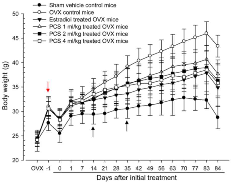
Significant decreases in body weight
were recorded in PCS-treated mice
Significant increases (P<0.05 or P<0.01) in
body weight were detected in all OVX mice compared with control
mice (red arrow), with significant (P<0.01) increases in body
weight gains during the 4-week OVX recovery/induction periods.
However, significant decreases in body weights were observed in the
estradiol group from 14 days after initial treatment, and from 35
days after initial treatment, for all three PCS dosages, compared
with OVX control mice (arrows; P<0.05 or P<0.01). In
addition, all test substance-treated mice exhibited significant
decreases in body weight gains during 84 days of treatment compared
with OVX controls (Fig. 1).
PCS induced significant decreases in
food consumption
OVX mice exhibited significant increases (P<0.01)
in food consumption compared with control mice at all six
measurement times (1, 3, 7, 28, 56 and 83 days after initial
administration). However, estradiol subcutaneously-treated mice
showed significant decreases (P<0.01) in food consumption from 7
days after initial treatment, from 28 days after initial
administration of 2 and 4 ml/kg PCS, and from 56 days after initial
administration of 1 ml/kg PCS compared with OVX mice until 83 days
after initial administration (Table
I).
 | Table I.Food consumptions in sham-operated or
OVX ddY mice. |
Table I.
Food consumptions in sham-operated or
OVX ddY mice.
|
| Food consumption
(g/24 h/mouse) on the indicated days after initial treatment |
|---|
|
|
|
|---|
| Groups | 1 | 3 | 7 | 28 | 56 | 83 |
|---|
| Controls |
|
|
|
|
|
|
|
Sham | 7.36±0.63 | 6.39±0.65 | 6.46±0.91 | 6.66±0.85 | 7.41±0.97 | 7.63±0.83 |
|
OVX |
11.55±1.33a |
11.20±0.93a |
11.43±1.05a |
11.63±1.04a |
12.92±1.45a |
14.94±1.46a |
|
Estradiol |
11.66±1.29a |
10.64±1.03a |
9.74±0.99a,b |
9.64±1.13a,b |
10.34±0.92a,b |
10.73±1.57a,b |
| PCS |
|
|
|
|
|
|
| 1
ml/kg |
11.73±0.97a |
10.87±0.78a |
10.69±0.92a |
10.82±0.77a |
11.06±0.87a,b |
12.50±0.79a,b |
| 2
ml/kg |
11.47±1.35a |
11.00±1.34a |
10.77±0.88a |
10.55±0.69a,c |
10.62±0.60a,b |
11.42±1.06a,b |
| 4
ml/kg |
11.66±1.53a |
11.18±1.18a |
10.85±0.90a |
10.36±0.73a,b |
10.47±0.88a,b |
11.31±1.65a,b |
Significant decreases of abdominal fat
pad weight was observed in PCS-treated mice
Significant increases in abdominal fat pad weights
deposited in the abdominal cavity, as well as in absolute and
relative weights, were observed in OVX mice compared with sham
control mice (P<0.01). However, significant decreases in
abdominal fat pad weights were observed in all test
substance-treated mice, including estradiol-treated OVX mice,
compared with OVX mice (P<0.01) (Table II). The absolute weights of
abdominal fat pads deposited into the abdominal cavity in OVX
controls were altered by 2,339.34% compared with the sham control,
and by −79.29, −42.14, −64.20 and −66.65% in estradiol- and 1-, 2-
and 4-ml/kg PCS-treated mice compared with OVX controls,
respectively.
| Table II.Abdominal fat pad, uerus and liver
weights in sham-operated or OVX ddY mice. |
Table II.
Abdominal fat pad, uerus and liver
weights in sham-operated or OVX ddY mice.
|
| Absolute wet-weight
of organ (g) | Relative wet-weight
of organ (% of body weight) |
|---|
|
|
|
|
|---|
| Groups | Abdominal fat
pad | Uterus | Liver | Abdominal fat
pad | Uterus | Liver |
|---|
| Controls |
|
|
|
|
|
|
|
Sham | 0.140±0.103 | 0.254±0.079 | 1.240±0.259 | 0.499±0.384 | 0.885±0.277 | 4.316±0.799 |
|
OVX |
3.403±0.486c |
0.029±0.008c | 1.232±0.193 |
7.821±0.802a |
0.068±0.018c |
2.842±0.434a |
|
Estradiol |
0.705±0.572d,g |
0.109±0.039c,g | 1.337±0.084 |
2.052±1.706b,e |
0.315±0.119c,g |
3.845±0.341e |
| PCS |
|
|
|
|
|
|
| 1
ml/kg |
1.969±0.754c,g |
0.041±0.008c,h | 1.302±0.076 |
5.207±1.873a,e |
0.109±0.023c,g |
3.461±0.261a,f |
| 2
ml/kg |
1.218±0.480c,g |
0.045±0.009c,g | 1.319±0.071 |
3.319±1.169a,e |
0.124±0.024c,g |
3.659±0.445b,e |
| 4
ml/kg |
1.135±0.198c,g |
0.044±0.011c,g | 1.312±0.057 |
3.249±0.894a,e |
0.125±0.033c,g |
3.719±0.517b,e |
Effects on uterus weights
Significant decreases in the uterus absolute and
relative wet-weights were observed in OVX mice compared with sham
control mice (P<0.01). However, significant increases in the
uterus weights were observed in all test substance-treated mice,
including 1 ml/kg PCS, compared with OVX control mice (P<0.01)
(Table II). Absolute uterine
weights of OVX controls were altered by −88.45% compared with the
sham controls, and by 272.34, 39.57, 52.77 and 51.06% in estradiol
and 1-, 2- and 4-ml/kg PCS treated mice, respectively, as compared
with OVX controls. Relative uterine weights of OVX were altered by
−92.33% compared with the sham controls, and by 364.57, 61.15,
82.57 and 83.77% in estradiol- and 1-, 2- and 4-ml/kg PCS treated
mice, respectively, as compared with OVX controls. Our results
indicated that PCS causes estrogenic activities.
Significant increases in liver weight
were observed in PCS-treated mice
Significant decreases in the liver relative
wet-weights were detected in OVX mice compared with sham control
mice (P<0.01); however, significant increases in the liver
relative weights were observed in all test substance-treated mice,
including all three different dosages of PCS, compared with OVX
mice (P<0.05 or P<0.01). Estradiol- and 1-, 2- and 4-ml/kg
PCS-treated mice did not exhibit any significant changes in
absolute liver weights compared with OVX control mice, or in OVX
mice compared with sham control mice (Table II). These data suggested that PCS
exerts hepatoprotective effects.
Significant increases in femur weight
were detected in PCS-treated mice
Significant decreases in the femur relative
wet-weights and absolute and relative dry and ash weights were
observed in OVX mice compared with sham control mice (P<0.01).
However, significant increases in the femur wet relative weights
and dry and ash absolute and relative weights were observed in all
test substance-treated mice, including estradiol treated mice,
compared with OVX mice (P<0.05 and P<0.01) (Table III). Our observations indicated
that PCS has anti-osteoporosis activities.
| Table III.Right femur weights in sham-operated
or OVX ddY mice. |
Table III.
Right femur weights in sham-operated
or OVX ddY mice.
|
| Absolute weight
(g) | Relative weight (%
of body weight) |
|---|
|
|
|
|
|---|
| Groups | Wet | Dry | Ash | Wet | Dry | Ash |
|---|
| Controls |
|
|
|
|
|
|
|
Sham | 0.094±0.006 | 0.065±0.004 | 0.039±0.003 | 0.327±0.025 | 0.229±0.024 | 0.137±0.019 |
|
OVX | 0.089±0.007 |
0.051±0.003a |
0.026±0.003a |
0.205±0.010b |
0.118±0.010a |
0.059±0.007a |
|
Estradiol | 0.095±0.010 |
0.059±0.003a,c |
0.034±0.004a,c |
0.273±0.038b,e |
0.170±0.012a,c |
0.098±0.013a,c |
| PCS |
|
|
|
|
|
|
| 1
ml/kg | 0.091±0.005 |
0.054±0.003a,d |
0.032±0.004a,c |
0.240±0.010b,e |
0.145±0.012a,c |
0.086±0.011a,c |
| 2
ml/kg | 0.095±0.008 |
0.056±0.003a,c |
0.033±0.004a,c |
0.263±0.024b,e |
0.156±0.017a,c |
0.091±0.009a,c |
| 4
ml/kg | 0.092±0.007 |
0.056±0.004a,c |
0.033±0.003a,c |
0.261±0.043b,e |
0.158±0.020a,c |
0.093±0.013a,c |
Changes of serum biochemistry indices
were induced by PCS-treatment
Significant increases in serum AST, ALT, TC, LDL and
TG levels and significant decreases in serum HDL levels were
observed in OVX control mice compared with sham control mice.
However, significant decreases in serum AST, ALT, TC, LDL and TG
levels and significant increases in serum HDL levels were observed
in all test material-treated mice, including 1 ml/kg PCS-treated
mice, compared with OVX mice (Table
IV). These results demonstrated that PCS causes
hepatoprotective and hypolipidemic effects.
| Table IV.Serum biochemistry: AST, ALT, TC,
LDL, HDL and TG Levels in sham-operated or OVX ddY mice. |
Table IV.
Serum biochemistry: AST, ALT, TC,
LDL, HDL and TG Levels in sham-operated or OVX ddY mice.
|
| Serum biochemical
values |
|---|
|
|
|
|---|
| Groups | AST (U/l) | ALT (U/l) | TC (mg/dl) | LDL (mg/dl) | HDL (mg/dl) | TG (mg/dl) |
|---|
| Controls |
|
|
|
|
|
|
|
Sham | 84.88±14.56 | 38.25±12.07 | 92.25±18.81 | 64.00±10.61 | 94.88±12.84 | 37.88±11.67 |
|
OVX |
162.75±15.78a |
77.38±9.07a |
181.63±19.00a |
182.50±18.62a |
46.25±11.94a |
154.00±24.68b |
|
Estradiol |
108.00±13.65a,c |
53.13±9.22a,c |
134.75±21.73a,c |
134.75±11.47a,c |
70.50±12.69a,c |
101.00±19.13b,e |
| PCS |
|
|
|
|
|
|
| 1
ml/kg |
141.38±8.93a,c |
62.38±10.03a,c |
152.50±15.74a,c |
156.00±6.32a,c |
61.88±8.17a,d |
123.50±11.34b,f |
| 2
ml/kg |
126.25±8.86a,c |
58.38±7.89a,c |
143.38±11.56a,c |
144.75±14.59a,c |
67.75±12.09a,c |
109.88±16.50b,e |
| 4
ml/kg |
125.88±11.24a,c |
58.50±9.27a,c |
142.50±13.67a,c |
144.38±19.38a,c |
68.25±14.89a,c |
109.00±25.48b,e |
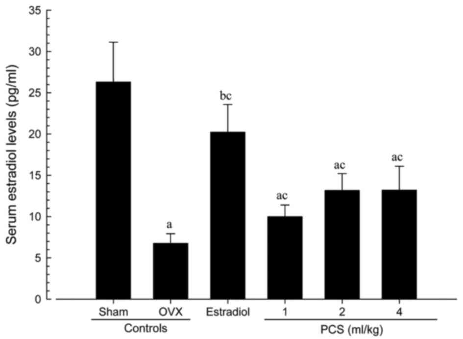
Significant decreases in serum
estradiol levels in OVX mice were observed compared with sham
control mice (P<0.01)
However, significant increases in serum estradiol
levels were observed in all test substance-treated mice, including
4 ml/kg PCS-treated mice, as compared with OVX mice (P<0.01)
(Fig. 2). Serum estradiol levels in
OVX were altered by −74.27% compared with sham controls, and by
199.08, 47.69, 94.64 and 95.19% in estradiol- and 1-, 2- and
4-ml/kg PCS-treated mice, respectively, compared with OVX controls.
Our results indicated that PCS causes estrogenic activities.
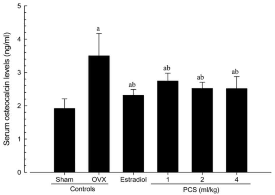
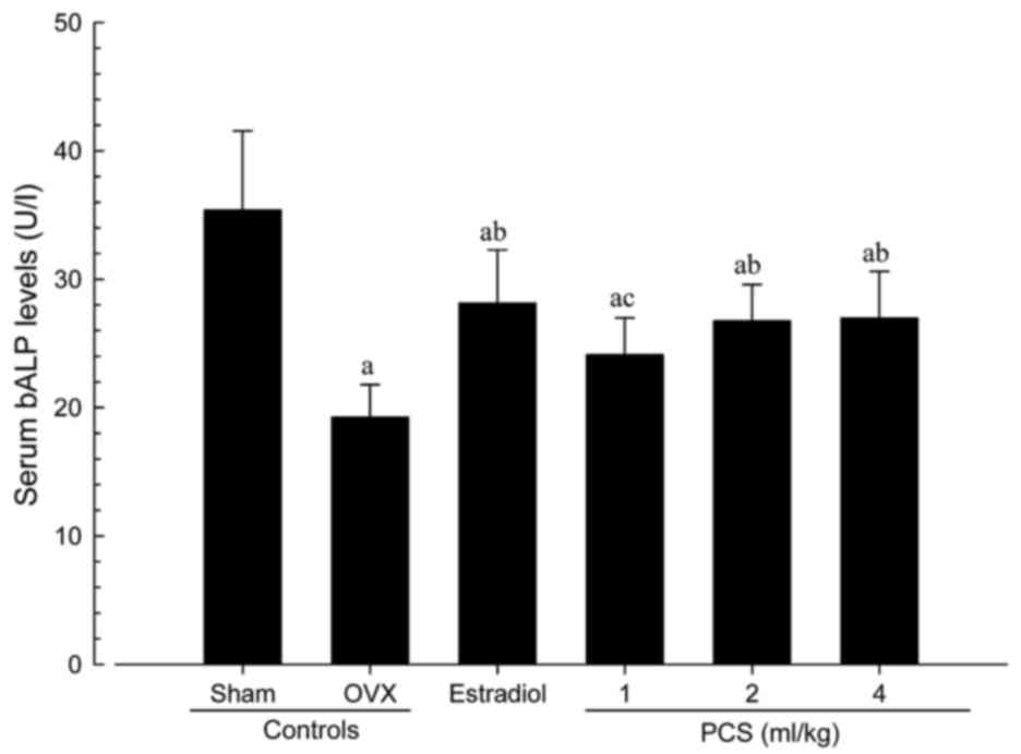
Significant increases in serum osteocalcin levels,
and significant decreases in serum bALP levels, were detected in
OVX mice compared with sham control mice (P<0.01). However,
significant decreases in serum osteocalcin and increases in bALP
levels were observed in all test material-treated mice, including
estradiol-treated mice, compared with OVX control mice (Figs. 3 and 4). Serum osteocalcin and bALP levels in OVX
were altered by 82.59 and −45.63% compared with sham controls, and
by −33.92, −21.60, −27.99 and −28.20% (for serum osteocalcin
levels) and 46.16, 25.32, 39.04 and 40.14% (for serum bALP levels)
in estradiol- and 1-, 2- and 4-ml/kg PCS-treated mice,
respectively, as compared with OVX controls. Our observations
indicated that PCS has anti-osteoporosis activities.
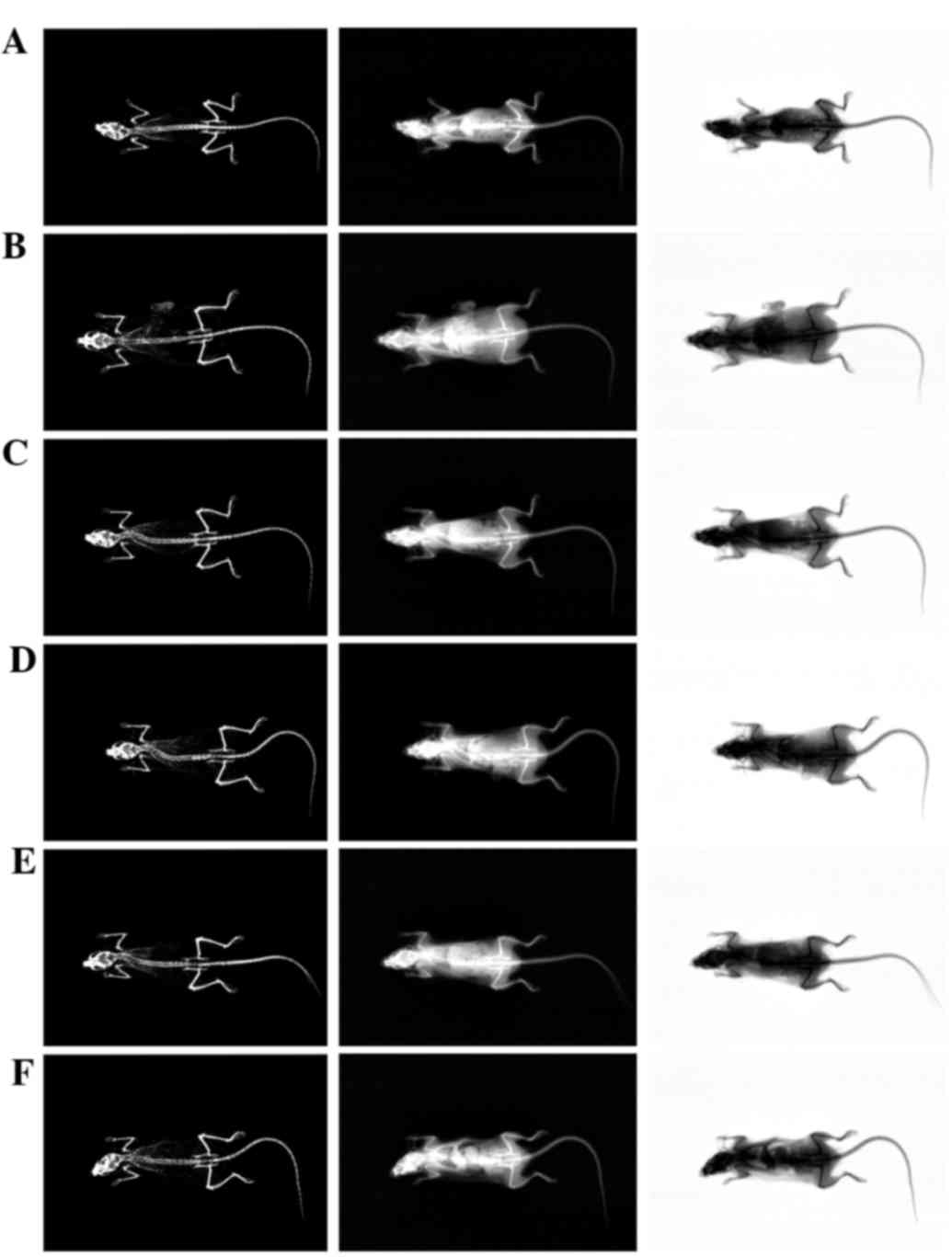
Significant increases of BMD were
recorded in PCS-treated mice
The total body and femur mean BMD of OVX mice were
significantly decreased compared with sham control mice
(P<0.01). However, significant increases in total body and femur
mean BMD were detected in estradiol- and PCS (all three different
dosages)-treated mice compared with OVX mice (P<0.01) (Table V; Fig.
5). The total body mean BMD of OVX controls were altered by
−14.80% compared with sham controls, and by 11.23, 5.26, 10.18 and
10.64% in estradiol- and 1-, 2-, and 4-ml/kg PCS treated mice,
respectively, as compared with OVX controls. The total femur mean
BMD of OVX was changed by −13.73% compared with sham controls, and
by 12.20, 3.51, 6.96 and 7.12% in estradiol- and 1-, 2-, and
4-ml/kg PCS-treated mice, respectively, as compared with OVX
controls. Our results showed that PCS exerts anti-osteoporosis
activities.
| Table V.Bone mineral density and body fat
density in sham-operated or OVX ddY mice. |
Table V.
Bone mineral density and body fat
density in sham-operated or OVX ddY mice.
| Variable | Bone mineral
density (g/cm2) | Fat density (% of
body mass) |
|---|
|
|
|
|
|---|
| Groups | Total body | Right femur | Total body | Abdominal
cavity |
|---|
| Controls |
|
|
|
|
|
Sham | 0.0251±0.0011 | 0.0269±0.0007 | 11.31±2.10 | 11.17±1.65 |
|
OVX |
0.0214±0.0006a |
0.0232±0.0005a |
35.27±3.47a |
41.91±4.40c |
|
Estradiol |
0.0238±0.0006a,b |
0.0260±0.0008b |
24.10±3.46a,b |
26.87±4.30c,d |
| PCS |
|
|
|
|
| 1
ml/kg |
0.0225±0.0004a,b |
0.0240±0.0004a |
29.16±2.89a,b |
30.05±7.21c,d |
| 2
ml/kg |
0.0236±0.0009a,b |
0.0248±0.0014a,b |
25.58±6.22a,b |
26.96±7.07c,d |
| 4
ml/kg |
0.0237±0.0009a,b |
0.0248±0.0013a,b |
25.52±5.90a,b |
26.94±9.07c,d |
Significant decreases of body fat
densities in PCS-treated mice
Total body and abdominal fat densities of OVX
control mice were significantly increased, as compared with sham
control mice (P<0.01). However, significant decreases in total
body and abdominal fat densities were detected in all test
substance-administrated mice, including subcutaneous
estradiol-treated mice, as compared with OVX control mice
(P<0.01) (Table V). The total
mean body fat densities of OVX controls were altered by 211.77%
compared with sham controls, and by −31.65, −17.33, −27.47 and
−27.64% in estradiol- and 1-, 2- and 4-ml/kg PCS-treated mice,
respectively, as compared with OVX controls. The mean abdominal fat
densities of OVX controls were altered by 275.16% compared with
sham controls, and by −35.88, −28.29, −35.68 and −35.73% in
estradiol- and 1-, 2- and 4-ml/kg PCS-treated mice, respectively,
as compared with OVX controls. These results indicated that PCS
exerts anti-obese actions.
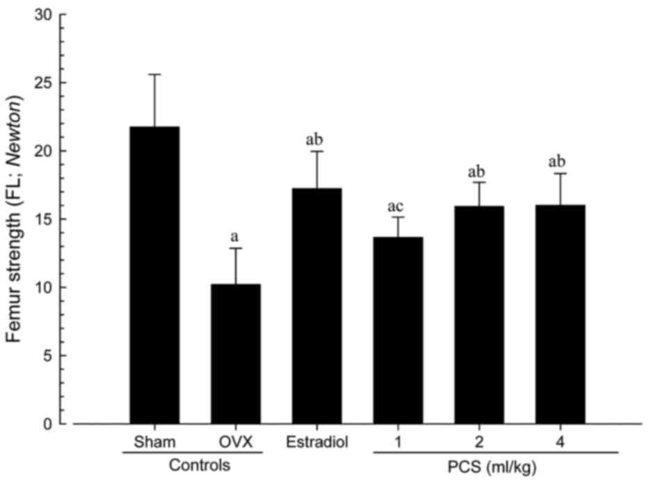
Significant increases of bone strength
in PCS-treated mice
The strengths of femur mid-shaft regions in OVX
control mice, determined as FL, were significantly decreased
compared with sham control mice (P<0.01); however, significant
increases in FL on the femur were detected in all test
substance-administrated mice including 1 ml/kg PCS-treated mice
compared with OVX control mice (P<0.05 or P<0.01) (Fig. 6). The FL in the femur mid-shaft
regions of OVX control were altered by −53.05% compared with sham
controls, and by 68.69, 33.74, 55.99, and 56.74% in estradiol- and
1-, 2- and 4-ml/kg PCS-treated mice, respectively, as compared with
OVX controls. Our data indicated that PCS causes anti-osteoporosis
activities.
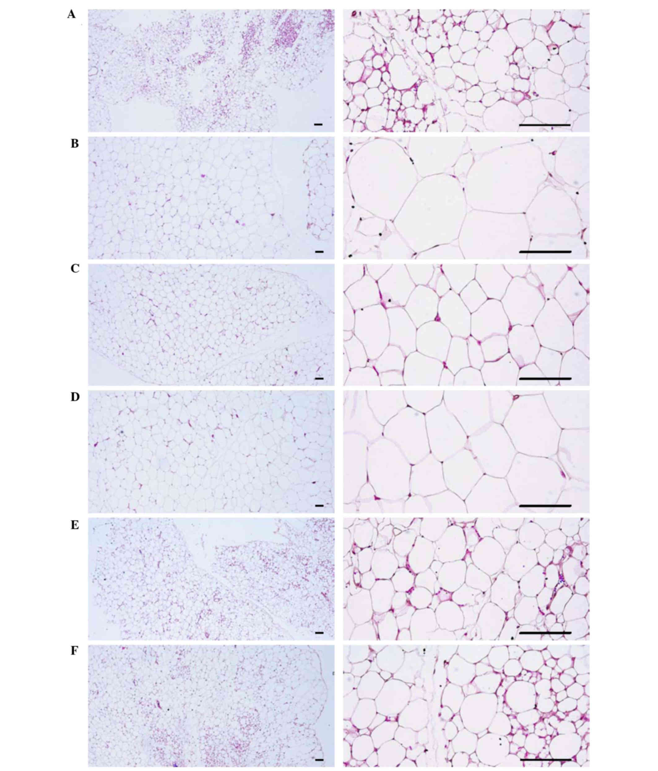
Changes in abdominal fat pad, uterus,
and liver histopathology
Significant increases in the thickness of abdominal
fat pads deposited into the abdominal cavity and the mean adipocyte
diameters were observed in OVX mice due to the deposition in
adipose tissues in the abdominal cavity and the hypertrophy of
adipocytes, respectively (P<0.01). However, significant
decreases in the thickness of abdominal fat pads and their mean
diameters of adipocytes were detected in all test
substance-administrated mice, including estradiol treated mice,
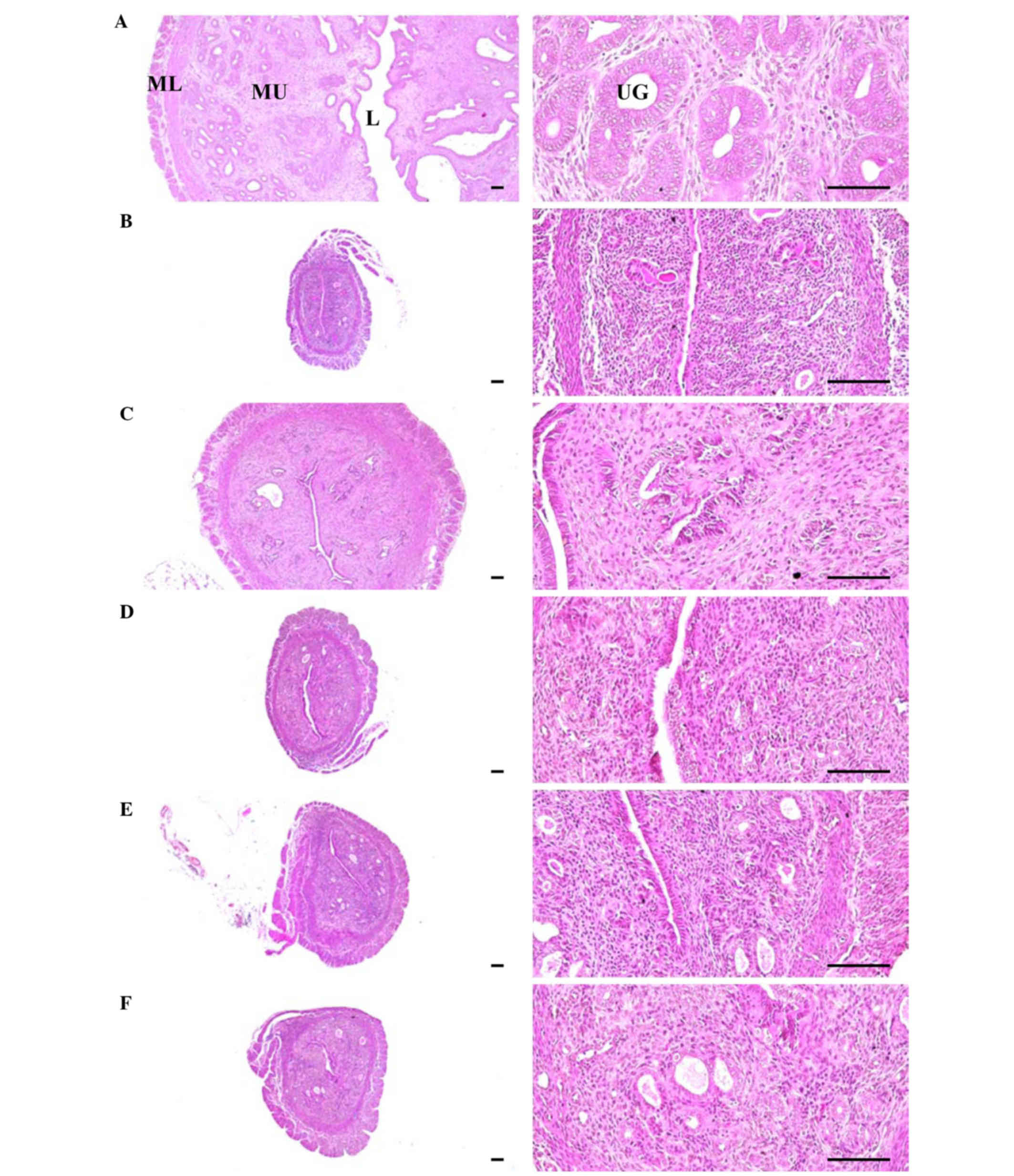
compared with OVX control mice (P<0.01) (Table VI; Fig.
7). Significant decreases in total, mucosa, and epithelial
thicknesses of the uterus, and in the percentages of uterine glands
in the mucosa, were observed in OVX control mice due to estrogen
depletion-related atrophic changes. However, significant increases
in total, mucosa, and epithelial thicknesses of the uterus, as well
as in the percentages of uterine glands in the mucosa, were
detected in estradiol- and 1-, 2- and 4-ml/kg PCS-treated mice,
respectively, as compared with OVX control mice (P<0.01)
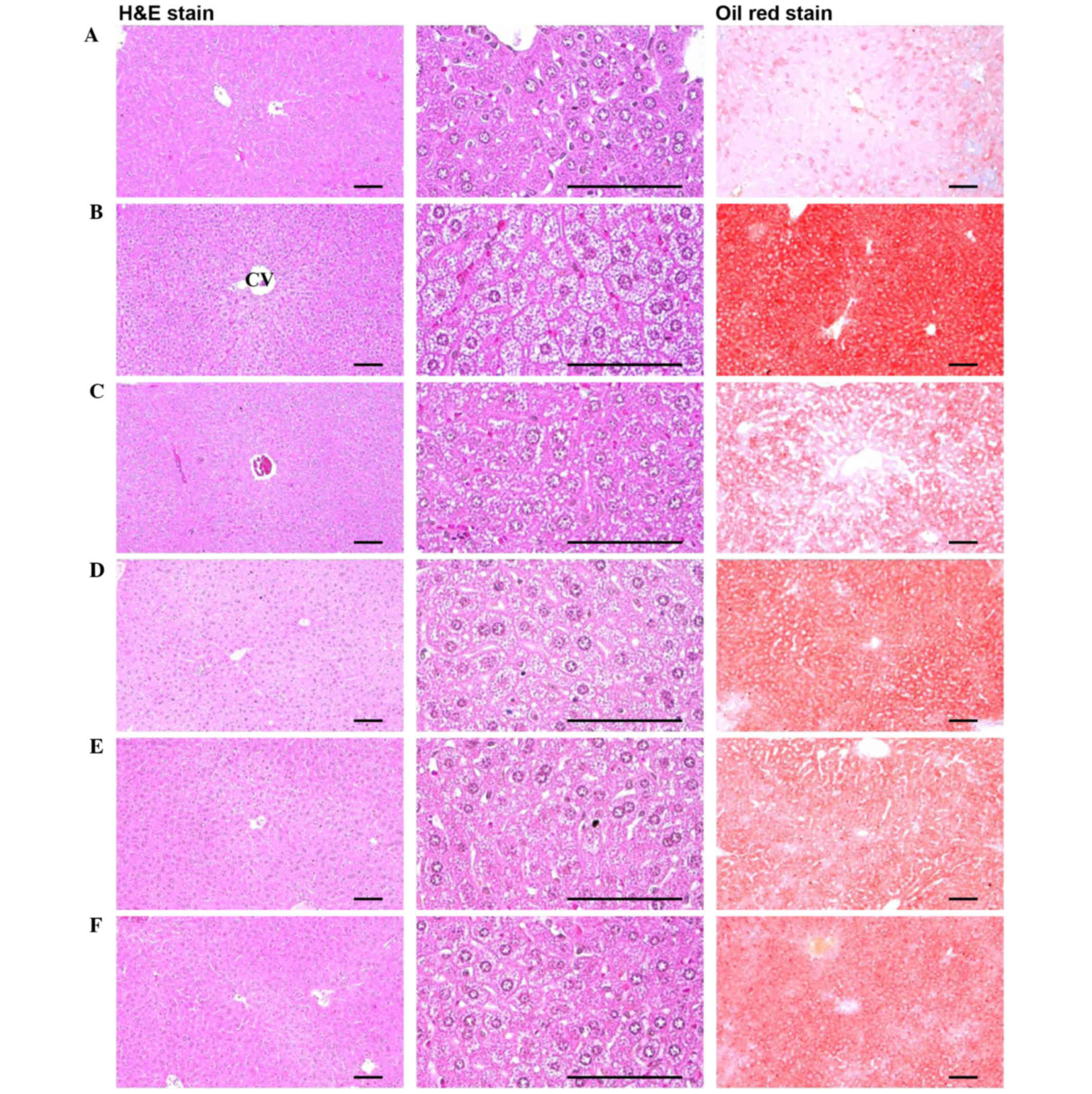
(Table VI; Fig. 8). Furthermore, significant increases
in the percentage of fatty change regions and the mean diameters of
hepatocytes were observed in OVX control mice (P<0.01). This was
thought to be due to the deposition of lipids into hepatocytes and
steatosis. However, significant decreases in the percentage of
fatty change regions and mean diameters of hepatocytes were
detected in all test substance-administered mice in the present
study, including estradiol-treated mice, as compared with OVX
control mice (Table VI; Fig. 9). These results suggested that PCS
exerts anti-obesity, estrogenic and hepatoprotective effects.
| Table VI.Histopathology-histomorphometry
analysis of the abdominal fat pads, uterus and liver in
sham-operated or OVX ddY mice. |
Table VI.
Histopathology-histomorphometry
analysis of the abdominal fat pads, uterus and liver in
sham-operated or OVX ddY mice.
|
| Control |
| PCS |
|---|
|
|
|
|
|
|---|
| Variable | Sham | OVX | Estradiol | 1 ml/kg | 2 ml/kg | 4 ml/kg |
|---|
| Abdominal fat
pads |
|
|
|
|
|
|
| Total
Th (mm) | 1.47±0.56 |
6.33±0.94b |
2.82±0.29b,e |
4.94±0.64b,e |
3.75±0.72b,e |
3.75±1.38b,e |
|
Adipocyte DM (µm) | 36.09±10.80 |
133.37±20.97a |
66.05±16.12a,d |
98.94±15.53a,d |
75.16±15.70a,d |
72.71±22.41a,d |
| Uterus |
|
|
|
|
|
|
| Total
Th (µm) | 2271.81±664.94 |
542.19±123.09b |
1609.39±364.98c,e |
757.88±89.21b,e |
908.06±83.37b,e |
910.73±155.32b,e |
| Epi Th
(µm) | 35.64±6.06 |
7.54±1.45b |
21.39±4.32b,e |
13.22±2.94b,e |
17.81±4.46b,e |
17.97±4.87b,e |
| Mucosa
Th (µm) | 991.09±243.96 |
194.15±52.74b |
627.08±163.07b,e |
306.49±59.11b,e |
424.09±91.22b,e |
426.09±101.39b,e |
| UG
percentage (%) | 55.25±11.50 |
12.29±3.27b |
36.70±4.67b,e |
22.41±4.06b,e |
30.66±5.61b,e |
30.83±8.35b,e |
| Liver |
|
|
|
|
|
|
| FC
region (%) | 13.06±3.18 |
79.55±6.78a |
42.53±8.11a,d |
64.28±11.93a,d |
49.28±13.38a,d |
47.81±13.06a,d |
|
Hepatocyte DM (µm) | 10.43±4.21 |
32.71±4.47a |
18.09±3.84a,d |
24.77±2.11a,d |
21.51±2.18a,d |
21.49±5.23a,d |
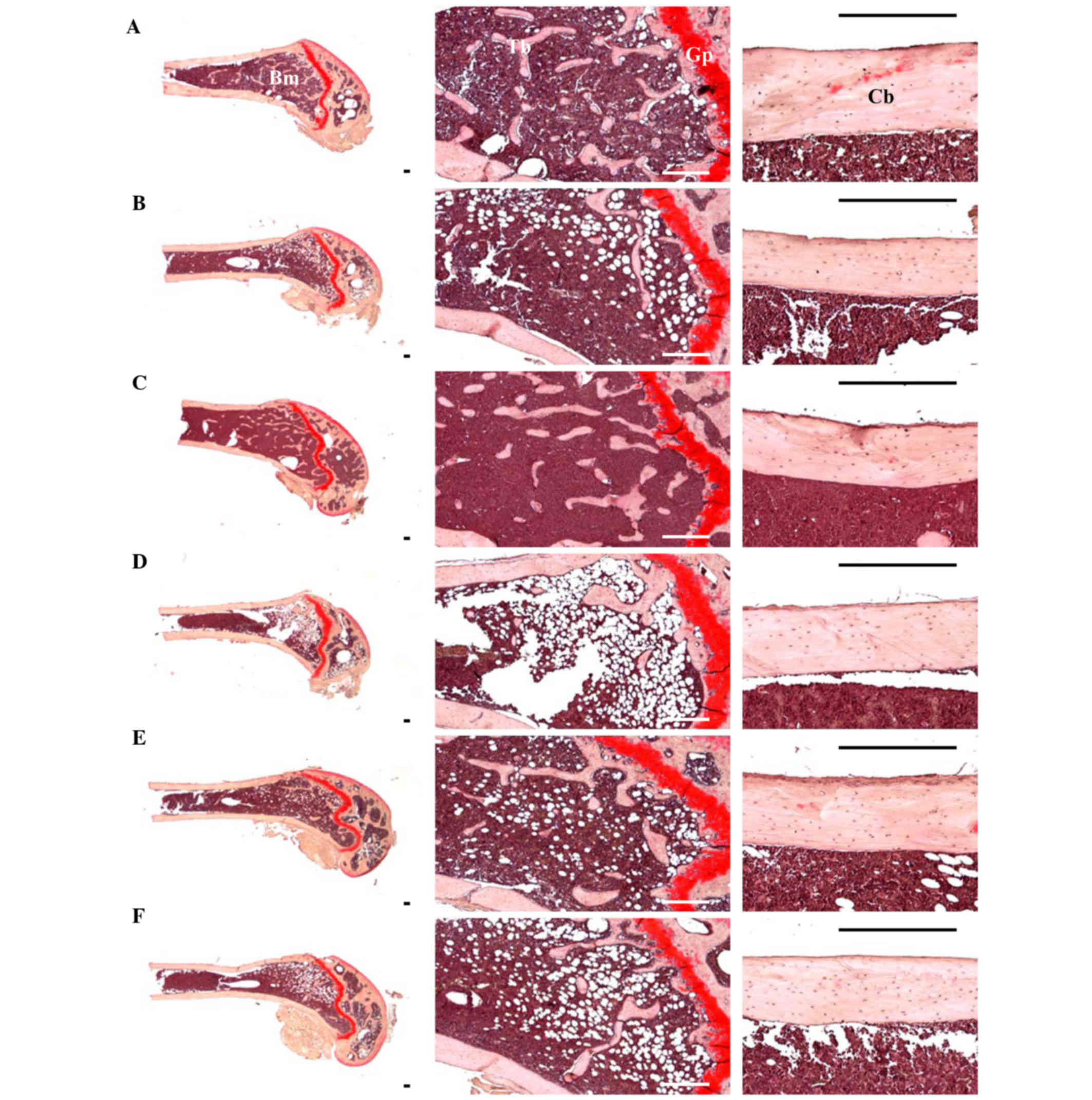
Effects on femur histopathology
Although relatively well-developed trabecular and
cortical bone were observed in the femur of sham control mice,
classical osteoporotic histological profiles were observed in OVX
control mice as significant decreases in trabecular and cortical
bone masses and increases in connective tissues in periosteum of
cortical bone resulting from resorption of osteoid tissues related
to osteoclast activation (P<0.01). However, significant
increases in bone mass and structures, of both trabecular and
cortical bones, were detected in all test substance-administered
mice including 1 ml/kg (phencyclidine) PCS-administered mice
compared with OVX control mice, which is related to their
inhibitory activities on osteoclast cell activities (P<0.05 or
P<0.01) (Table VII; Fig. 10).
| Table VII.Histopathology-histomorphometry
analysis of the femur in sham-operated or OVX ddY mice. |
Table VII.
Histopathology-histomorphometry
analysis of the femur in sham-operated or OVX ddY mice.
|
| Control |
| PCS |
|---|
|
|
|
|
|
|---|
| Variable | Sham | OVX | Estradiol | 1 ml/kg | 2 ml/kg | 4 ml/kg |
|---|
| Bone mass and
structure |
|
|
|
|
|
|
| TBV,
BV/TV | 36.16±4.85 |
16.83±3.49a |
30.82±5.56a,d |
22.25±2.56a,e |
26.17±2.71a,d |
26.28±4.59a,d |
|
Tbn | 12.88±1.89 |
4.88±0.99b |
10.25±1.98c,f |
6.75±1.28b,f |
9.38±1.60b,f |
9.63±2.50c,f |
|
Tbl | 1036.43±149.31 |
453.98±90.73a |
927.20±177.64d |
683.74±86.34a,d |
826.77±112.94a,d |
828.78±144.54a,d |
|
Tbt | 74.06±12.38 |
31.60±7.22a |
57.86±10.87a,d |
41.26±5.08a |
49.90±11.18a,d |
50.34±13.29a,d |
|
Cbt-shaft | 214.27±18.29 |
149.13±14.34b |
187.92±11.49b,f |
169.70±10.87b,g |
179.43±21.13b,f |
180.04±18.52b,g |
| Bone
resorption |
|
|
|
|
|
|
|
Ocn | 5.38±0.92 |
17.38±2.45a |
7.38±1.41d |
13.50±1.77a,d |
11.25±2.60a,d |
11.13±2.23a,d |
|
OS/BS | 8.29±1.45 |
21.61±3.60b |
12.86±1.77b,f |
17.71±1.83b,g |
14.15±3.00b,f |
14.09±3.96b,f |
Significant decreases in TV/BV, Tbn, Tbt, Tbl and
Cbt were detected in OVX control mice compared with sham-operated
control mice in the femur (P<0.01). However, these decreases in
bone mass and structures were significantly inhibited by treatment
with estradiol and 1, 2 and 4 ml/kg PCS, respectively, as compared
with OVX control mice in the present study (P<0.05 or P<0.01)
(Table VII; Fig. 10). Significant increases in Ocn and
OS/BS were detected in OVX control mice compared with sham control
mice, in the femur (P<0.01). However, these activations and
increases in osteoclast cells were significantly inhibited by
treatment with all test substances, including estradiol, as
compared with OVX control mice (P<0.05 or P<0.01) (Table VII; Fig. 10). These results indicated that PCS
has anti-osteoporosis activity.
Discussion
In the present study, PCS effectively inhibited or
refined climacterium symptoms, including obesity, hyperlipidemia,
hepatic steatosis, and osteoporosis, induced by OVX in ddY mice.
The results of PCS use in the present study were consistent with
the results of the use of ellagic acid and other organic materials
including flavonoids and polyphenols (47–50).
Previous studies have explored alternative therapies, such as the
use of phytoestrogens, to relieve menopausal symptoms.
Phytohormones can be extracted from plants and, when purified,
exhibit enhanced activity in the body as well as improved
bioavailability (18).
Phytoestrogens are polyphenolic non-steroid plant compounds with
estrogenic-like effects. Previous results have shown that
pomegranate seed oil prevents bone loss in OVX mice through
osteoblastic stimulation, osteoclastic inhibition, and decreased
inflammatory status (51). In
addition, pomegranate seed extract exhibits therapeutic potential
for avoidance memories, which is most likely related at least in
part to its phytoestrogenic and antioxidative actions (52,53). The
present study demonstrated that dried pomegranate concentrate
powder enhanced the anti-climacteric effects of red clover in OVX
rats. Therefore, we suggest that PCS is an attractive ingredient
with anti-OVX benefits.
Firstly, to clarify the anti-obesity effect of PCS,
food consumption, body weight and gains, and abdominal fat
depositions were investigated. As a result, OVX-induced changes,
including noticeable increases in food consumption, body weight and
gains, and abdominal fat depositions with adipocyte hypertrophy,
were significantly inhibited by treatment with estradiol and 1, 2,
and 4 ml/kg PCS. Estrogen depletion in an OVX animal model was
observed along with significant increases in food intake and
changes in body fat depositions, especially in the abdominal cavity
(54–56). In addition, obesity-induced OVX mice
exhibited an accumulation of fat deposition and cellular
hypertrophy through the expansion of intra-abdominal adipose tissue
(57,58). Estradiol has been shown to regulate
eating and body weight by controlling the potency of the feedback
signals that control meal size (59,60). The
correlation between cholecystokinin (CCK) and estradiol is
well-documented (61,62). Similar mechanisms may be in operation
for glucagon as the effects of glucagon and glucagon antibodies, on
decreased and increased meal size, respectively, were both enhanced
by estradiol in a previous study of OVX animal models (59). In the absence of estradiol, food
consumption and body weight are increased (63–65).
These observations are of clinical relevance as estradiol levels
decrease in postmenopausal women and, notably, postmenopausal women
account for a high proportion of the obese population (55). It has been assumed that the
anti-obesity effects of PCS may be mediated by estrogenic food
intake effects, but more complex mechanisms are involved in the
anti-obesity effects of PCS. Typically, increased digestive
motility leads to stimulated fecal excretions, resulting in a
reduction in body weight in rodents (66,67).
Diuretics are able to decrease body weight (68,69)
along estrogenic suppression, by enhancing the satiating potency of
CCK (62) and glucagon (59). More detailed mechanistic studies are
required to explore the anti-obesity effects of PCS.
Secondly, the present results showed that
OVX-induced groups significantly increased serum TC, LDL and TG
levels, but decreased serum HDL contents. In contrast, OVX-induced
hyperlipidemia was significantly inhibited by treatment with oral
1, 2 and 4 ml/kg PCS and estradiol. This finding is similar to
previous findings of a significant increase in TC, LDL, and TG, and
low HDL levels, in postmenopausal women (70). Similar trends in serum lipids were
observed in OVX mice (40). The
effects of estradiol on serum lipid profiles are believed to be
mediated by inhibiting the activity of 3-hydroxy-3-methylglutaryl
coenzyme A reductase (HMG-CoA) (71). Since HMG-CoA is the rate-limiting
enzyme involved in cholesterol synthesis, these effects may occur
through the elevation of HMG-CoA activity, which is associated with
cholesterol synthesis (71).
Thirdly, OVX-induced liver steatosis was observed in
the present study, whereas 1-, 2- and 4-ml/kg PCS-treated ddY mice
were significantly inhibited in OVX-induced hepatic steatosis.
These findings supported the favorable hepatoprotective activity of
PCS. Since the liver is the main target organ of HMG-CoA reductase
(45,72), hypertrophy and fatty change in
hepatocytes are accompanied by increased AST and ALT activities
(73,74), which are related to estrogen
deficiency-mediated obese and hyperlipidemia (45,74,75).
Estrogen deficiency is associated with an atherogenic lipid profile
characterized by HDL-cholesterol, LDL-cholesterol, triglyceride
levels (11), central adiposity
(12), increased diastolic pressure
(13), and increased insulin
resistance (14).
Fourthly, the present study demonstrated, via
histopathological and histomorphometrical analysis, that
osteocalcin levels and bone resorption markers (Ocn and OS/BS) were
significantly increased, accompanied by decreases in femur weights
and serum bALP levels, in OVX control mice. In addition, bone mass
and structures of the femur were decreased in OVX control mice
compared with sham-operated control mice. However, these
estrogen-deficient osteoporosis were effectively inhibited by 1, 2,
and 4 ml/kg PCS, respectively. These results support the notion
that PCS has favorable and potent anti-osteoporotic activities, as
reported previously (31,51). Bone loss is accelerated in menopausal
women due to the loss of estrogen. Osteoporosis is a common
disorder related to the imbalance between bone resorption and bone
formation, which leads to bone loss and the structural
deterioration of bone (76).
Increased bone weight is considered a good indicator of
anti-osteoporotic activities (77,78),
despite the fact that changes in bone weight are not an important
marker for evaluating anti-osteoporotic agents, with the exception
of ash bone weight (79). For an
osteoporosis-related OVX model, serum bALP content and osteocalcin
levels are typically accepted as bone turnover markers (80–82), and
BMD is used as a major determinant of osteoporosis (83–85). As
microscopic observation of bone can provide good evidence regarding
bone morphology (35,37,38,86),
trabecular and cortical bone is significantly changed in
osteoporotic animals. In addition, several histomorphometrical
indices for bone mass and bone formations are clearly reduced,
whereas histomorphometrical indices for bone resorption are
increased (37,38,87).
Therefore, the histology of bones has previously been evaluated to
examine the efficacy of various anti-osteoporosis agents (37,38,86). In
this respect, PCS exhibited anti-osteoporotic activities similar to
previous findings (31,51).
Lastly, OVX mice exhibited a significant decrease in
uterine weights along with marked decreases in serum estradiol
levels and associated uterine atrophic changes, including decreases
in total, mucosa and epithelial thicknesses, and uterine glands in
the mucosa. However, these estrogen-deficient uterine atrophies
were significantly inhibited by PCS treatment. As estrogens are
shown to act on numerous female target organs, such as the uterus,
vagina, and skeletal and cardiovascular systems (88,89),
menopausal women may experience climacterium symptoms due to lack
of estrogen (90,91). Loss of estrogen is accompanied by
atrophy of the uterus and vagina (56,91).
Phytoestrogenic effects of isoflavonoids cause increases in uterine
masses via uterine water imbibitions and/or a cell proliferation
(88,92), which are mediated through ERα
(93–95).
In conclusion, the present results indicated that
PCS effectively inhibited climacterium symptoms including obesity,
hyperlipidemia, hepatic steatosis, and osteoporosis in OVX-ddY
mice. Therefore, PCS may be a promising novel protective agent for
relieving climacterium symptoms in menopausal women.
Acknowledgements
The present work was supported by the National
Research Foundation of Korea grant funded by the Korea government
(grant no. 2012R1A5A2A42671316) and the Basic Science Research
Program provided by the Ministry of Education, Science and
Technology (grant no. NRF-2012R1A1A2043886).
References
|
1
|
del Giorno C, Fonseca AM, Bagnoli VR,
Assis JS, Soares JM Jr and Baracat EC: Effects of Trifolium
pratense on the climacteric and sexual symptoms in postmenopause
women. Rev Assoc Med Bras. 56:558–562. 2010.(In English and
Portuguese). PubMed/NCBI
|
|
2
|
Speroff L: The menopause: A signal for the
futureTreatment of the postmenopausal woman. Lobo RA: Raven Press;
New York: pp. 1–7. 2005
|
|
3
|
Wolff LP, Martins MR, Bedone AJ and
Monteiro IM: Endometrial evaluation in menopausal women after six
months of isoflavones. Rev Assoc Med Bras. 52:419–423. 2006.(In
Protuguese). View Article : Google Scholar : PubMed/NCBI
|
|
4
|
Dennerstein L, Lehert P and Guthrie J: The
effects of the menopausal transition and biopsychosocial factors on
well-being. Arch Womens Ment Health. 5:15–22. 2002. View Article : Google Scholar : PubMed/NCBI
|
|
5
|
van Seumeren I: Weight gain and hormone
replacement therapy: Are women's fears justified? Maturitas. 34
Suppl 1:S3–S8. 2000. View Article : Google Scholar : PubMed/NCBI
|
|
6
|
Choi JS, Koh IU and Song J: Genistein
reduced insulin resistance index through modulating lipid
metabolism in ovariectomized rats. Nutr Res. 32:844–855. 2012.
View Article : Google Scholar : PubMed/NCBI
|
|
7
|
Liang YQ, Akishita M, Kim S, Ako J,
Hashimoto M, Iijima K, Ohike Y, Watanabe T, Sudoh N, Toba K, et al:
Estrogen receptor beta is involved in the anorectic action of
estrogen. Int J Obes Relat Metab Disord. 26:1103–1109. 2002.
View Article : Google Scholar : PubMed/NCBI
|
|
8
|
Saengsirisuwan V, Pongseeda S,
Prasannarong M, Vichaiwong K and Toskulkao C: Modulation of insulin
resistance in ovariectomized rats by endurance exercise training
and estrogen replacement. Metabolism. 58:38–47. 2009. View Article : Google Scholar : PubMed/NCBI
|
|
9
|
Kannel WB, Hjortland MC, McNamara PM and
Gordon T: Menopause and risk of cardiovascular disease: The
Framingham study. Ann Intern Med. 85:447–452. 1976. View Article : Google Scholar : PubMed/NCBI
|
|
10
|
Liu Y, Ding J, Bush TL, Longenecker JC,
Nieto FJ, Golden SH and Szklo M: Relative androgen excess and
increased cardiovascular risk after menopause: A hypothesized
relation. Am J Epidemiol. 154:489–494. 2001. View Article : Google Scholar : PubMed/NCBI
|
|
11
|
Warren MP and Halpert S: Hormone
replacement therapy: Controversies, pros and cons. Best Pract Res
Clin Endocrinol Metab. 18:317–332. 2004. View Article : Google Scholar : PubMed/NCBI
|
|
12
|
Augoulea A, Mastorakos G, Lambrinoudaki I,
Christodoulakos G and Creatsas G: Role of postmenopausal hormone
replacement therapy on body fat gain and leptin levels. Gynecol
Endocrinol. 20:227–235. 2005. View Article : Google Scholar : PubMed/NCBI
|
|
13
|
Reckelhoff JF: Basic research into the
mechanisms responsible for postmenopausal hypertension. Int J Clin
Pract Suppl. 13–19. 2004.PubMed/NCBI
|
|
14
|
Wu SI, Chou P and Tsai ST: The impact of
years since menopause on the development of impaired glucose
tolerance. J Clin Epidemiol. 54:117–120. 2001. View Article : Google Scholar : PubMed/NCBI
|
|
15
|
Han KK, Soares JM Jr, Haidar MA, de Lima
GR and Baracat EC: Benefits of soy isoflavone therapeutic regimen
on menopausal symptoms. Obstet Gynecol. 99:389–394. 2002.
View Article : Google Scholar : PubMed/NCBI
|
|
16
|
Beral V, Bull D and Reeves G: Million
Women Study Collaborators: Endometrial cancer and
hormone-replacement therapy in the Million Women Study. Lancet.
365:1543–1551. 2005. View Article : Google Scholar : PubMed/NCBI
|
|
17
|
Kaari C, Haidar MA, Júnior JM, Nunes MG,
Quadros LG, Kemp C, Stavale JN and Baracat EC: Randomized clinical
trial comparing conjugated equine estrogens and isoflavones in
postmenopausal women: A pilot study. Maturitas. 53:49–58. 2006.
View Article : Google Scholar : PubMed/NCBI
|
|
18
|
Knight DC and Eden JA: A review of the
clinical effects of phytoestrogens. Obstet Gynecol. 87:897–904.
1996.PubMed/NCBI
|
|
19
|
Setchell KD: Phytoestrogens: The
biochemistry, physiology, and implications for human health of soy
isoflavones. Am J Clin Nutr. 68 6 Suppl:1333S–1346S.
1998.PubMed/NCBI
|
|
20
|
Bachmann GA: Vasomotor flushes in
menopausal women. Am J Obstet Gynecol. 180:S312–S316. 1999.
View Article : Google Scholar : PubMed/NCBI
|
|
21
|
Hedelin M, Klint A, Chang ET, Bellocco R,
Johansson JE, Andersson SO, Heinonen SM, Adlercreutz H, Adami HO,
Grönberg H and Bälter KA: Dietary phytoestrogen, serum
enterolactone and risk of prostate cancer: The cancer prostate
Sweden study (Sweden). Cancer Causes Control. 17:169–180. 2006.
View Article : Google Scholar : PubMed/NCBI
|
|
22
|
Taku K, Umegaki K, Sato Y, Taki Y, Endoh K
and Watanabe S: Soy isoflavones lower serum total and LDL
cholesterol in humans: A meta-analysis of 11 randomized controlled
trials. Am J Clin Nutr. 85:1148–1156. 2007.PubMed/NCBI
|
|
23
|
Ma DF, Qin LQ, Wang PY and Katoh R: Soy
isoflavone intake increases bone mineral density in the spine of
menopausal women: Meta-analysis of randomized controlled trials.
Clin Nutr. 27:57–64. 2008. View Article : Google Scholar : PubMed/NCBI
|
|
24
|
Sreeja S, Kumar TR Santhosh, Lakshmi BS
and Sreeja S: Pomegranate extract demonstrate a selective estrogen
receptor modulator profile in human tumor cell lines and in vivo
models of estrogen deprivation. J Nutr Biochem. 23:725–732. 2012.
View Article : Google Scholar : PubMed/NCBI
|
|
25
|
Elfalleh W, Tlili N, Nasri N, Yahia Y,
Hannachi H, Chaira N, Ying M and Ferchichi A: Antioxidant
capacities of phenolic compounds and tocopherols from Tunisian
pomegranate (Punica granatum) fruits. J Food Sci. 76:C707–C713.
2011. View Article : Google Scholar : PubMed/NCBI
|
|
26
|
Zahin M, Aqil F and Ahmad I: Broad
spectrum antimutagenic activity of antioxidant active fraction of
punica granatum L. peel extracts. Mutat Res. 703:99–107. 2010.
View Article : Google Scholar : PubMed/NCBI
|
|
27
|
Landete JM: Ellagitannins, ellagic acid
and their derived metabolites: A review about source, metabolism,
functions and health. Food Res Int. 44:1150–1160. 2011. View Article : Google Scholar
|
|
28
|
Wasila H, Li X, Liu L, Ahmad I and Ahmad
S: Peel effects on phenolic composition, antioxidant activity, and
making of pomegranate juice and wine. J Food Sci. 78:C1166–C1172.
2013. View Article : Google Scholar : PubMed/NCBI
|
|
29
|
Lansky EP and Newman RA: Punica granatum
(pomegranate) and its potential for prevention and treatment of
inflammation and cancer. J Ethnopharmacol. 109:177–206. 2007.
View Article : Google Scholar : PubMed/NCBI
|
|
30
|
Singh RP, Murthy KN Chidambara and
Jayaprakasha GK: Studies on the antioxidant activity of pomegranate
(Punica granatum) peel and seed extracts using in vitro models. J
Agric Food Chem. 50:81–86. 2002. View Article : Google Scholar : PubMed/NCBI
|
|
31
|
Mori-Okamoto J, Otawara-Hamamoto Y, Yamato
H and Yoshimura H: Pomegranate extract improves a depressive state
and bone properties in menopausal syndrome model ovariectomized
mice. J Ethnopharmacol. 92:93–101. 2004. View Article : Google Scholar : PubMed/NCBI
|
|
32
|
Tominaga K, Yamauchi A, Shuto H, Niizeki
M, Makino K, Oishi R and Kataoka Y: Ovariectomy aggravates
convulsions and hippocampal gamma-aminobutyric acid inhibition
induced by cyclosporin A in rats. Eur J Pharmacol. 430:243–249.
2001. View Article : Google Scholar : PubMed/NCBI
|
|
33
|
Iwamoto J, Seki A, Takeda T, Sato Y,
Yamada H and Yeh JK: Comparative therapeutic effects of alendronate
and alfacalcidol on cancellous and cortical bone mass and
mechanical properties in ovariectomized osteopenic rats. J Nutr Sci
Vitaminol (Tokyo). 52:1–8. 2006. View Article : Google Scholar : PubMed/NCBI
|
|
34
|
Cheng K and Tian SL: Effects of
preventive-electroacupuncture of ‘Guanyuan’ (CV 4) and ‘Sanyinjiao’
(SP 6) on hypothalamus-pituitary-ovary axis in ovariectomized rats.
Zhen Ci Yan Jiu. 37:15–19, 45. 2012.(In Chinese). PubMed/NCBI
|
|
35
|
Yamaguchi K, Yada M, Tsuji T, Kuramoto M
and Uemura D: Suppressive effect of norzoanthamine hydrochloride on
experimental osteoporosis in ovariectomized mice. Biol Pharm Bull.
22:920–924. 1999. View Article : Google Scholar : PubMed/NCBI
|
|
36
|
Ku SK and Lee HS: Distribution and
frequency of endocrine cells in the pancreas of the ddY mouse: An
immunohistochemical study. Eur J Histochem. 49:125–130.
2005.PubMed/NCBI
|
|
37
|
Han SY, Lee JR, Kwon YK, Jo MJ, Park SJ,
Kim SC, Lee HS and Ku SK: Ostreae Testa prevent ovariectomy-induced
bone loss in mice by osteoblast activations. J Ethnopharmacol.
114:400–405. 2007. View Article : Google Scholar : PubMed/NCBI
|
|
38
|
Shin HD, Yang KJ, Park BR, Son CW, Jang HJ
and Ku SK: Antiosteoporotic effect of Polycan, beta-glucan from
Aureobasidium, in ovariectomized osteoporotic mice. Nutrition.
23:853–860. 2007. View Article : Google Scholar : PubMed/NCBI
|
|
39
|
US Food and Drug Administration, .
Guidelines for preclinical and clinical evaluation of agents for
the treatment or prevention of postmenopausal osteoporosis.
Rockville, MA: 1994
|
|
40
|
Chiba H, Uehara M, Wu J, Wang X, Masuyama
R, Suzuki K, Kanazawa K and Ishimi Y: Hesperidin, a citrus
flavonoid, inhibits bone loss and decreases serum and hepatic
lipids in ovariectomized mice. J Nutr. 133:1892–1897.
2003.PubMed/NCBI
|
|
41
|
Murakami A, Song M, Katsumata S, Uehara M,
Suzuki K and Ohigashi H: Citrus nobiletin suppresses bone loss in
ovariectomized ddY mice and collagen-induced arthritis in DBA/1J
mice: Possible involvement of receptor activator of NF-kappaB
ligand (RANKL)-induced osteoclastogenesis regulation. Biofactors.
30:179–192. 2007. View Article : Google Scholar : PubMed/NCBI
|
|
42
|
Tousen Y, Abe F, Ishida T, Uehara M and
Ishimi Y: Resistant starch promotes equol production and inhibits
tibial bone loss in ovariectomized mice treated with daidzein.
Metabolism. 60:1425–1432. 2011. View Article : Google Scholar : PubMed/NCBI
|
|
43
|
Kang SJ, Lee JE, Lee EK, Jung DH, Song CH,
Park SJ, Choi SH, Han CH, Ku SK and Lee YJ: Fermentation with
Aquilariae Lignum enhances the anti-diabetic activity of green tea
in type II diabetic db/db mouse. Nutrients. 6:3536–3571. 2014.
View Article : Google Scholar : PubMed/NCBI
|
|
44
|
Jung YM, Lee SH, Lee DS, You MJ, Chung IK,
Cheon WH, Kwon YS, Lee YJ and Ku SK: Fermented garlic protects
diabetic, obese mice when fed a high-fat diet by antioxidant
effects. Nutr Res. 31:387–396. 2011. View Article : Google Scholar : PubMed/NCBI
|
|
45
|
Lim DW, Kim JG and Kim YT: Effects of
dietary isoflavones from Puerariae radix on lipid and bone
metabolism in ovariectomized rats. Nutrients. 5:2734–2746. 2013.
View Article : Google Scholar : PubMed/NCBI
|
|
46
|
Choi JS, Kim JW, Kim KY, Cho HR, Choi IS
and Ku SK: Antiosteoporotic effects of Polycan in combination with
calcium lactate-gluconate in ovariectomized rats. Exp Ther Med.
8:957–967. 2014.PubMed/NCBI
|
|
47
|
Kang SJ, Choi BR, Kim SH, Yi HY, Park HR,
Kim DC, Choi SH, Han CH, Park SJ, et al: Dried pomegranate
potentiates anti-osteoporotic and anti-obesity activities of red
clover dry extracts in ovariectomized rats. Nutrients. 7:2622–2647.
2015. View Article : Google Scholar : PubMed/NCBI
|
|
48
|
Occhiuto F, Pasquale RD, Guglielmo G,
Palumbo DR, Zangla G, Samperi S, Renzo A and Circosta C: Effects of
phytoestrogenic isoflavones from red clover (Trifolium pratense L.)
on experimental osteoporosis. Phytother Res. 21:130–134. 2007.
View Article : Google Scholar : PubMed/NCBI
|
|
49
|
Sung MJ, Davaatseren M, Hur HJ, Kim HJ,
Ryu SY, Choi YH, Cha MR and Kwon DY: Antiosteoporotic activity of
Saururus chinensis extract in ovariectomized rats. Phytother Res.
26:1182–1188. 2012. View Article : Google Scholar : PubMed/NCBI
|
|
50
|
Kaume L, Gilbert W, Smith BJ and Devareddy
L: Cyanidin 3-O-b-D-Glucoside Improves Bone Indices. J Med Food.
18:690–697. 2015. View Article : Google Scholar : PubMed/NCBI
|
|
51
|
Spilmont M, Léotoing L, Davicco MJ,
Lebecque P, Mercier S, Miot-Noirault E, Pilet P, Rios L, Wittrant Y
and Coxam V: Pomegranate and its derivatives can improve bone
health through decreased inflammation and oxidative stress in an
animal model of postmenopausal osteoporosis. Eur J Nutr.
53:1155–1164. 2014. View Article : Google Scholar : PubMed/NCBI
|
|
52
|
Sarkaki A, Farbood Y, Hashemi S and
Rafieirad M: Pomegranate seed hydroalcoholic extract improves
memory deficits in ovariectomized rats with permanent cerebral
hypoperfusion/ischemia. Avicenna J Phytomed. 5:43–55.
2015.PubMed/NCBI
|
|
53
|
Sarkaki A, Rezaiei M, Naseri M Gharib and
Rafieirad M: Improving active and passive avoidance memories
deficits due to permanent cerebral ischemia by pomegranate seed
extract in female rats. Malays J Med Sci. 20:25–34. 2013.PubMed/NCBI
|
|
54
|
Lee SH, Jin N, Paik DJ, Kim DY, Chung IM
and Park Y: Consumption of legumes improves certain bone markers in
ovariectomized rats. Nutr Res. 31:397–403. 2011. View Article : Google Scholar : PubMed/NCBI
|
|
55
|
Lutz TA: Amylin may offer (more) help to
treat postmenopausal obesity. Endocrinology. 152:1–3. 2011.
View Article : Google Scholar : PubMed/NCBI
|
|
56
|
Ateba SB, Njamen D, Medjakovic S, Hobiger
S, Mbanya JC, Jungbauer A and Krenn L: Eriosema laurentii De Wild
(Leguminosae) methanol extract has estrogenic properties and
prevents menopausal symptoms in ovariectomized Wistar rats. J
Ethnopharmacol. 150:298–307. 2013. View Article : Google Scholar : PubMed/NCBI
|
|
57
|
Morange PE, Lijnen HR, Alessi MC, Kopp F,
Collen D and Juhan-Vague I: Influence of PAI-1 on adipose tissue
growth and metabolic parameters in a murine model of diet-induced
obesity. Arterioscler, Thromb Vasc Biol. 20:1150–1154. 2000.
View Article : Google Scholar
|
|
58
|
Kim CM, Yi SJ, Cho IJ and Ku SK: Red-koji
fermented red ginseng ameliorates high fat diet-induced metabolic
disorders in mice. Nutrients. 5:4316–4332. 2013. View Article : Google Scholar : PubMed/NCBI
|
|
59
|
Geary N and Asarian L: Estradiol increases
glucagon's satiating potency in ovariectomized rats. Am J Physiol
Regul Integr Comp Physiol. 281:R1290–R1294. 2001.PubMed/NCBI
|
|
60
|
Asarian L and Geary N: Modulation of
appetite by gonadal steroid hormones. Philos Trans R Soc Lond B
Biol Sci. 361:1251–1263. 2006. View Article : Google Scholar : PubMed/NCBI
|
|
61
|
Asarian L and Geary N: Estradiol enhances
cholecystokinin-dependent lipid-induced satiation and activates
estrogen receptor-alpha-expressing cells in the nucleus tractus
solitarius of ovariectomized rats. Endocrinology. 148:5656–5666.
2007. View Article : Google Scholar : PubMed/NCBI
|
|
62
|
Thammacharoen S, Lutz TA, Geary N and
Asarian L: Hindbrain administration of estradiol inhibits feeding
and activates estrogen receptor-alpha-expressing cells in the
nucleus tractus solitarius of ovariectomized rats. Endocrinology.
149:1609–1617. 2008. View Article : Google Scholar : PubMed/NCBI
|
|
63
|
Eckel LA: Estradiol: A rhythmic,
inhibitory, indirect control of meal size. Physiol Behav. 82:35–41.
2004. View Article : Google Scholar : PubMed/NCBI
|
|
64
|
Geary N: The estrogenic inhibition of
eatingNeurobiology of food and fluid intake. Stricker EM and Woods
S: Springer; US New York: pp. 307–345. 2004, View Article : Google Scholar
|
|
65
|
Butera PC: Estradiol and the control of
food intake. Physiol Behav. 99:175–180. 2010. View Article : Google Scholar : PubMed/NCBI
|
|
66
|
Bertrand RL, Senadheera S, Markus I, Liu
L, Howitt L, Chen H, Murphy TV, Sandow SL and Bertrand PP: A
Western diet increases serotonin availability in rat small
intestine. Endocrinology. 152:36–47. 2011. View Article : Google Scholar : PubMed/NCBI
|
|
67
|
Snedeker SM and Hay AG: Do interactions
between gut ecology and environmental chemicals contribute to
obesity and diabetes? Environ Health Perspect. 120:332–339. 2012.
View Article : Google Scholar : PubMed/NCBI
|
|
68
|
Dagnault A, Deshaies Y and Richard D:
Involvement of type I corticosteroid receptor in the effects of
ovariectomy on energy balance. Am J Physiol. 270:R199–R206.
1996.PubMed/NCBI
|
|
69
|
Verlander JW, Tran TM, Zhang L, Kaplan MR
and Hebert SC: Estradiol enhances thiazide-sensitive NaCl
cotransporter density in the apical plasma membrane of the distal
convoluted tubule in ovariectomized rats. J Clin Invest.
101:1661–1669. 1998. View
Article : Google Scholar : PubMed/NCBI
|
|
70
|
Kim HM, Park J, Ryu SY and Kim J: The
effect of menopause on the metabolic syndrome among Korean women:
The Korean National Health and Nutrition Examination Survey, 2001.
Diabetes Care. 30:701–706. 2007. View Article : Google Scholar : PubMed/NCBI
|
|
71
|
Di Croce L, Bruscalupi G and Trentalance
A: Independent behavior of rat liver LDL receptor and HMGCoA
reductase under estrogen treatment. Biochem Biophys Res Commun.
224:345–350. 1996. View Article : Google Scholar : PubMed/NCBI
|
|
72
|
Wang JF, Guo YX, Niu JZ, Liu J, Wang LQ
and Li PH: Effects of Radix Puerariae flavones on liver lipid
metabolism in ovariectomized rats. World J Gastroenterol.
10:1967–1970. 2004. View Article : Google Scholar : PubMed/NCBI
|
|
73
|
Radi ZA, Koza-Taylor PH, Bell RR, Obert
LA, Runnels HA, Beebe JS, Lawton MP and Sadis S: Increased serum
enzyme levels associated with kupffer cell reduction with no signs
of hepatic or skeletal muscle injury. Am J Pathol. 179:240–247.
2011. View Article : Google Scholar : PubMed/NCBI
|
|
74
|
Kanaya N, Vonderfecht S and Chen S:
Androgen (dihydrotestosterone)-mediated regulation of food intake
and obesity in female mice. J Steroid Biochem Mol Biol.
138:100–106. 2013. View Article : Google Scholar : PubMed/NCBI
|
|
75
|
Zhang L, Zhou M, Fang G, Tang Y, Chen Z
and Liu X: Hypocholesterolemic effect of capsaicinoids by increased
bile acids excretion in ovariectomized rats. Mol Nutr Food Res.
57:1080–1088. 2013. View Article : Google Scholar : PubMed/NCBI
|
|
76
|
Sakai A, Nishida S, Okimoto N, Okazaki Y,
Hirano T, Norimura T, Suda T and Nakamura T: Bone marrow cell
development and trabecular bone dynamics after ovariectomy in ddy
mice. Bone. 23:443–451. 1998. View Article : Google Scholar : PubMed/NCBI
|
|
77
|
Puel C, Mathey J, Kati-Coulibaly S,
Davicco MJ, Lebecque P, Chanteranne B, Horcajada MN and Coxam V:
Preventive effect of Abelmoschus manihot (L.) Medik. on bone loss
in the ovariectomised rats. J Ethnopharmacol. 99:55–60. 2005.
View Article : Google Scholar : PubMed/NCBI
|
|
78
|
Xie F, Wu CF, Zhang Y, Yao XS, Cheung PY,
Chan AS and Wong MS: Increase in bone mass and bone strength by
Sambucus williamsii HANCE in ovariectomized rats. Biol Pharm Bull.
28:1879–1885. 2005. View Article : Google Scholar : PubMed/NCBI
|
|
79
|
Yamamoto M, Fisher JE, Gentile M, Seedor
JG, Leu CT, Rodan SB and Rodan GA: The integrin ligand echistatin
prevents bone loss in ovariectomized mice and rats. Endocrinology.
139:1411–1419. 1998. View Article : Google Scholar : PubMed/NCBI
|
|
80
|
Cui Y, Bhandary B, Marahatta A, Lee GH, Li
B, Kim DS, Chae SW, Kim HR and Chae HJ: Characterization of Salvia
miltiorrhiza ethanol extract as an anti-osteoporotic agent. BMC
Complement Altern Med. 11:1202011. View Article : Google Scholar : PubMed/NCBI
|
|
81
|
Farley JR, Chesnut CH III and Baylink DJ:
Improved method for quantitative determination in serum of alkaline
phosphatase of skeletal origin. Clin Chem. 27:2002–2007.
1981.PubMed/NCBI
|
|
82
|
Li JY, Tawfeek H, Bedi B, Yang X, Adams J,
Gao KY, Zayzafoon M, Weitzmann MN and Pacifici R: Ovariectomy
disregulates osteoblast and osteoclast formation through the T-cell
receptor CD40 ligand. Proc Natl Acad Sci USA. 108:768–773. 2011.
View Article : Google Scholar : PubMed/NCBI
|
|
83
|
Bilston LE, Little DG, Smith NC, Williams
P and Briody J: Zoledronic acid improves the mechanical properties
of normal and healing bone. Clin Biomech (Bristol, Avon).
17:716–718. 2002. View Article : Google Scholar : PubMed/NCBI
|
|
84
|
Diez F: Guidelines for the diagnosis of
osteoporosis by densitometric methods. J Manipulative Physiol Ther.
25:403–415. 2002. View Article : Google Scholar : PubMed/NCBI
|
|
85
|
Syed Z and Khan A: Bone densitometry:
Applications and limitations. J Obstet Gynaecol Can. 24:476–484.
2002. View Article : Google Scholar : PubMed/NCBI
|
|
86
|
Heikkinen T, Puolivali J and Tanila H:
Effects of long-term ovariectomy and estrogen treatment on maze
learning in aged mice. Exp Gerontol. 39:1277–1283. 2004. View Article : Google Scholar : PubMed/NCBI
|
|
87
|
Glatt M, Pataki A, Evans GP, Hornby SB and
Green JR: Loss of vertebral bone and mechanical strength in
estrogen-deficient rats is prevented by long-term administration of
zoledronic acid. Osteoporos Int. 15:707–715. 2004. View Article : Google Scholar : PubMed/NCBI
|
|
88
|
Couse JF and Korach KS: Estrogen receptor
null mice: What have we learned and where will they lead us? Endocr
Rev. 20:358–417. 1999. View Article : Google Scholar : PubMed/NCBI
|
|
89
|
Korach KS, Migliaccio S and Davis VL:
EstrogensPrinciples of Pharmacology: Basic Concepts and Clinical
Applications. Munson PL, Mueller RA and Breese GR: Chapman &
Hall; New York: pp. 809–825. 1995
|
|
90
|
Turner RT, Riggs BL and Spelsberg TC:
Skeletal effects of estrogen. Endocr Rev. 15:275–300. 1994.
View Article : Google Scholar : PubMed/NCBI
|
|
91
|
Versi E, Harvey MA, Cardozo L, Brincat M
and Studd JW: Urogenital prolapse and atrophy at menopause: A
prevalence study. Int Urogynecol J Pelvic Floor Dysfunct.
12:107–110. 2001. View Article : Google Scholar : PubMed/NCBI
|
|
92
|
Hewitt SC, Deroo BJ, Hansen K, Collins J,
Grissom S, Afshari CA and Korach KS: Estrogen receptor-dependent
genomic responses in the uterus mirror the biphasic physiological
response to estrogen. Mol Endocrinol. 17:2070–2083. 2003.
View Article : Google Scholar : PubMed/NCBI
|
|
93
|
Akiyama T, Ishida J, Nakagawa S, Ogawara
H, Watanabe S, Itoh N, Shibuya M and Fukami Y: Genistein, a
specific inhibitor of tyrosine-specific protein kinases. J Biol
Chem. 262:5592–5595. 1987.PubMed/NCBI
|
|
94
|
Kuiper GG, Carlsson B, Grandien K, Enmark
E, Häggblad J, Nilsson S and Gustafsson JA: Comparison of the
ligand binding specificity and transcript tissue distribution of
estrogen receptors alpha and beta. Endocrinology. 138:863–870.
1997. View Article : Google Scholar
|
|
95
|
Cornwell T, Cohick W and Raskin I: Dietary
phytoestrogens and health. Phytochemistry. 65:995–1016. 2004.
View Article : Google Scholar : PubMed/NCBI
|