Introduction
Hepatocellular carcinoma (HCC) is among the most
prevalent forms of cancer and is the third most common cause of
cancer-associated mortality worldwide, resulting in ~600,000
mortalities per year (1,2). Inhibitors that target their
corresponding pathways in HCC have achieved promising results
(3), however the detailed molecular
mechanisms of HCC tumorigenesis remain unknown.
During cell cycle progression, signaling pathways
monitor the upstream events that occur prior to proceeding to the
next phase. These regulatory switches are known as cell cycle
checkpoints (4). Forkhead Box M1
(FoxM1) is a mammalian transcription factor that regulates mitotic
entry and subsequent execution of the mitotic program by
controlling the expression of a cluster of G2/M target genes
(5). The human FOXM1 gene consists
of 10 exons, including exon Va and exon VIIa that are alternatively
spliced. These splices give rise to three distinct isoforms of
FOXM1:FOXM1a, FOXM1b, and FOXM1c (6–8). FOXM1a
harbors exon Va and exon VIIa and is transcriptionally inactive
owing to disruption of its transactivation domain (TAD) by exon
VIIa. FOXM1b contains neither of these two exons and FOXM1c only
contains exon Va. These two isoforms are transcriptionally active
and activate expression of their target genes via different
mechanisms (7). In mice, FoxM1 has
been reported to regulate the expression of cell division cycle 25
(Cdc25) B, Aurora B kinase, survivin, polo-like kinase (PLK)1,
centromere proteins (CENP) A and B and cyclin B1 (9).
PLK was first identified in the polo gene of
Drosophila melanogaster and functional mutations of PLK
cause various defects in mitosis (10). Five mammalian PLK family members have
been identified so far: PLK1, 2, 3, 4 and 5 (11,12).
PLK1 is the most investigated member of the family and has been
widely pursued as an oncology target (13,14). The
PLK1 protein activates Cdc25c phosphatase, resulting in the removal
of inhibitory phosphorylations from the cell cycle regulatory
protein cyclin dependent kinase 1 (CDK1)/cyclin B (15).
A phosphorylated peptide motif has been detected in
PLK1 substrates including Cdc25c, which regulates the activation of
CDK1/cyclin B, and the crystal structures of peptides based on this
motif and in complex with the Polo box domain have been determined
(16,17).
Ubiquitin specific peptidase 39 (Usp39) has been
identified as a novel factor that maintains the spindle checkpoint
and supports successful cytokinesis. It has been demonstrated that
USP39 is an important part of the spliceosome, which is composed of
the small nuclear ribonucleoproteins (snRNPs) U1, U2, U4, U5 and
U6, as well as >200 polypeptides (18,19). A
specific reduction in Aurora B mRNA levels following the depletion
of USP39 has been observed, however, forced expression of Aurora B
is not sufficient to reverse the damage to the checkpoints caused
by USP39 depletion in U2OS cells (20). In addition, the zebrafish USP39
mutation induces G1/S arrest by a retinoblastoma 1 (rb1) splicing
defect and transcription factor e2f4 (e2f4) is the target of USP39
(21). USP39 mutations contribute to
the adenohypophyseal sensitivity of rb1 and e2f4 that causes
pituitary tumorigenesis (21). USP39
may act as an oncogenic factor in breast cancer since the
downregulation of USP39 induces the apoptosis of MCF-7 cells in
vitro. It was demonstrated that the inhibition of USP39 induced
G0/G1phase arrest and the apoptosis of breast cancer cells
(22).
These results suggest that USP39 may be a potential
molecular target for cancer therapy. However, the roles of USP39 in
HCC are not well studied. The present study aimed to investigate
the potential functions of USP39 in HCC. It has been determined
that levels of USP39 expression are higher in liver cancer cells,
indicating that USP39 may act as a novel target in liver cancer
(23). To test this hypothesis,
USP39 expression was attenuated by small interfering RNA (siRNA)
and USP39 expression was upregulated in the HCC cell line SMMC7721.
It was found that knockdown of USP39 decreased cell proliferation
in vitro and inhibited tumor growth in vivo, while
overexpression of USP39 promoted the growth of HCC. Additionally,
it was demonstrated that the inhibition of USP39 suppressed the
tumorigenesis of HCC by inhibiting FoxM1 pre-mRNA splicing.
Materials and methods
Antibodies and cell culture
Antibodies against GAPDH (cat. no. sc-166574), FoxM1
(cat. no. sc-376471), cyclin B1 (cat. no. sc-7393), CENPA (cat. no.
sc-22787), PLK1 (cat. no. sc-55504; dilution, 1:1,000 for all) for
western blot analysis and against proliferating cell nuclear
antigen (PCNA; cat. no. sc-56; dilution, 1:500) for
immunohistochemistry (IHC) were purchased from Santa Cruz
Biotechnology, Inc. (Dallas, TX, USA). Antibodies against USP39
(cat. no. ab131332; dilution, 1:5,000) were purchased from Abcam
(Cambridge, UK).
The human HCC cell line SMMC-7721 was purchased from
the Shanghai Institutes for Biological Sciences. Cells were
cultured in RPMI-1640 medium (cat. no. 11875) supplemented with 10%
fetal bovine serum (cat. no. 10099; both, Thermo Fisher Scientific,
Inc., Waltham, MA, USA), 100 U/ml penicillin and 100 µg/ml
streptomycin (complete medium) and maintained at 37°C and 5%
CO2.
Transfection
The siRNAs targeting USP39 knockdown (KD): ACC AAG
TTG CCT CCA TAT CTA], (KD#: CCA GAC AAC TAT GAG ATC ATC GAT T) and
control siRNA negative control (NC): TTC TCC GAA CGT GTC ACG T)
were transformed into short hairpin RNA (shRNA) (stem-loop-stem
structure) and cloned into the pGCSIL-GFP lentiviral vector
(GeneChem Co. Ltd., Shanghai, China) following
AgeI/EcoRI digestion. The recombinant plasmid and two
virus packaging plasmids (GeneChem Co., Ltd) were transfected into
human 293T cells, which were purchased from the Shanghai Institutes
for Biological Sciences (Shanghai, China) using Lipofectamine™ 3000
(Invitrogen; Thermo Fisher Scientific, Inc.) following the
manufacturer's protocol. After 3 days incubation, at 37°C and in an
atmosphere containing 5% CO2, the lentivirus from the
complete medium was collected. For stable infection, HCC cell lines
were cultured in 6-well plates and infected with USP39
shRNA-expressing lentivirus (USP39-shRNA) or non-silencing
shRNA-expressing lentivirus (control) with a multiplicity of
infection (MOI) of 10. Five days after infection, cells were
observed using fluorescence microscopy (DMI4000B; Leica
Microsystems GmbH, Wetzlar, Germany). For USP39 overexpression,
full length USP39 cDNA was amplified by reverse transcription (RT)
polymerase chain reaction (PCR). Total RNA was extracted from
SMMC-7721 cell lines using an RNAsimple Total RNA kit (Tiangen
Biotech Co., Ltd., Beijing, China). Total RNA was converted into
cDNA using an RT kit (Takara Biotechnology Co., Ltd., Dalian,
China), using the following reaction mixture: 5X PrimeScript
buffer, 2 µl; PrimeScript Enzyme mix, 0.5 µl; Oligo dT Primer, 0.5
µl; random 6 mers, 0.5 µl; total RNA, 500 µg; RNase-free
dH2O, ≤10 µl. RT conditions were as follows: 37°C, 15
min; 85°C, 5 sec; and a final hold at 4°C. The USP39 primers used
were: forward 5′-CCGCTCGAGATGTCCGGCCGGTCTAAGC-3′, and reverse
5′-CGGAATTCGAGCCCCCTGCTGGTTGGTTTC-3′, which contains restriction
endonuclease XhoI and EcoRI sites. The endonuclease
used was purchased from Tiangen Biotech Co., Ltd. PCR cycling
conditions were as follows: 95°C for 5 min, 95°C for 30 sec, 55°C
for 30 sec, 72°C for 2 min (30 cycles), 72°C for 10 min, and a
final hold at 4°C. The products of PCR reaction and the empty
vectors pEGFP-N2 (BD Biosciences, Franklin Lakes, NJ, USA) were
joined together and transformed into DH5α E. coli (GeneChem
Co., Ltd.), and the recombinant plasmids were subsequently
extracted using a plasmid extraction kit (Tiangen Biotech Co.,
Ltd.). Recombinant plasmids were transfected into cells using
Lipofectamine 3000 (Invitrogen; Thermo Fisher Scientific, Inc.)
following the manufacturer's protocol.
Cell growth
Cell growth was measured by multiparametric high
content screening (HCS). SMMC-7721 cells infected with either NC
lentivirus or USP39 siRNA lentivirus were seeded at 2,000
cells/well in 96-well plates and incubated for 5 days in complete
medium at 37°C. Plates were processed with the ArrayScan High
Content Platform (cat. no. ASN00004F; Thermo Fisher Scientific,
Inc.) and kept at 4°C for up to 24 h prior to daily analysis. The
system identified stained cells and reported the intensity and
distribution of fluorescence in each cell. In each well, ≥800 cells
were analyzed. Images and data were stored in a Microsoft SQL
database.
MTT assay
SMMC-7721 cells (1×10⁵ in 0.2 ml/well)
were seeded in three replicate 96-well plates and cultured in
complete medium at 37°C for one to five days. A total of 100 µl (5
mg/ml) MTT solution was added to each well and incubated at 37°C
for 4 h. Supernatant was removed and 150 µl dimethyl sulfoxide was
added to each well. The plate was oscillated for 30 min at room
temperature. Absorbance at 490 nm was measured via microplater
reader (ELx800; BioTek Instruments, Inc., Winooski, VT, USA) and
the values were determined following background subtraction. All
MTT experiments were repeated a minimum of three times.
Tumor xenografts
A total of 20 female BALB/c nude mice aged 4–6 weeks
old (weight, 20–22 g) were obtained from the Laboratory Animal
Center of the Affiliated Drum Tower Hospital of Nanjing University
Medical School (Nanjing, China) and maintained in standard
pathogen-free conditions (temperature, 18–22°C; humidity, 50–60%;
12-h light/dark cycle). Mice were fed a diet that consisted of
flour (40%), maize meal (25%), bran (20%) and beans (15%)
supplemented with fish meal, egg, yeast, bone meal and cod-liver
oil. Mice were provided with ad libitum access to food and
water. A total of 2×10⁶ tumor cells in 0.2 ml serum-free
RPMI-1640 medium were subcutaneously injected into the flank of
each mouse. Some mice were injected with tumor cells containing
USP39 shRNA, while the control group was injected with tumor cells
containing non-silencing RNA. The experimental and control group
each consisted of 10 mice and three mice within the control groups
succumbed by day 8. Tumor growth was maintained for 22 days and
monitored by measuring the length (L) and width (W) of tumors using
a caliper and tumor size was calculated by the formula L ×
W2 × (π/6). Mice were then sacrificed via cervical
dislocation and tumors were isolated from the mice and embedded in
paraffin with 4% paraformaldehyde fixative at room temperature. All
experiments were approved by the Institutional Animal Care and Use
Committee of Nanjing University (Nanjing, China).
RT-quantitative PCR (RT-qPCR)
Total RNA was extracted from HCC cell lines using
TRIzol (Invitrogen; Thermo Scientific, Inc.) following the
manufacturer's protocol. RT-qPCR was performed as reported
previously (23). A total of 1 µg
RNA was transcribed using random primers and Primescript reverse
transcriptase (Takara Biotechnology Co., Ltd., Dalian, China). qPCR
for USP39 and GAPDH was conducted using a SYBR green qPCR kit
(Takara Biotechnology Co., Ltd.) on a fluorescent temperature
cycler (Mx3000P Real Time PCR system; Agilent Technologies, Inc.,
Santa Clara, CA, USA). The following primers were used to detect
the expression of USP39: Forward, 5′-CCAGCGATGGCAACTAC-3′ and
reverse, 5′-ACCACAACGGAAACACG-3′; GAPDH: Forward,
5′-TGACTTCAACAGCGACACCCA-3′ and reverse,
5′-CACCCTGTTGCTGTAGCCAAA-3′). PCR was performed with the following
parameters: Denaturation at 95°C for 5 min, followed by 45 cycles
at 95°C for 15 sec and 60°C for 1 min. Using GAPDH as a loading
control, relative gene expression was determined using the
∆∆Cq method (23). Gene expression was analyzed with
MxPro version 1.0 software (Agilent Technologies, Inc.).
Experiments were repeated a minimum of three times.
Flow cytometry
Cells transfected with lentivirus were harvested,
washed twice with cold phosphate-buffered saline (PBS), fixed at
4°C with cold 70% ethanol overnight and resuspended in PBS. The
suspension was filtrated through a 400-mesh membrane. The cells
were stained with propidium iodide or Annexin V-APC (eBioscience,
Inc., San Diego, CA, USA) and analyzed using a BD FACSCalibur Flow
Cytometer with Kaluza Analysis software (version 1.3; BD
Biosciences). Experiments were repeated a minimum of three
times.
Cell colony formation
SMMC-7721 cells were resuspended and seeded into
6-well plates at a final concentration of 200 cells/well and
cultured at 37°C for 14 days. The medium was replaced every 3 to 4
days. Following the incubation period, the cells were washed with
PBS twice and fixed with 4% paraformaldehyde at room temperature
for 1 h. The cells were washed with PBS twice, stained with Giemsa
(Sigma-Aldrich; Merck Millipore, Darmstadt, Germany) for 10 min and
washed with ddH2O three times. The plates were
photographed with a digital camera. Experiments were repeated a
minimum of three times.
Immunohistochemistry
Following deparaffinization of tumor tissue, antigen
retrieval was performed in citrate buffer (Nanjing KeyGen Biotech
Co., Ltd., Nanjing, China). Non-specific binding was blocked with
0.3% H2O2 (Nanjing KeyGen Biotech Co., Ltd.)
for 10 min, and with 10% goat serum (Nanjing KeyGen Biotech Co.,
Ltd.) at room temperature for 30 min. Samples were then incubated
at 4°C overnight with anti-PCNA (dilution, 1:500) and anti-USP39
antibodies (dilution, 1:200). Samples were washed in buffer and
incubated with the goat anti-mouse IgG-HRP secondary antibody (cat.
no. KGAA37; dilution, 1:1,000; Nanjing KeyGen Biotech Co., Ltd.)
for 45 min, followed by the diaminobenzidine-peroxidase reaction
and counterstaining with hematoxylin. Negative control sections
were stained under identical conditions by omitting the primary
antibody, and incubating with PBS. Stained sections were
subsequently observed with light microscopy (Leica Microsystems
GmbH) and evaluated using Image-Pro Plus 6.0 (Media Cybernetics,
Inc., Rockville, MD, USA).
Western blotting
Total protein was extracted from tumor cell lines
using radioimmunoprecipitation assay buffer (Nanjing KeyGen Biotech
Co., Ltd.) containing fresh protease and phosphatase inhibitors.
Protein concentration was determined using the BCA assay (Pierce;
Thermo Fisher Scientific, Inc.). A total of 50 µg protein per lane
was separated on 10% SDS-PAGE and transferred onto a PVDF membrane.
The membrane was blocked with 3% bovine serum albumin in 10 mM
Tris-HCl (pH 7.4) containing 0.05% Tween-20 and incubated with
primary antibodies for USP39, FoxM1, PLK1, cyclin B1 and CENPA at
4°C for 12 h. Following 3 washes with Tris-HCl buffer, the membrane
was incubated with a corresponding peroxidase-conjugated secondary
antibody (cat no. sc-2005; dilution, 1:8,000; Santa Cruz
Biotechnology, Inc.) and developed in Super-Signal West Pico
Chemiluminescent Substrate (Pierce Protein Biology; Thermo Fisher
Scientific, Inc.). The protein was visualized by autoradiography
and quantified by densitometric analysis using a Versadoc Imaging
system model 3000 (Bio-Rad Laboratories, Inc., Hercules, CA, USA).
Experiments were repeated a minimum of three times.
Statistical analyses
The data shown are presented as the mean ± standard
deviation of three independent experiments. All statistical
analyses were performed using the SPSS 11.0 software (SPSS Inc.,
Chicago, IL, USA). The xenografted tumor was analyzed with the
paired t-test. P<0.05 was considered to indicate a statistically
significant difference.
Results
Downregulation and upregulation of
USP39 in SMMC-7721 cells
To verify the roles of USP39 in tumorigenesis of HCC
in vitro, USP39 expression was inhibited in SMMC-7721 cells
by shRNA targeting of USP39. The expression of USP39 mRNA was
significantly attenuated in SMMC-7721/KD (P<0.01) and
SMMC-7721/KD# (P<0.05) compared with SMMC-7721/NC (Fig. 1A). The expression of USP39 protein
was also decreased in SMMC-7721/KD and SMMC-7721/KD# cells compared
with the control (Fig. 1B). However,
USP39 protein expression was markedly increased in SMMC-7721/USP39
cells compared with SMMC-7721/vector cells at the protein level
(Fig. 1C), indicating that
SMMC-7721/USP39 cells successfully overexpress USP39.
USP39 knockdown inhibits growth of
SMMC-7721 cells and USP39 upregulation enhances growth of SMMC-7721
cells
To explore the function of USP39 on cell growth,
SMMC-7721 cells transfected with either USP39-siRNA lentivirus or
NC lentivirus were monitored by cellomics assay. SMMC-7721 cells
containing USP39-siRNA and NC were seeded in 96-well plates and
cell growth was assayed every day for 5 days (Fig. 2A). The results demonstrated that cell
growth was inhibited when USP39 was knocked down (P<0.01;
Fig. 2B). MTT assays also indicated
that growth of SMMC-7721 cells was significantly increased after
USP39 was overexpressed (P<0.01; Fig.
2C). These results indicated that USP39 knockdown suppressed
the growth of HCC cells and USP39 overexpression promoted HCC cell
proliferation.
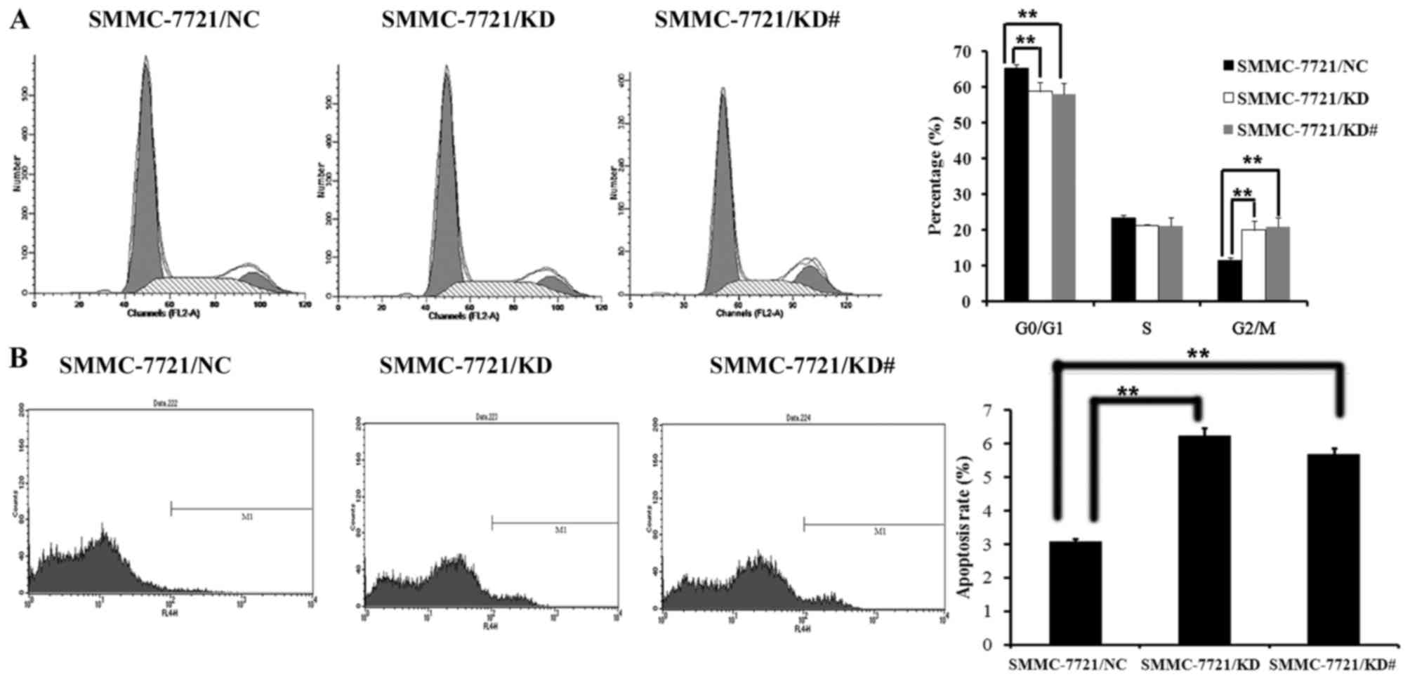
USP39 induces G2/M arrest and
suppresses cell colony formation
It has been demonstrated that USP39 regulates the
cell cycle checkpoint (20–22). Therefore, changes in the cell cycle
were examined using flow cytometry and the colony formation
capacity of SMMC-7721 was determined. It was observed that
SMMC-7721 cells with USP39 knockdown exhibited a reduced cell
population in the G0/G1 stage and a greater proportion of cells in
the G2/M stage (P<0.01; Fig. 3A).
SMMC-7721/KD cells and SMMC-7721/KD# cells also showed an increased
rate of apoptosis compared with control cells (P<0.01; Fig. 3B). Furthermore, the colony formation
capacity of SMMC-7721 cells after USP39 knockdown was investigated.
HCC cells were grown for 14 days to form colonies. The number of
SMMC-7721 colonies was significantly decreased following USP39
knockdown compared with control cells (P<0.05; Fig. 4).
Effect of USP39 on xenograft tumor
growth
To investigate the effects of USP39 on xenograft
tumor growth, SMMC-7721 cells containing USP39 shRNA or
non-silencing RNA were injected into nude mice. USP39 knockdown
significantly decreased tumor growth in SMMC-7721 cells (P<0.05;
Fig. 5A and B). In addition, cell
proliferation in SMMC-7721/KD tumors was decreased as demonstrated
by anti-PCNA staining (Fig. 5C).
These results indicate that USP39 contributes to HCC tumor growth
in vivo.
USP39-induced G2/M arrest depends on
FoxM1
To further investigate the underlying molecular
mechanisms by which USP39 regulates HCC cell proliferation, the
expression of important cell cycle proteins was examined. Since
USP39 is important in pre-mRNA splicing and FoxM1 is regulated by
mRNA splicing, it was hypothesized that FoxM1 was involved in
pre-mRNA splicing induced by USP39. The expression of FoxM1 was
determined at the mRNA and protein levels (Fig. 6A). The expression of FoxM1 mRNA was
significantly decreased following USP39 knockdown (P<0.01). The
expression of genes downstream of FoxM1 including PLK1, cyclin B1
and CENPA was also decreased following USP39 knockdown (Fig. 6B).
Discussion
HCC is one of the most prevalent tumors worldwide
and has a high mortality rate, particularly in China (24). Gene therapy has attracted increasing
attention in recent years. Meanwhile, RNA interference (RNAi) has
also developed rapidly over the past few decades. During RNAi,
double stranded siRNA degrades target mRNA, subsequently inhibiting
protein synthesis of the associated target gene (25). Identification of novel target genes
for further research is in progress. In the current study, it was
demonstrated that USP39 knockdown by siRNA causes cell cycle arrest
in the G2/M phase of HCC cells.
Stable lentiviral vectors were constructed,
containing two siRNA sequences for the knockdown of endogenous
USP39 and a construct containing USP39 cDNA was created to
overexpress USP39. It was demonstrated that USP39 knockdown
significantly blocked the growth and colony formation of SMMC-7721
cells. By contrast, USP39 overexpression promoted the growth of
SMMC-7721 cells. Moreover, USP39 knockdown resulted in G2/M arrest
and induced apoptosis in SMMC-7721 cells. These results imply that
USP39 knockdown inhibited HCC growth, possibly by inducing G2/M
arrest. Furthermore, tumor growth was decreased in SMMC-7721/KD
engrafted mice compared with control mice. Thus, USP39 knockdown
inhibited the growth of HCC cells in vitro and in
vivo.
Cell-cycle checkpoints control the proper timing of
cell-cycle events by enforcing dependency of later events on the
completion of earlier events in the cycle (26). Consequently, checkpoint blockage
results in cell-cycle arrest and alters cell proliferation. It has
been indicated that the downregulation of USP39 may inactivate the
G0/G1 arrest in the zebrafish in vivo by splicing the rb1
protein, which serves an important role in the transition from G0
to G1 phase (21). A similar effect
has been observed in human breast cancer cells (22). The results of the present study
showed that USP39 knockdown induced G2/M arrest in SMMC-7721 cells.
Levels of FoxM1 were decreased following USP39 knockdown in
SMMC-7721 cells and a similar downregulatory effect on some of the
downstream factors of FoxM1 including PLK1, CENPA and cyclin B1,
was observed. It is well known that these genes are all involved in
the G2/M transition (27).
Therefore, USP39 knockdown may induce G2/M arrest, leading to
suppression of HCC growth, possibly through the modulation of FoxM1
mRNA splicing.
In conclusion, using in vitro and in
vivo approaches, the present study provides evidence that USP39
knockdown inhibits tumor growth in human HCC by inducing G2/M
arrest. This may be partly due to blockage of FoxM1 splicing,
suggesting that USP39 may be considered as a promising molecular
target to treat HCC.
Acknowledgements
The present study was supported by the National
Natural Science Foundation of China (grant no. 31300103), the
Clinical Medical Center for Hepatobiliary Disease of Jiangsu
Province (grant no. ZX201105) and the Clinical Medical Center for
Digestive Disease of Jiangsu Province (grant no. BL2012001).
References
|
1
|
Wang G, Chen JH, Qiang Y, Wang DZ and Chen
Z: Decreased STAT4 indicates poor prognosis and enhanced cell
proliferation in hepatocellular carcinoma. World J Gastroenterol.
21:3983–3993. 2015. View Article : Google Scholar : PubMed/NCBI
|
|
2
|
Villanueva A, Minguez B, Forner A, Reig M
and Llovet JM: Hepatocellular carcinoma: Novel molecular approaches
for diagnosis, prognosis and therapy. Annu Rev Med. 61:317–328.
2010. View Article : Google Scholar : PubMed/NCBI
|
|
3
|
Huynh H: Molecularly targeted therapy in
hepatocelluar carcinoma. Biochem Pharmacol. 80:550–560. 2010.
View Article : Google Scholar : PubMed/NCBI
|
|
4
|
Fisher D, Krasinska L, Coudreuse D and
Novák B: Phosphorylation network dynamics in the control of cell
cycle transitions. J Cell Sci. 125:4703–4711. 2012. View Article : Google Scholar : PubMed/NCBI
|
|
5
|
Fu Z, Malureanu L, Huang J, Wang W, Li H,
van Deursen JM, Tindall DJ and Chen J: Plk1-dependent
phosphorylation of FoxM1 regulates a transcriptional programme
required for mitotic progression. Nat Cell Biol. 10:1076–1082.
2008. View
Article : Google Scholar : PubMed/NCBI
|
|
6
|
Korver W, Roose J and Clevers H: The
winged-helix transcription factor Trident is expressed in cycling
cells. Nucleic Acids Res. 25:1715–1719. 1997. View Article : Google Scholar : PubMed/NCBI
|
|
7
|
Ye H, Kelly TF, Samadani U, Lim L, Rubio
S, Overdier DG, Roebuck KA and Costa RH: Hepatocyte nuclear factor
3/fork head homolog 11 is expressed in proliferating epithelial and
mesenchymal cells of embryonic and adult tissues. Mol Cell Biol.
17:1626–1641. 1997. View Article : Google Scholar : PubMed/NCBI
|
|
8
|
Yao KM, Sha M, Lu Z and Wong GG: Molecular
analysis of a novel winged helix protein, WIN. Expression pattern,
DNA binding property, and alternative splicing within the DNA
binding domain. J Biol Chem. 272:19827–19836. 1997. View Article : Google Scholar : PubMed/NCBI
|
|
9
|
Yoshida Y, Wang IC, Yoder HM, Davidson NO
and Costa RH: The forkhead box M1 transcription factor contributes
to the development and growth of mouse colorectal cancer.
Gastroenterology. 132:1420–1431. 2007. View Article : Google Scholar : PubMed/NCBI
|
|
10
|
Nagata A, Igarashi M, Jinno S, Suto K and
Okayama H: An additional homolog of the fission yeast cdc25+ gene
occurs in humans and is highly expressed in some cancer cells. New
Biol. 3:959–968. 1991.PubMed/NCBI
|
|
11
|
Galaktionov K and Beach D: Specific
activation of cdc25 tyrosine phosphatases by B-type cyclins:
Evidence for multiple roles of mitotic cyclins. Cell. 67:1181–1194.
1991. View Article : Google Scholar : PubMed/NCBI
|
|
12
|
Molinari M, Mercurio C, Dominguez J,
Goubin F and Draetta GF: Human Cdc25A inactivation in response to S
phase inhibition and its role in preventing premature mitosis. EMBO
Rep. 1:71–79. 2000. View Article : Google Scholar : PubMed/NCBI
|
|
13
|
Karlsson C, Katich S, Hagting A, Hoffmann
I and Pines J: Cdc25B and Cdc25C differ markedly in their
properties as initiators of mitosis. J Cell Biol. 146:573–584.
1999. View Article : Google Scholar : PubMed/NCBI
|
|
14
|
De Souza CP, Ellem KA and Gabrielli BG:
Centrosomal and cytoplasmic Cdc2/cyclin B1 activation precedes
nuclear mitotic events. Exp Cell Res. 257:11–21. 2000. View Article : Google Scholar : PubMed/NCBI
|
|
15
|
Hoffmann I, Clarke PR, Marcote MJ,
Karsenti E and Draetta G: Phosphorylation and activation of human
cdc25-C by cdc2-cyclin B and its involvement in the
self-amplification of MPF at mitosis. EMBO J. 12:53–63.
1993.PubMed/NCBI
|
|
16
|
Perdiguero E and Nebreda AR: Regulation of
cdc25c activity during the meiotic G2/M transtition. Cell Cycle.
3:733–737. 2004. View Article : Google Scholar : PubMed/NCBI
|
|
17
|
Schmitt A and Nebreda AR: Signalling
pathways in oocyte meiotic maturation. J Cell Sci. 115:2457–2459.
2002.PubMed/NCBI
|
|
18
|
Qian Y, Erikson E, Taieb FE and Maller JL:
The polo-like kinase p1×1 is required for activation of the
phosphatase Cdc25C and cyclin B-Cdc2 in Xenopus oocytes. Mol Biol
Cell. 12:1791–1799. 2001. View Article : Google Scholar : PubMed/NCBI
|
|
19
|
Reyes-Turcu FE, Ventii KH and Wilkinson
KD: Regulation and cellular roles of ubiquitin-specific
deubiquitinating enzymes. Annu Rev Biochem. 78:363–397. 2009.
View Article : Google Scholar : PubMed/NCBI
|
|
20
|
van Leuken RJ, Luna-Vargas MP, Sixma TK,
Wolthuis RM and Medema RH: Usp39 is essential for mitotic spindle
checkpoint integrity and controls mRNA-levels of aurora B. Cell
Cycle. 7:2710–2719. 2008. View Article : Google Scholar : PubMed/NCBI
|
|
21
|
Ríos Y, Melmed S, Lin S and Liu NA:
Zebrafish usp39 mutation leads to rb1 mRNA splicing defect and
pituitary lineage expansion. PLoS Genet. 7:e10012712011. View Article : Google Scholar : PubMed/NCBI
|
|
22
|
Wang H, Ji X, Liu X, Yao R, Chi J, Liu S,
Wang Y, Cao W and Zhou Q: Lentivirus-mediated inhibition of USP39
suppresses the growth of breast cancer cells in vitro. Oncol Rep.
30:2871–2877. 2013.PubMed/NCBI
|
|
23
|
Yuan X, Sun X, Shi X, Jiang C, Yu D, Zhang
W, Guan W, Zhou J, Wu Y, Qiu Y and Ding Y: USP39 promotes the
growth of human hepatocellular carcinoma in vitroin vivo. Oncol
Rep. 34:823–832. 2015.PubMed/NCBI
|
|
24
|
Sun XT, Yuan XW, Zhu HT, Deng ZM, Yu DC,
Zhou X and Ding YT: Endothelial precursor cells promote
angiogenesis in hepatocellular carcinoma. World J Gastroenterol.
18:4925–4933. 2012. View Article : Google Scholar : PubMed/NCBI
|
|
25
|
Lindon C and Pines J: Ordered proteolysis
in anaphase inactivates Plk1 to contribute to proper mitotic exit
in human cells. J Cell Biol. 164:233–241. 2004. View Article : Google Scholar : PubMed/NCBI
|
|
26
|
Makarova OV, Makarov EM and Luhrmann R:
The 65 and 110 kDa SR-related proteins of the U4/U6.U5 tri-snRNP
are essential for the assembly of mature spliceosomes. EMBO J.
20:2553–2563. 2001. View Article : Google Scholar : PubMed/NCBI
|
|
27
|
Hanahan D and Weinberg RA: Hallmarks of
cancer: The next generation. Cell. 144:646–674. 2011. View Article : Google Scholar : PubMed/NCBI
|