Introduction
Spinal cord injury (SCI) includes primary and
secondary SCI (1). Presently, there
is no ideal treatment for SCI, creating a heavy burden for
patients, their families, and the society (2,3). The
pathological and physiological responses to SCI are very complex.
They include a sequence of cascade reactions, such as spinal cord
edema, ischemia, ion imbalance, electrolyte disturbances,
excitatory amino acid toxicity, inflammation, cell necrosis, and
apoptosis (4).
Neurotrophic factors (NFs) are a family of
polypeptide factors that support cell survival, promote cell growth
and differentiation, and maintain normal cell function. NFs are
important for spinal cord recovery, neuronal survival and
regeneration, synaptic regeneration, and limb motor function after
SCI (5–8). However, the effect of NFs after SCI has
not been well defined. The mechanisms underlying NF-mediated
neuronal recovery after SCI need to be explored further.
Materials and methods
Materials
Taq Master Mix (SinoBio USA, Walpole, MA, USA),
agarose (Biowest, Nuaillé, France), sterile double-distilled water,
anti-phospho-Trk receptor B (1:1,000; Cell Signaling Technology,
Inc., Beverly, MA, USA), Trk receptor B antibody (1:5,000;
Invitrogen, Carlsbad, CA, USA), 0.9% sterile normal saline (Otsuka
Pharmaceutical Co., Ltd., Tokyo, Japan), and NFs.
Instruments
PCR amplification and transilluminator (both from
Bio-Rad, Berkeley, CA, USA), electrophoresis apparatus (Beijing
Liuyi Biotechnology Co., Ltd., Beijing, China), centrifuge and
micro-pipette (both from Eppendorf AG, Hamburg), Haier ice maker,
western blotting electrophoresis apparatus (Bio-Rad), −80°C freezer
(Thermo Fisher Scientific, Waltham, MA, USA), 10 ml syringe, 5 ml
syringe (Tianjin Hanaco Medical Co., Ltd., Tianjin, China),
surgical instruments for use on laboratory animals (Beijing Medical
Device Factory, Beijing, China), NanoDrop2000 photometric analyzer
(Thermo Fisher Scientific, Wilmington, DE, USA), Eppendorf tube
(Eppendorf AG), water bath (Beijing Medical Device Factory), and
pathology microtome (Leica, Mannheim, Germany).
Research methods
Animals and surgical procedure
Laboratory animal grouping: Seventy-two adult
Sprague-Dawley rats were randomly assigned to three groups
regardless of gender: Sham-operated, model, and NFs/Trk groups.
There were 24 rats in each group. For each group, four subgroups
(1, 3, 5 and 7 days) were created with respect to the survival
time. Sham-operated rats were operated with a T10 laminectomy
without SCI. The rats were given normal saline via gavage after
analepsia. Motor function was measured using the Basso, Beattie and
Bresnahan (BBB) scale 1, 3, 5 and 7 days after the operation in all
groups. Rats from all groups were sacrificed for experiments after
motor function was assessed. Rats in the model group received a
laminectomy and SCI was induced using Allen's method (9). In the NF/Trk group, rats were operated
in the same way as the model group, but received NF/Trk by gavage
with normal saline after analepsia. This study was approved by the
Animal Ethics Committee of Guizhou Provincial People's
Hospital.
Evaluation of neuronal morphology and NT
expression
Changes in neuronal morphology were observed by
hematoxylin and eosin staining and Nissl staining. Expression of
brain-derived NF (BNDF) and neurotrophin-3 (NT-3) was detected by
immunofluorescence staining and the number of positive cells was
calculated based on semi-quantitative analysis. Expression of Trk
receptor B and C was tested by western blotting.
BBB locomotor scale
The BBB locomotor scale is a scoring system that
tests the motor function of rats with SCI. Scores range from 1 to
21, and the higher the score, the better the motor function of the
lower limb.
Statistical analysis
Statistical analysis was performed using SPSS 19.0
(IBM, Armonk, NY, USA). Qualitative data were analyzed by analysis
of variance (ANOVA) and χ2 test, and data that did not
meet the conditions for a 2×2 table were analyzed by Fisher's exact
test. Quantitative data were compared by ANOVA, and a P-value of
<0.05 was considered to indicate a statistically significant
difference.
Results
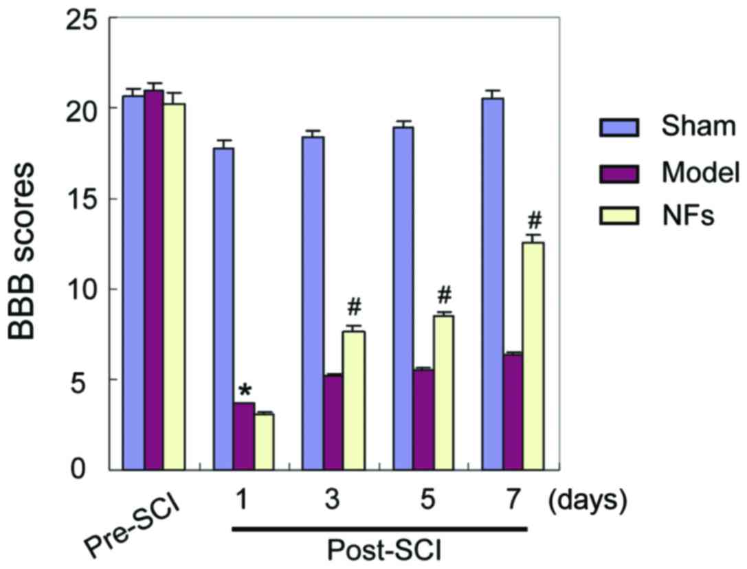
BBB locomotor scale
The BBB locomotor scale was used to assess the limb
motor function of rats with SCI. The results indicated no
significant difference in limb motor function between the three
groups before treatment. In the sham-operated group, there was no
significant difference in motor function before and after the
operation. In the groups of model and NFs/Trk rats, the BBB scale
scores decreased significantly after the operation (P<0.05).
However, as shown in Fig. 1, the BBB
scale scores were significantly higher in the NF/Trk group compared
with those in the model group at all tested time-points. In
addition, rats in the NFs/Trk group recovered significantly faster
after the operation (Fig. 1)
(P<0.05).
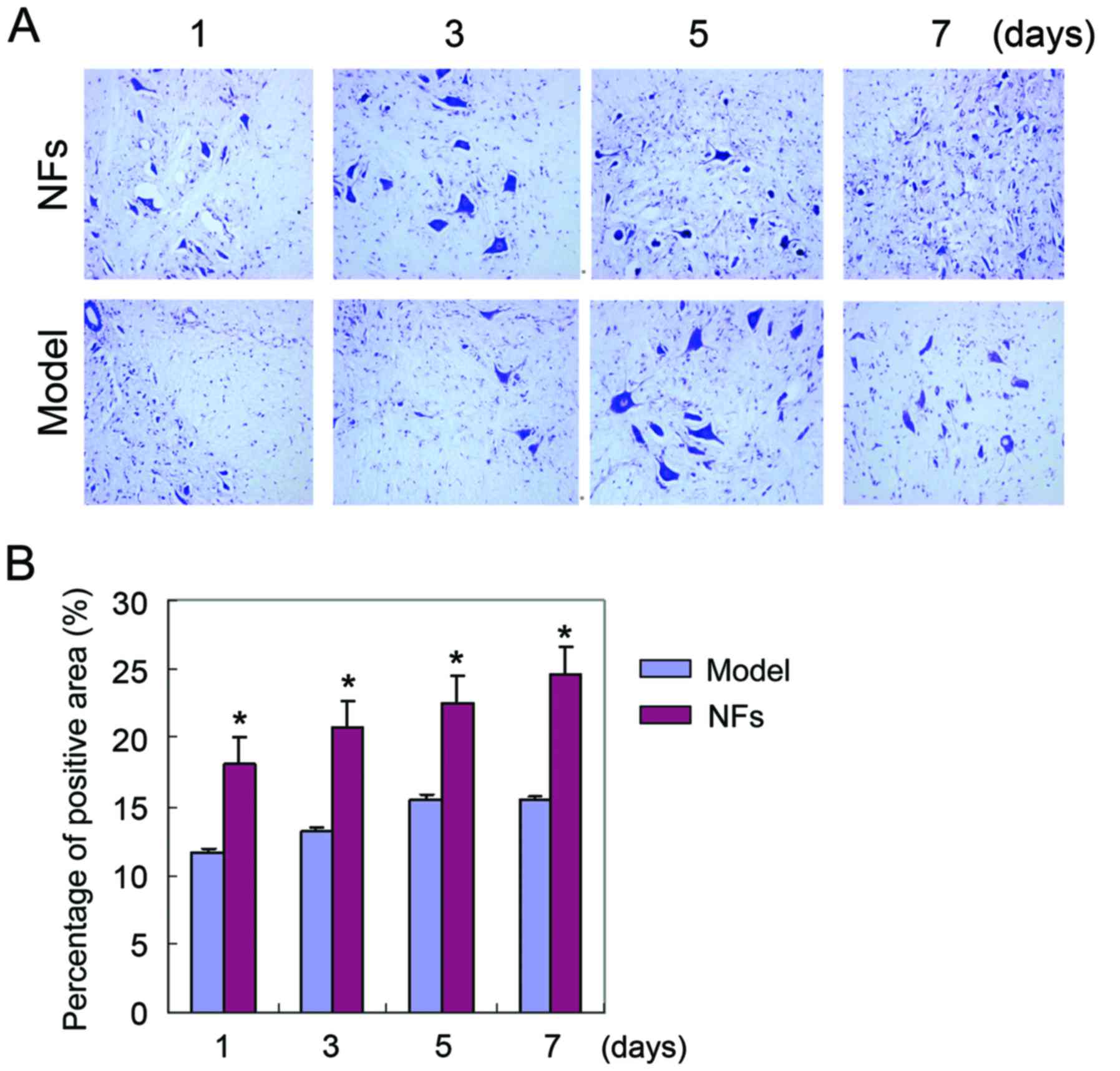
Nissl staining of spinal cord
tissue
Nissl staining was used to examine the injury of
spinal cord tissue in SCI rats. Nissl bodies were stained with
purple-blue, which identified the basic structure of spinal cord
neurons. Large and numerous Nissl bodies indicate strong protein
synthesis in neural cells, while Nissl bodies are reduced or absent
in injured neurons. Nissl staining revealed that varying degrees of
neuronal injury in the spinal cord of NF/Trk and model rats after
SCI was induced. However, neuronal injury in the NFs/Trk group was
significantly less severe than that in the model group (Fig. 2) (P<0.05).
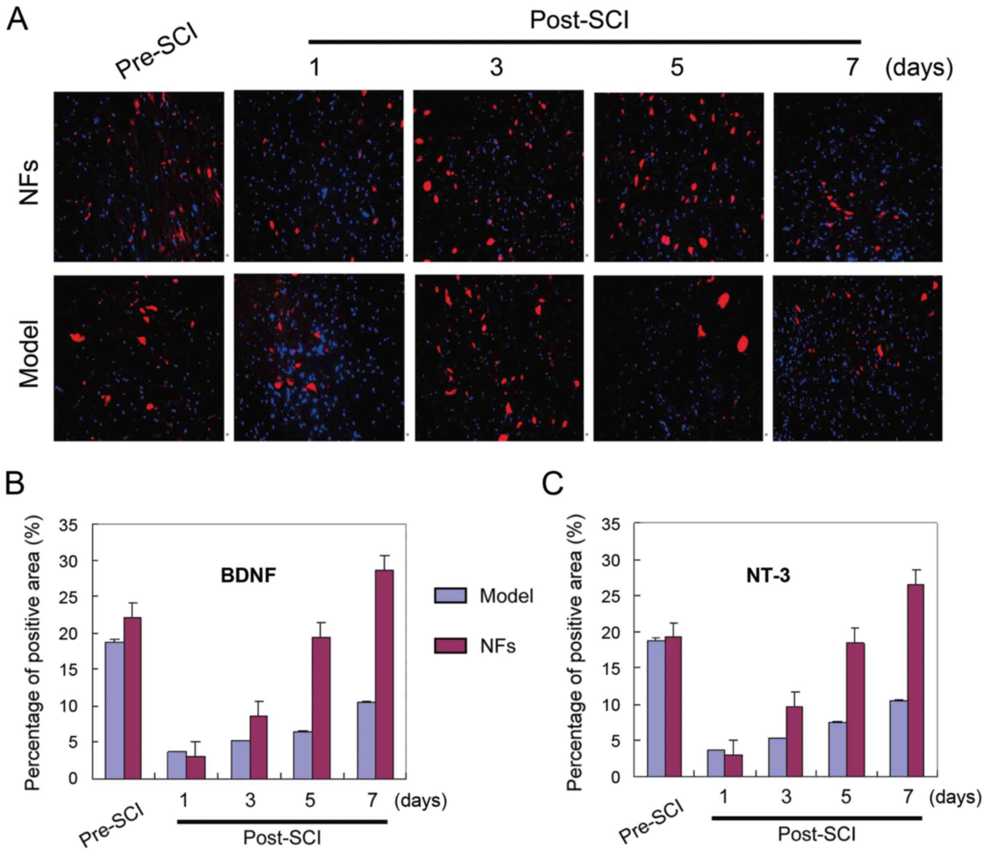
BNDF and NT-3 expression in rat spinal
cord tissue
BNDF and NT-3 staining were visualized in the spinal
cord tissue of model and NFs/Trk rats. Immunofluorescence
experiments showed significantly more BDNF and NT-3-positive cells
in the spinal cord of NFs/Trk rats compared to those in model rats
(Fig. 3) (P<0.05). BDNF and NT-3
were mainly localized in neurons of spinal cord tissue.
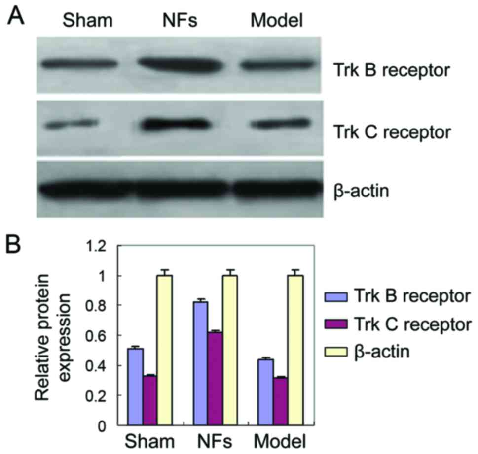
Trk B and Trk C receptor expression in
rat spinal cord tissue
To investigate the role of NF signaling in SCI
recovery, the expressions of Trk B and Trk C receptors were
analyzed by western blotting in the injured rat spinal cord with
and without NF/Trk treatment. The expressions of Trk B and Trk C
receptors were significantly higher in the spinal cord of NFs/Trk
rats than model group rats (Fig. 4)
(P<0.05). This finding indicates that NFs promote recovery
following SCI by activating the expressions of Trk B and Trk C
receptors in injured rat spinal cord tissue.
Discussion
The spinal cord can be directly or indirectly
injured by external force, causing motor, sense, and sphincter
dysfunction, abnormal muscle tone, and pathological changes
(10–14). The degree and clinical
characteristics of SCI depend on the primary source of the injury.
The high incidence of traumatic SCI in young adults often results
in limb motor dysfunction and multiple associated injuries. This
can make treatment difficult and lead to many complications and
disability. SCI represents a serious physical and psychological
injury that creates an enormous burden for patients, their
families, and society. Therefore, preclinical research of trauma
surgery is both important and challenging (15–17).
Primary and secondary SCI induces a sequence of
cellular and molecular pathological events. Secondary injury
includes an inflammatory response, which increases the severity of
SCI. Both primary and secondary SCI can cause paralysis or death.
SCI can trigger pathological and physical events. Correlations
between the primary mechanical injury and the secondary injury
promote pathological changes (4,5). SCI is
often followed by local and systemic inflammation, which increases
the pathogenesis of the injury (6).
In addition, the expression of certain genes is increased, which
may mediate the inflammatory response. These genes belong to
signaling pathways including NF-κB and adenosine
monophosphate-activated protein kinase.
Nerve growth factor (NGF) is a regulator of nerve
growth and mediates neurological nutrition and neurite outgrowth.
NGF is often released after SCI. NGF was the first NF to be
described and has been thoroughly studied. It has important
regulatory effects on the development, differentiation, growth,
regeneration, and function of central and peripheral neurons. The
NF family is very large and includes NGF, BDNF, basic fibroblast
growth factor (bFGF), ciliary NF, and NT-3 (18–21).
NFs can promote recovery after SCI by multiple
mechanisms. The main effects and mechanisms are: i) Inhibition of
c-fos and c-jun expressions in the spinal cord, thereby reducing
apoptosis of spinal cord nerve cells; ii) decreasing TNF-α, IL-1β,
and IL-8 expressions in the spinal cord, inhibiting the
inflammatory response; and iii) reduction of inflammation and
spinal cord ischemia-reperfusion injury by inhibiting NF-κB and
VCAM-1.
In this study, we induced SCI in rats using the
Allen's method, and investigated the effect of NF treatment on SCI
recovery. We demonstrated that limb motor function was
significantly improved after SCI by NF treatment 3 days after
injury. This suggests that NFs can effectively improve motor neuron
function, preserve uninjured spinal cord tissues, and accelerate
functional recovery in rats following SCI. Nissl staining of
injured spinal cord tissue indicated that neuronal injuries were
less severe following NF treatment compared with those after saline
treatment. These findings demonstrated that NFs could effectively
protect neuronal tissues after injury.
To explore the mechanism of NF action after SCI, we
examined expression of BNDF and NT-3 in spinal cord tissue after NF
and saline treatment. NF treatment significantly increased the
number of BDNF and NT-3-positive cells in rat spinal cord tissue
after injury. This finding is consistent with previous published
results. For example, Zhang et al treated rabbits with NFs
after ischemia-reperfusion injury and observed satisfactory
recovery (22).
BDNF and NT-3 were predominantly expressed in spinal
cord neurons. BDNF and NT-3 are members of the NGF family and are
weakly expressed under normal conditions. Following neuronal
injury, multiple inflammatory factors are stimulated and the
expression of BDNF, NT-3, and other NGFs increases, initiating
neuronal protective mechanisms through multiple upstream and
downstream signaling pathways. This protects uninjured neurons and
promotes the growth and repair of injured nerve tissue. Clinical
findings have shown that NGF concentrations correlate positively
with the recovery of nerve functions. This is because the extent of
self-recovery of nerve tissues was limited. Increasing NGF levels
can promote self-recovery and improve prognosis, which is
consistent with the present findings.
We demonstrated that the neuroprotective effect of
NFs was caused by enhanced expression of Trk B and Trk C receptors
in the injured rat spinal cord. The Trk receptor family is a family
of receptor tyrosine kinase that regulates the strength and
plasticity of synapses in the mammalian nervous system (1). The activation of Trk receptors affects
the survival, differentiation, and function of neurons through
multiple signaling pathways. NF is a common ligand of Trk receptors
and plays a critical role in the nervous system (2,23). The
binding between NFs and Trk receptors is highly specific. Each NF
has a corresponding Trk receptor, with a different affinity. The
signaling pathway that is induced when NF binds to a Trk receptor
regulates cell survival and function. Therefore, NFs improve
recovery of rat spinal cord tissue after injury by promoting Trk
receptor expression, thereby increasing NGF binding to Trk
receptors and enhancing the effect of NGF.
In conclusion, we have demonstrated that NFs can
promote recovery of motor function after acute SCI in rats through
NF/Trk pathways. NFs support the survival, regeneration, and axonal
repair of neurons and improve motor function after SCI by promoting
BDNF, NGF, and NT-3 expression and activation of their
high-affinity receptors.
Acknowledgements
This study was supported by the National Key
Technology R&D Program (2014BAI05B05), the Foundation of
Science and Technology Department of Guizhou Province (Qiankehe SY
zi[2012]3090), the Foundation of Science and Technology Department
of Guizhou Province (Qiankehe SY zi[2013]3063), and the Foundation
of Science and Technology Department of Guizhou Province (Qiankehe
LH zi[2014]7021).
References
|
1
|
Tashiro S, Nishimura S, Iwai H, Sugai K,
Zhang L, Shinozaki M, Iwanami A, Toyama Y, Liu M, Okano H, et al:
Functional recovery from neural stem/progenitor cell
transplantation combined with treadmill training in mice with
chronic spinal cord injury. Sci Rep. 6:308982016. View Article : Google Scholar : PubMed/NCBI
|
|
2
|
Baaj AA, Uribe JS, Nichols TA, Theodore N,
Crawford NR, Sonntag VK and Vale FL: Health care burden of cervical
spine fractures in the United States: analysis of a nationwide
database over a 10-year period. J Neurosurg Spine. 13:61–66. 2010.
View Article : Google Scholar : PubMed/NCBI
|
|
3
|
Pickelsimer E, Shiroma EJ and Wilson DA:
Statewide investigation of medically attended adverse health
conditions of persons with spinal cord injury. J Spinal Cord Med.
33:221–231. 2010. View Article : Google Scholar : PubMed/NCBI
|
|
4
|
Volarevic V, Erceg S, Bhattacharya SS,
Stojkovic P, Horner P and Stojkovic M: Stem cell-based therapy for
spinal cord injury. Cell Transplant. 22:1309–1323. 2013. View Article : Google Scholar : PubMed/NCBI
|
|
5
|
Kazim SF and Iqbal K: Neurotrophic factor
small-molecule mimetics mediated neuroregeneration and synaptic
repair: emerging therapeutic modality for Alzheimer's disease. Mol
Neurodegener. 11:502016. View Article : Google Scholar : PubMed/NCBI
|
|
6
|
Fernandes BS, Molendijk ML, Köhler CA,
Soares JC, Leite CM, Machado-Vieira R, Ribeiro TL, Silva JC, Sales
PM, Quevedo J, et al: Peripheral brain-derived neurotrophic factor
(BDNF) as a biomarker in bipolar disorder: a meta-analysis of 52
studies. BMC Med. 13:2892015. View Article : Google Scholar : PubMed/NCBI
|
|
7
|
Cook DJ, Nguyen C, Chun HN, L Llorente I,
Chiu AS, Machnicki M, Zarembinski TI and Carmichael ST:
Hydrogel-delivered brain-derived neurotrophic factor promotes
tissue repair and recovery after stroke. J Cereb Blood Flow Metab.
May 12–2016.(Epub ahead of print). PubMed/NCBI
|
|
8
|
Zhong Z, Gu H, Peng J, Wang W, Johnstone
BH, March KL, Farlow MR and Du Y: GDNF secreted from
adipose-derived stem cells stimulates VEGF-independent
angiogenesis. Oncotarget. 7:36829–36841. 2016.PubMed/NCBI
|
|
9
|
Koozekanani SH, Vise WM, Hashemi RM and
McGhee RB: Possible mechanisms for observed pathophysiological
variability in experimental spinal cord injury by the method of
Allen. J Neurosurg. 44:429–434. 1976. View Article : Google Scholar : PubMed/NCBI
|
|
10
|
Yang L, Ge Y, Tang J, Yuan J, Ge D, Chen
H, Zhang H and Cao X: Schwann cells transplantation improves
locomotor recovery in rat models with spinal cord injury: a
systematic review and meta-analysis. Cell Physiol Biochem.
37:2171–2182. 2015. View Article : Google Scholar : PubMed/NCBI
|
|
11
|
Yüksekkaya M, Tutar N, Büyükoğlan H,
Dündar M, Yılmaz İ, Gülmez İ, Oymak FS, Balta B, Korkmaz K and
Demir R: The association of brain-derived neurotrophic factor gene
polymorphism with obstructive sleep apnea syndrome and obesity.
Lung. 194:839–846. 2016. View Article : Google Scholar : PubMed/NCBI
|
|
12
|
Wang X, Peng B, Xu C, Gao Z, Cao Y, Liu Z
and Liu T: BDNF-ERK1/2 signaling pathway in ketamine-associated
lower urinary tract symptoms. Int Urol Nephrol. 48:1387–1393. 2016.
View Article : Google Scholar : PubMed/NCBI
|
|
13
|
Adhikary S, Li H, Heller J, Skarica M,
Zhang M, Ganea D and Tuma RF: Modulation of inflammatory responses
by a cannabinoid-2-selective agonist after spinal cord injury. J
Neurotrauma. 28:2417–2427. 2011. View Article : Google Scholar : PubMed/NCBI
|
|
14
|
Chiu WT, Lin HC, Lam C, Chu SF, Chiang YH
and Tsai SH: Review paper: epidemiology of traumatic spinal cord
injury: comparisons between developed and developing countries.
Asia Pac J Public Health. 22:9–18. 2010. View Article : Google Scholar : PubMed/NCBI
|
|
15
|
Cuzzocrea S, Riley DP, Caputi AP and
Salvemini D: Antioxidant therapy: a new pharmacological approach in
shock, inflammation, and ischemia/reperfusion injury. Pharmacol
Rev. 53:135–159. 2001.PubMed/NCBI
|
|
16
|
Kernie SG and Parent JM: Forebrain
neurogenesis after focal ischemic and traumatic brain injury.
Neurobiol Dis. 37:267–274. 2010. View Article : Google Scholar : PubMed/NCBI
|
|
17
|
Freund P, Weiskopf N, Ward NS, Hutton C,
Gall A, Ciccarelli O, Craggs M, Friston K and Thompson AJ:
Disability, atrophy and cortical reorganization following spinal
cord injury. Brain. 134:1610–1622. 2011. View Article : Google Scholar : PubMed/NCBI
|
|
18
|
Boyce VS, Tumolo M, Fischer I, Murray M
and Lemay MA: Neurotrophic factors promote and enhance locomotor
recovery in untrained spinalized cats. J Neurophysiol.
98:1988–1996. 2007. View Article : Google Scholar : PubMed/NCBI
|
|
19
|
Garraway SM, Woller SA, Huie JR, Hartman
JJ, Hook MA, Miranda RC, Huang YJ, Ferguson AR and Grau JW:
Peripheral noxious stimulation reduces withdrawal threshold to
mechanical stimuli after spinal cord injury: role of tumor necrosis
factor alpha and apoptosis. Pain. 155:2344–2359. 2014. View Article : Google Scholar : PubMed/NCBI
|
|
20
|
Huie JR, Stuck ED, Lee KH, Irvine KA,
Beattie MS, Bresnahan JC, Grau JW and Ferguson AR: AMPA receptor
phosphorylation and synaptic colocalization on motor neurons drive
maladaptive plasticity below complete spinal cord injury. eNeuro.
2:pii: ENEURO.0091–15.2015. 2015. View Article : Google Scholar
|
|
21
|
Liu NK and Xu XM: Neuroprotection and its
molecular mechanism following spinal cord injury. Neural Regen Res.
7:2051–2062. 2012.PubMed/NCBI
|
|
22
|
Zhang S, Tobaru T, Zivin JA and
Shackelford DA: Activation of nuclear factor-kappaB in the rabbit
spinal cord following ischemia and reperfusion. Brain Res Mol Brain
Res. 63:121–132. 1998. View Article : Google Scholar : PubMed/NCBI
|
|
23
|
Paterniti I, Esposito E, Mazzon E,
Bramanti P and Cuzzocrea S: Evidence for the role of
PI(3)-kinase-AKT-eNOS signalling pathway in secondary inflammatory
process after spinal cord compression injury in mice. Eur J
Neurosci. 33:1411–1420. 2011. View Article : Google Scholar : PubMed/NCBI
|