Introduction
Ovarian cancer is a common malignant tumor type of
the female reproductive system with a high mortality rate. The
5-year survival rate of patients with ovarian cancer is 25–30%
(1). Although progress has been made
in the treatment of ovarian cancer, the mortality rate remains
high. The majority of ovarian cancer patients have tumor metastasis
at the time-point of diagnosis, and tumor invasion and metastasis
are important causes of treatment failure (2). Therefore, investigation of the
molecular mechanisms involved in tumor proliferation, adhesion and
invasion is of great importance for improving the therapeutic
effectiveness of ovarian cancer treatments.
CD44 is a transmembrane protein that mediates
cell-cell interactions as well as cell adhesion and migration. It
is widely expressed on the cell surface (3). CD44 participates in numerous biological
processes such as tumor metastasis, hematopoiesis, lymphocyte
activation, recirculation and homing. CD44 is a receptor for
hyaluronic acid and interacts with other ligands, such as matrix
metalloproteinases, collagens and osteopontin (3). Positive CD44 expression was
demonstrated to be a prognostic indicator of shorter survival time
in patients with ovarian cancer (4–7). CD44
expression was also reported to contribute to the development of
carboplatin resistance in advanced serous epithelial ovarian
cancer, and contribute to a worse prognosis for these patients
(7). In addition, epithelial
cadherin (E-cadherin) is a calcium-dependent cell-cell adhesion
glycoprotein. It interacts with the cytoskeleton, forming complexes
with an anchoring effect. Genetic variants of the E-cadherin gene
were reported to be a prognostic marker to identify patients at
increased risk of invasive or metastatic cancer (8). E-cadherin fragmentation was revealed to
promote the progression of intraperitoneal epithelial ovarian
cancer (9). However, the effects of
CD44 and E-cadherin overexpression on cell proliferation, adhesion
and invasion of ovarian cancer cells have remained elusive.
Therefore, the present study aimed to investigate
the effects of CD44 and E-cadherin overexpression on the
proliferation, adhesion and invasion of SKOV-3 and OVCAR-3 ovarian
cancer cells.
Materials and methods
Cells and reagents
The ovarian cancer cell lines SKOV-3 and OVCAR-3
were obtained from the Cell Bank of the Chinese Academy of Science
(Shanghai, China). OVCAR-3 cells were cultured in RPMI-1640 (Thermo
Fisher Scientific Inc., Waltham, MA, USA) supplemented with 20%
fetal bovine serum (FBS; Thermo Fisher Scientific Inc.), 1%
penicillin and streptomycin, and 10 µg/ml insulin (Beyotime
Institute of Biotechnology, Shanghai, China). SKOV-3 cells were
cultured in McCoy's 5A medium (Multicell Technologies Inc.,
Woonsocket, RI, USA) supplemented with 10% FBS. Cells were cultured
at 37°C in a humidified atmosphere of 5% CO2 and 95%
air.
Reagents for vector construction and identification
were as follows: Endonuclease EcoRI/BamHI, T4 DNA
ligase, GeneRuler DNA ladder (Fermentas; Thermo Fisher Scientific
Inc.); primer Oligo, 0.05% Trypsin, Lipofectamine® 2000
(Invitrogen; Thermo Fisher Scientific Inc.); gel extraction kit
(AP-GX-50), plasmid extraction kit (AP-MN-P-50; Corning Inc., New
York, NY, USA); packaging mix, 293T cells, cloning vector
pcDNA3.1(+)/enhanced green fluorescence protein (EGFP)/internal
ribosome entry site (IRES), lentiviral vector pL/GFP/IRES/MCS-BSD
(Novobio Inc., Shanghai, China); Escherichia coli (E.
coli) DH5α competent cells (Takara Biotechnology Inc., Dalian,
China).
The following antibodies were used: Anti-GAPDH
(1:5,000 dilution; bs-0755R, Biosscn Inc., Beijing, China); CD44
(1:1,000 dilution; bs-4916R), horseradish peroxidase-conjugated
anti-rabbit immunoglobulin (Ig)G (1:5,000 dilution; bs-0295G-HRP),
E-cadherin (1:1,000 dilution; bs-1519R).
Reagents used for cell proliferation, migration and
adhesion assays were as follows: Bicinchoninic acid (BCA) kit
(P0011, Beyotime Institute of Biotechnology); Cell Counting Kit-8
(CCK-8; CK04, Dojindo Molecular Technologies Inc., Rockville, MD,
USA); Transwell plate (8 µm pore width; Corning Inc.); crystal
violet (Sigma-Aldrich; Merck KGaA, Darmstadt, Germany); 0.05%
trypsin (Invitrogen; Thermo Fisher Scientific Inc.).
Construction of recombinant plasmids
and lentiviral packaging
SKOV-3 and OVCAR-3 ovarian cancer cells were divided
into 4 groups, respectively: Blank group, untransfected SKOV-3 or
OVCAR-3 cells; negative control group, cells were transfected with
empty viral vectors; CD44 overexpression group, cells were
transfected with CD44 expression vector; and E-cadherin
overexpression group, cells were transfected with E-cadherin
expression vector.
The CD44 and E-cadherin genes were synthesized,
individually cloned into pcDNA3.1(+)/EGFP/IRES cloning vector and
amplified. The recombinant plasmids were extracted and cleaved by
endonuclease. They were then inserted into the similarly digested
lentiviral vector pL/GFP/IRES/MCS-BSD with T4 DNA ligase, and
transformed into E. coli DH5α competent cells for
amplification. The resulting recombinant lentiviral plasmids
carrying CD44 and E-cadherin genes were named
pL/to/v5/GFP/IRES/MCS-V2-CD44 and pL/to/v5/GFP/IRES/MCS-V2-Ecad,
respectively. Packaging mix (9 µg) and recombinant lentiviral
plasmids (3 µg) were added into Opti-Minimum Essential Medium (MEM;
1.5 ml; Invitrogen; Thermo Fisher Scientific, Inc.) and mixed.
Lipofectamine® 2000 (36 µl) was mixed with Opti-MEM (1.5
ml), and incubated at room temperature for 5 min. The plasmid
solution and diluted Lipofectamine® 2000 were then
mixed, and incubated at room temperature for 5 min. The mixture was
added into a culture dish with 293 T cells, and cells were cultured
for 48 h. Cell supernatant was then collected, centrifuged at 1,500
× g for 10 min at room temperature and filtered. The virus solution
was then condensed by centrifuging at 50,000 × g for 2 h at 4°C,
and re-suspended in Dulbecco's modified Eagle's medium (Gibco;
Thermo Fisher Scientific, Inc.).
Western blot analysis
Protein expression of CD44 and E-cadherin in SKOV-3
and OVCAR-3 ovarian cancer cells in the 4 groups was detected by
western blot analysis. Cells were lysed in lysis buffer (P0013,
Beyotime Institute of Biotechnology) with protease and phosphatase
inhibitors (P1045, Beyotime Institute of Biotechnology) at 4°C. The
lysis mixture was centrifuged at 10,000 × g for 10 min at 4°C, and
the supernatant containing cellular proteins was used in following
experiments. The protein concentration was determined using a BCA
kit. Proteins were separated by 10% SDS-PAGE (120 V) (40 µg/lane).
The separated proteins were then transferred to polyvinylidene
fluoride membranes (FFP24, Beyotime Institute of Biotechnology; 100
V for 120 min). After being blocked with 5% non-fat milk for 1 h,
the membranes were incubated with primary antibodies against CD44
and E-cadherin at 4°C overnight. Membranes were then washed with
Tris-buffered saline containing Tween 20 and incubated with
HRP-conjugated goat anti-rabbit secondary antibody at room
temperature for 1 h. Membranes were washed and incubated in
enhanced chemiluminescence solution (P0018A, Beyotime Institute of
Biotechnology), and images were captured on film (FF057, Beyotime
Institute of Biotechnology) in a dark room. Experiments were
repeated for 3 times.
Cell proliferation assay
The proliferation of SKOV-3 and OVCAR-3 ovarian
cancer cells in the 4 groups was measured by a CCK-8 cell
proliferation assay kit at 0, 24 and 48 h after ovarian cancer
cells were transfected with the viruses. CCK-8 solution was added
to each well, followed by incubation for 4 h. The absorbance was
measured using a microplate reader at 490 nm. Relative tumor cell
proliferation rate was calculated by dividing the reading of each
group at 24 or 48 h by the baseline reading at 0 h. Experiments
were repeated for 3 times.
Cell adhesion assay
Endothelial cells (2E4 cells; H-001F, HUVEC,
Allcells, Alameda, CA, USA) were cultured in 96-well plates and
grown as a monolayer, and SKOV-3 and OVCAR-3 cells
(2×10⁴ cells) from the 4 groups were individually added
to the cell culture. SKOV-3 and OVCAR-3 cells were stained with
Hoechst 33342 before the adhesion assay. Hoechst 33342 (1 µg/ml)
was incubated with SKOV-3 and OVCAR-3 cells at room temperature for
10 min, respectively. Cells were then washed with PBS twice. The
cell mixture was then centrifuged at 1,000 × g for 15 min, and
washed with PBS 3 times. The percentage of cells that adhered to
the endothelial layer was evaluated under a microscope. The number
of tumor cells remaining in each well was counted. The relative
tumor cell adhesion ability was calculated by dividing the number
of remaining tumor cells in each group by that in the blank group.
Experiments were repeated for 3 times.
Transwell invasion assay
The membrane of the upper compartment was coated
with Matrigel (1 g/l; 50 µl), which was allowed to solidify by
incubation at 37°C for 1 h SKOV-3 or OVCAR-3 cell suspension
(2×10⁵ cells/ml, 200 µl) was added to the upper
compartment of each Transwell, while 800 µl culture medium
containing FBS was added to the lower compartment. Cells were
incubated at 37°C for 24 h. Subsequently, 4% paraformaldehyde was
utilized to fix cells on the microporous membrane for 30 min. Cells
on the lower side of the membrane were stained with 1% crystal
violet for 10 min and washed with PBS twice. Cells were then
observed under a microscope (magnification, ×400), and the number
of cells that had transgressed through the membrane was counted.
Relative tumor cell invasion was calculated by dividing the average
number of cells that invaded through the membrane in the
experimental groups by that in the blank group. Experiments were
repeated for 3 times.
Statistical analysis
The statistical data were analyzed by GraphPad Prism
5.0 software (GraphPad Software Inc., La Jolla, CA, USA). Values
are expressed as the mean ± standard error of the mean. Differences
among 3 or more groups were compared by analysis of variance,
followed by the Bonferroni post-hoc test for multiple comparisons.
P≤0.05 was considered to indicate a statistically significant
difference.
Results
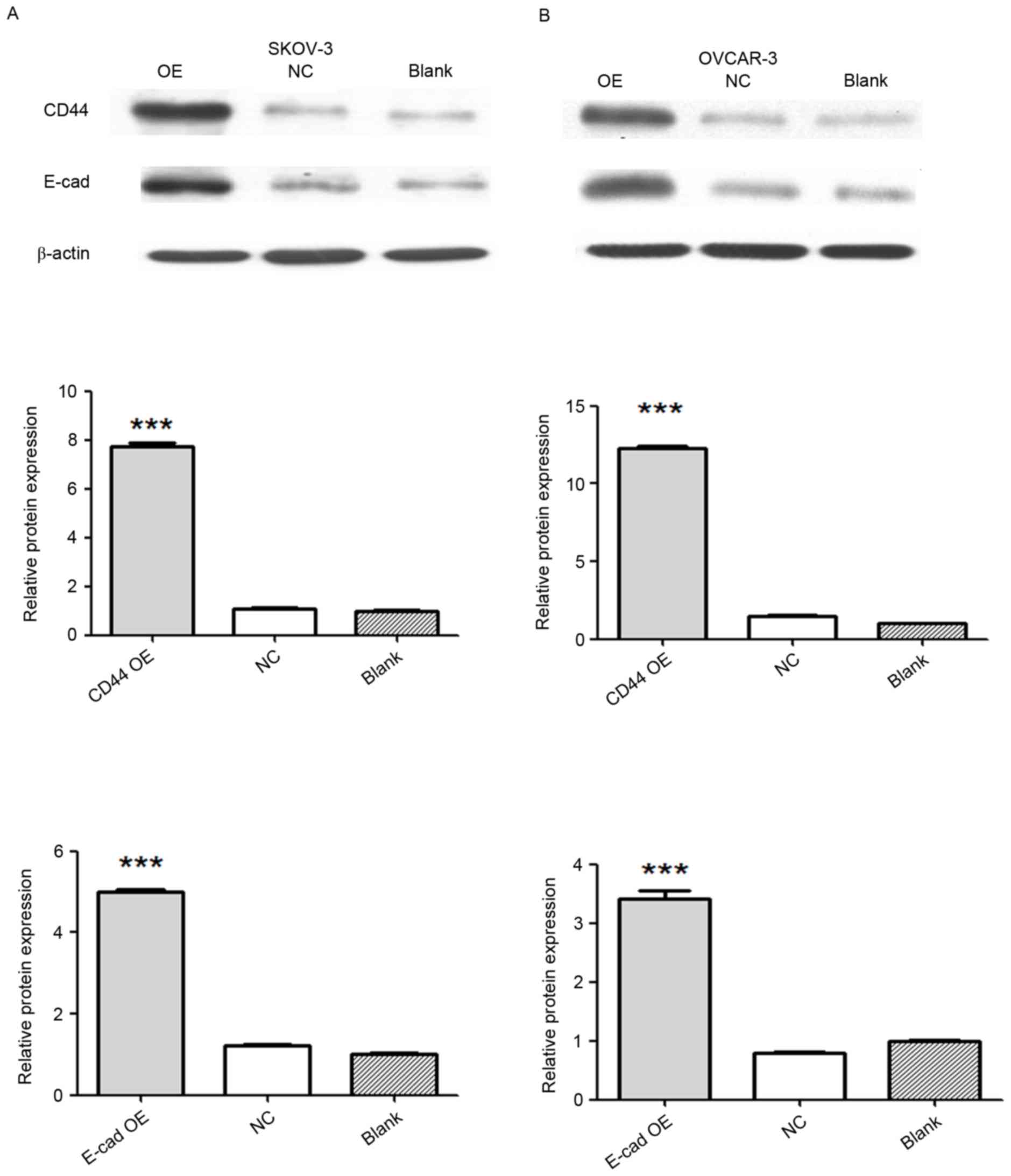
Confirmation of CD44 and E-cadherin
overexpression by western blot
SKOV-3 and OVCAR-3 ovarian cancer cells were divided
into 4 groups as described above. Overexpression of CD44 and
E-cadherin was achieved by transfecting SKOV-3 and OVCAR-3 cells
with viruses carrying the CD44 or E-cadherin gene. Expression of
CD44 and E-cadherin was detected by western blot analysis. Compared
with that in the negative control and blank groups, the protein
expression of CD44 in the CD44 overexpression group was
significantly increased (P<0.001), and the protein expression of
E-cadherin markedly increased in the E-cadherin overexpression
group (P<0.001; Fig. 1).
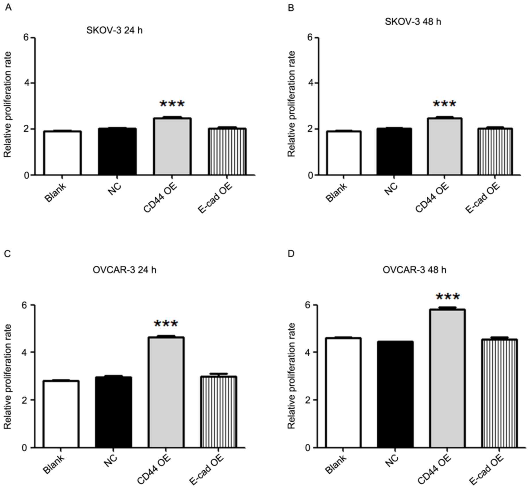
Overexpression of CD44 increases the
proliferation of ovarian cancer cells
The proliferation of SKOV-3 and OVCAR-3 ovarian
cancer cells in the 4 groups was determined by a CCK-8 cell
proliferation assay at 0, 24 and 48 h after viral transfection. The
results demonstrated that compared with the blank or negative
control group, the CD44 overexpression group had an increased cell
proliferation rate at 24 and 48 h in SKOV-3 (P<0.001) as well as
in OVCAR-3 cells (P<0.001; Fig.
2). However, overexpression of E-cadherin did not alter the
proliferation of SKOV-3 and OVCAR-3 cells as compared with that in
the blank or negative control groups.
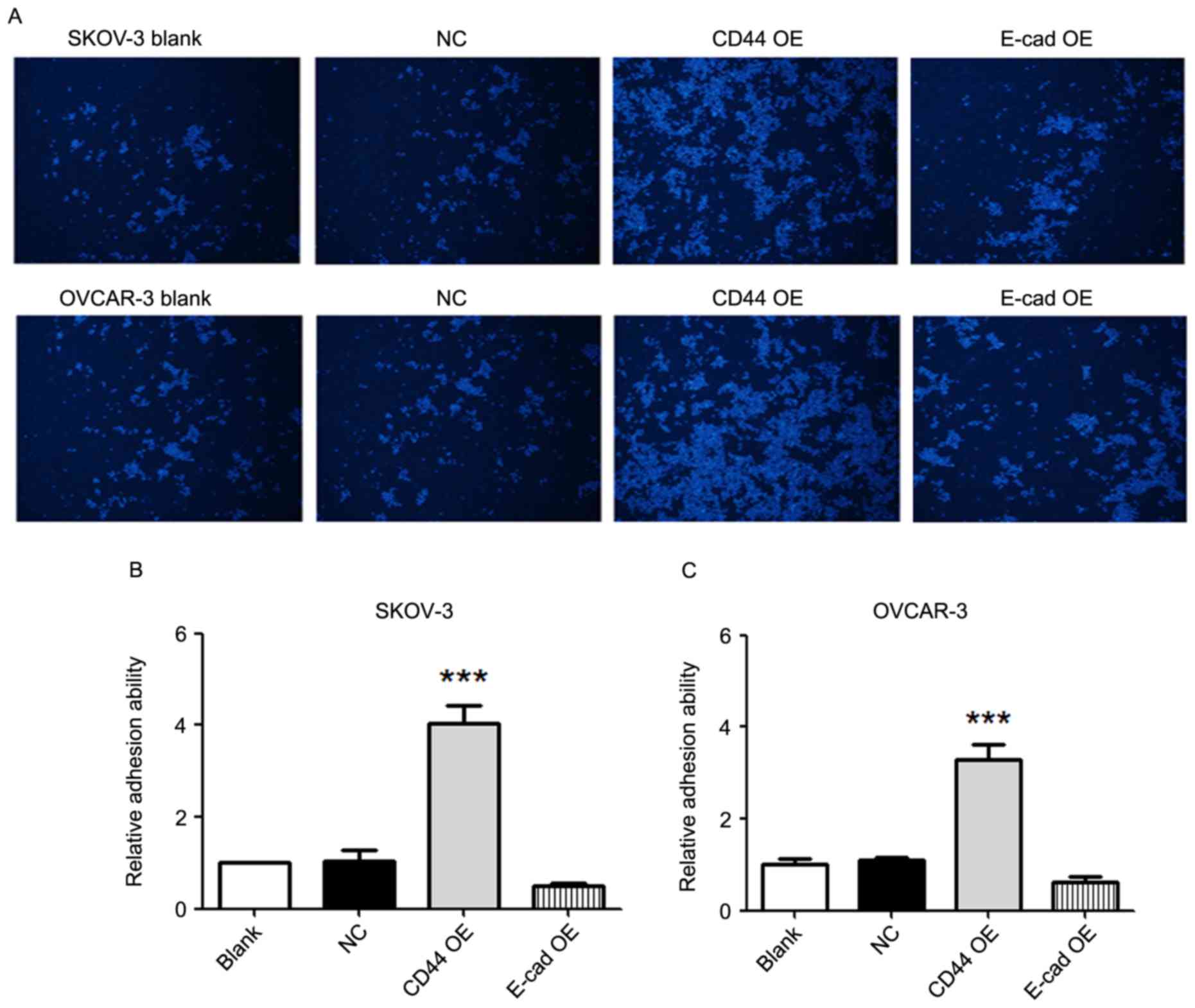
CD44 overexpression increases the
adhesion ability of ovarian cancer cells
The adhesion ability of SKOV-3 and OVCAR-3 cells to
an endothelial layer was detected. Compared with that in the blank
and negative control groups, the adhesion ability in the CD44
overexpression group was significantly higher in SKOV-3 as well as
OVCAR-3 cells (P<0.001; Fig. 3).
There were no significant differences in adhesion ability between
the E-cadherin overexpression groups and the blank/negative control
groups.
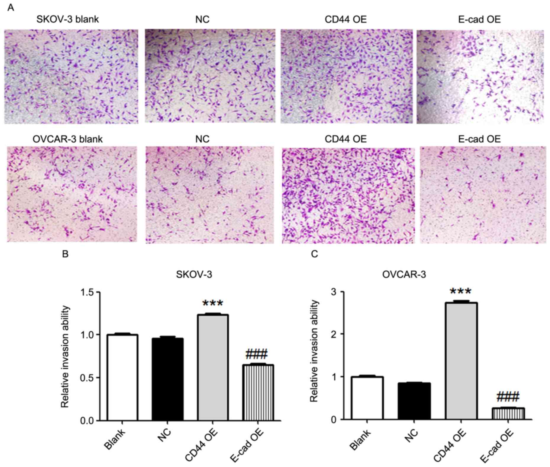
Effects of overexpression of CD44 and
E-cadherin on invasion of ovarian cancer cells
A Transwell invasion assay was utilized to detect
the tumor invasion ability. Compared with that in the blank or
negative control groups, overexpression of CD44 in SKOV-3 and
OVCAR-3 cells increased the tumor invasion ability (P<0.001;
Fig. 4). In addition, overexpression
of E-cadherin in SKOV-3 and OVCAR-3 cells decreased the tumor
invasion ability compared with that in the blank or negative
control groups (P<0.001; Fig.
4).
Discussion
The present study demonstrated that overexpression
of CD44 increased the proliferation, adhesion and invasion of
ovarian cancer cells. Overexpression of E-cadherin only decreased
the invasion of ovarian cancer cells.
A large number of studies have shown that
proliferation, adhesion, invasion and metastasis are the most
important biological behaviors of malignant tumors. Tumor cells
from the original site invade into surrounding tissue, penetrate
the vascular basement membrane or lymph duct, and seed into distal
organs via the blood or lymph circulation (10). During invasion and metastasis, tumor
cells interact with the extracellular matrix and host cells via
adhesion molecules. Alteration of adhesion molecules may change the
proliferation, adhesion and invasion of tumor cells (11,12).
CD44 was reported to be involved in the
epithelial-to-mesenchymal transition of ovarian cancer cells
(13). Furthermore, the CD44 splice
variant v8-10 was reported to be a marker of serous ovarian cancer
prognosis (6). An ovarian cancer
tissue microarray analysis of paired primary, metastatic and
recurrent tumor tissues from 26 patients, demonstrated that the
metastatic as well as recurrent ovarian cancer tissues expressed
higher levels of CD44 than the primary tumor (14). In addition, a meta-analysis unveiled
that patients with CD44 was associated with poor prognosis in
ovarian cancer patients, and that a CD44+ status was
associated with common clinicopathological features and poor
prognostic factors (5).
In line with the abovementioned studies, the present
study also revealed that overexpression of CD44 increased the
proliferation, adhesion and invasion of ovarian cancer cells.
Increased interaction of osteopontin with CD44 was reported to be
correlated with increased mitosis and in vitro
proliferation-enhancing effects in gastrointestinal stromal tumor
cells (15). M2 macrophages-secreted
osteoactivin/glycoprotein non-metastatic melanoma protein B was
reported to enhance the viability, proliferation and migration of
mesenchymal stem cells through extracellular signal-regulated
kinase (ERK) and AKT signaling pathways via CD44 (16). In addition, biological changes
induced by CD44 silencing were revealed to be mediated by
cumulative downregulation of c-Jun, Sp1 and c-Src in human breast
cancer cells (17). The molecular
mechanisms underlying the effects of CD44 overexpression on ovarian
cancer cells remain to be fully clarified. Whether the altered
proliferation, adhesion and invasion ability of ovarian cancer
cells are associated with osteopontin, the ERK and AKT signaling
pathway, c-Jun, Sp1 and c-Src requires further investigation.
In addition, the present study demonstrated that
overexpression of E-cadherin decreased the invasion of ovarian
cancer cells. A previous study unveiled that E-cadherin expression
was a predictor of better responses to first-line platinum-based
chemotherapy, platinum sensitivity and favorable clinical outcome
in patients with advanced-stage ovarian cancer (18). Furthermore, transforming growth
factor a was revealed to induce the invasion of human ovarian
cancer cells by downregulating E-cadherin (19). Betacellulin induced ovarian cancer
migration and Slug-dependent downregulation of E-cadherin via
epidermal growth factor receptor-mediated mitogen-activated protein
kinase kinase/ERK and phosphoinositide-3 kinase/Akt signaling
(20). Further research efforts are
required to elucidate the key molecular pathways triggered by
E-cadherin overexpression.
In conclusion, the present study highlighted the
crucial role of CD44 in promoting the proliferation, adhesion and
invasion of ovarian cancer cells. Overexpression of E-cadherin
decreased the invasion of ovarian cancer cells. Although further
studies on the underlying molecular mechanisms are required, the
present results may provide a foundation for possible
pharmaceutical targeting of ovarian cancer.
Acknowledgements
This study was funded by the Natural Science
Foundation of Ningbo (grant nos. 2013A610225, 2013A610228 and
2015A610228) and the Medical and Health Plan of Zhengjiang (grant
no. 2015KYA198).
References
|
1
|
Auersperg N, Wong AS, Choi KC, Kang SK and
Leung PC: Ovarian surface epithelium: Biology, endocrinology, and
pathology. Endocr Rev. 22:255–288. 2001. View Article : Google Scholar : PubMed/NCBI
|
|
2
|
Siegel R, Naishadham D and Jemal A: Cancer
statistics, 2013. CA Cancer J Clin. 63:11–30. 2013. View Article : Google Scholar : PubMed/NCBI
|
|
3
|
Jia Q, Feng M, Wang Y and Xue S: Gastric
cancer cells in collagen gel matrix: Three-dimensional growth and
differential expression of adhesion molecules (CD44s, CD54,
E-cadherin). J Biomed Mater Res A. 84:917–925. 2008. View Article : Google Scholar : PubMed/NCBI
|
|
4
|
Karan Križanac D, Krasić Arapović A,
Skočibušić S, Pintarić I, Trgo G and Tomić S: CD44 Immunoexpression
is unfavorable predictor in ovarian serous cancer. Appl
Immunohistochem Mol Morphol. Aug 3–2016.(Epub ahead of print).
|
|
5
|
Shi YY and Jiang H: Prognostic role of the
cancer stem cell marker CD44 in ovarian cancer: A meta-analysis.
Genet Mol Res. doi: 10.4238/gmr.15038325.
|
|
6
|
Sosulski A, Horn H, Zhang L, Coletti C,
Vathipadiekal V, Castro CM, Birrer MJ, Nagano O, Saya H, Lage K, et
al: CD44 Splice Variant v8-10 as a marker of serous ovarian cancer
prognosis. PLoS One. 11:e01565952016. View Article : Google Scholar : PubMed/NCBI
|
|
7
|
Elzarkaa AA, Sabaa BE, Abdelkhalik D,
Mansour H, Melis M, Shaalan W, Farouk M, Malik E and Soliman AA:
Clinical relevance of CD44 surface expression in advanced stage
serous epithelial ovarian cancer: A prospective study. J Cancer Res
Clin Oncol. 142:949–958. 2016. View Article : Google Scholar : PubMed/NCBI
|
|
8
|
Juan W, Shan K, Na W, Rong-Miao Z and Yan
L: The associations of genetic variants in E-cadherin gene with
clinical outcome of epithelial ovarian cancer. Int J Gynecol
Cancer. 26:1601–1607. 2016. View Article : Google Scholar : PubMed/NCBI
|
|
9
|
Trillsch F, Kuerti S, Eulenburg C, Burandt
E, Woelber L, Prieske K, Eylmann K, Oliveira-Ferrer L,
Milde-Langosch K and Mahner S: E-Cadherin fragments as potential
mediators for peritoneal metastasis in advanced epithelial ovarian
cancer. Br J Cancer. 114:213–220. 2016. View Article : Google Scholar : PubMed/NCBI
|
|
10
|
Henic E, Noskova V, Høyer-Hansen G,
Hansson S and Casslén B: Estradiol attenuates EGF-induced rapid
uPAR mobilization and cell migration via the G-protein-coupled
receptor 30 in ovarian cancer cells. Int J Gynecol Cancer.
19:214–222. 2009. View Article : Google Scholar : PubMed/NCBI
|
|
11
|
Banerjee S and Kaye SB: New strategies in
the treatment of ovarian cancer: Current clinical perspectives and
future potential. Clin Cancer Res. 19:961–968. 2013. View Article : Google Scholar : PubMed/NCBI
|
|
12
|
Lengyel E: Ovarian cancer development and
metastasis. Am J Pathol. 177:1053–1064. 2010. View Article : Google Scholar : PubMed/NCBI
|
|
13
|
Vos MC, Hollemans E, Ezendam N, Feijen H,
Boll D, Pijlman B, van der Putten H, Klinkhamer P, van Kuppevelt
TH, van der Wurff AA and Massuger LF: MMP-14 and CD44 in
Epithelial-to-Mesenchymal Transition (EMT) in ovarian cancer. J
Ovarian Res. 9:532016. View Article : Google Scholar : PubMed/NCBI
|
|
14
|
Gao Y, Foster R, Yang X, Feng Y, Shen JK,
Mankin HJ, Hornicek FJ, Amiji MM and Duan Z: Up-regulation of CD44
in the development of metastasis, recurrence and drug resistance of
ovarian cancer. Oncotarget. 6:9313–9326. 2015. View Article : Google Scholar : PubMed/NCBI
|
|
15
|
Hsu KH, Tsai HW, Lin PW, Hsu YS, Shan YS
and Lu PJ: Osteopontin expression is an independent adverse
prognostic factor in resectable gastrointestinal stromal tumor and
its interaction with CD44 promotes tumor proliferation. Ann Surg
Oncol. 17:3043–3052. 2010. View Article : Google Scholar : PubMed/NCBI
|
|
16
|
Yu B, Sondag GR, Malcuit C, Kim MH and
Safadi FF: Macrophage-Associated Osteoactivin/GPNMB Mediates
Mesenchymal stem cell survival, proliferation, and migration via a
CD44-dependent mechanism. J Cell Biochem. 117:1511–1521. 2016.
View Article : Google Scholar : PubMed/NCBI
|
|
17
|
Nam K, Oh S, Lee KM, Yoo SA and Shin I:
CD44 regulates cell proliferation, migration, and invasion via
modulation of c-Src transcription in human breast cancer cells.
Cell Signal. 27:1882–1894. 2015. View Article : Google Scholar : PubMed/NCBI
|
|
18
|
Miše BP, Telesmanić VD, Tomić S, Šundov D,
Čapkun V and Vrdoljak E: Correlation between E-cadherin
Immunoexpression and efficacy of first line platinum-based
chemotherapy in advanced high grade serous ovarian cancer. Pathol
Oncol Res. 21:347–356. 2015. View Article : Google Scholar : PubMed/NCBI
|
|
19
|
Qiu X, Cheng JC, Klausen C, Fan Q, Chang
HM, So WK and Leung PC: Transforming growth factor-α induces human
ovarian cancer cell invasion by down-regulating E-cadherin in a
Snail-independent manner. Biochem Biophys Res Commun. 461:128–135.
2015. View Article : Google Scholar : PubMed/NCBI
|
|
20
|
Zhao J, Klausen C, Qiu X, Cheng JC, Chang
HM and Leung PC: Betacellulin induces Slug-mediated down-regulation
of E-cadherin and cell migration in ovarian cancer cells.
Oncotarget. 7:28881–28890. 2016. View Article : Google Scholar : PubMed/NCBI
|