Introduction
Post-traumatic knee osteoarthritis (PTKO) is a
common disease presenting joint degenerative changes (1). PTKO is most frequently detected on the
elderly population, with an estimated 15 million cases in China
(2). Survey data in the United
States also demonstrated that PTKO is the second most common cause
of disability in males >50 years old, after cardiovascular
disease (3). Thus, prevention and
treatment of PTKO have become one of the most difficult issues in
clinical practice at present (3).
Transforming growth factor β1 (TGF-β1) is a member
of the TGF-β superfamily and a type of multifunctional polypeptide
growth factor (4). It participates
in the proliferation and differentiation processes of diversified
cell types, such as chondrocytes, epithelial cells and fibroblasts,
and has essential regulating effects in the developing process of
embryo, organs and cartilage. Studies have demonstrated that, in
the chondrocytes of rabbits and cattle, TGF-β1 can regulate the
synthesis of proteoglycan (4,5). In
addition, TGF-β1 has the ability to promote synthesis of type II
collagen by reducing consumption of proteoglycan in order to repair
the cartilage (6). Therefore, it can
be seen that TGF-β1 serves an important regulatory role in the
process of maintaining chondrocytes and cartilage (7).
PTKO is a chronic autoimmune disease characterized
by joint synovitis, and its early lesion mainly presents on the
synovial membrane (8). Synovial
cells are subject to rapid proliferation due to autoimmune response
resulting from an external stimulus, such as infection or trauma
(9); thus, the infiltration of
diversified inflammatory cells on the synovial membrane is enhanced
(9). In addition, a large number of
cytokines, inflammatory proteins and various antibodies jointly
mediate the inflammation-induced tissue damage in PTKO (10). During the pathological process of
PTKO, interleukin (IL)-1β, tumor necrosis factor (TNF)-α, IL-6 and
other proinflammatory cytokines, as well as the inflammatory
mediator prostaglandin E2 (PGE2), serve an important role (11).
Pharmacological and clinical studies in China and
abroad have revealed that myrtol standardized, a natural
phytomedicine used for the therapy of acute or chronic sinusitis
and bronchitis, has high safety, reduced adverse reactions and good
clinical efficacy (12).
Furthermore, it has extensive clinical efficacy on respiratory
system diseases. Domestic clinical studies have shown that its
clinical efficacy is similar to the total curative effect of the
import gel myrtol forte group (12).
Furthermore, pharmacological and pharmacodynamics studies have
demonstrated that myrtol standardized has expectorant
anti-inflammatory effects, relieving cough and asthma, and reported
reduced toxicity and side-effects (13).
The present study investigated whether myrtol
improves PTKO through regulation of reactive oxygen species (ROS),
TGF-β1 and apoptosis in mice.
Materials and methods
Animals
A total of 60 C57BL/6N female mice (20±1 g;
8-weeks-old) were obtained from Laboratory Animal Center of Anhui
Medical University (Hefei, China) and divided into five cages. All
mice were kept under controlled environmental conditions
(temperature, 23±1°C; 50–60% relative humidity; 12 h alternate
light-dark cycles). All experiments have been approved by the
Ethics Committee of Anhui Provincial Hospital (Hefei, China).
PTKO model and myrtol
administration
All mice were randomly divided into five groups,
including the control (8 mice), PTKO (16 mice), low dose
myrtol-treated (12 mice), medium dose myrtol-treated (12 mice) and
high dose myrtol-treated (12 mice) groups. This loading protocol
causes alendronate rupture with associated avulsion fracture from
the distal femur (14). Mice were
anesthetized with intraperitoneal injection of 3% sodium
pentobarbital (Sigma-Aldrich, St. Louis, MO, USA). Knee injury was
induced by an electromagnetic materials testing system (ELF 3200;
Bose, Eden Prairie, MN, USA), with 1 mm/sec loading rate of a
single dynamic overload cycle to a target compressive load of 12 N.
Knee injury in mice was performed by the release of compressive
force during loading and by an audible click. Mice in the low,
medium and high myrtol dose groups were administered with 150, 300
or 450 mg/kg myrtol (G.Pohl-Boskamp GmbH, Hohenlockstedt, Germany),
respectively, every day for 8 weeks following knee injury. In the
control group, rats were anesthetized only. The control and PTKO
groups were given equal volume of saline.
Hematoxylin and eosin staining
At 8 weeks following myrtol treatment, rats were
sacrificed, and knee tissue samples were acquired and fixed in 10%
paraformaldehyde for 24 h at room temperature. Tissues were
paraffin-embedded and stored at 4°C until required. The tissue
samples from each group were then cut into 4 µm sections using a
paraffin slicing machine (RM2235; Leica, Wetzlar, Germany), stained
with hematoxylin and eosin, and observed under light microscopy
(BH3-MJL; Olympus Corp., Tokyo, Japan). Osteoarthritis score was
determined using the modified Mankin scoring system (15). Next, the synovial extension score was
determined as absence (score 0) and presence (score 1) of synovial
tissue outgrowth parallel to the surface of the meniscus (score 2),
the meniscus adjacent to the femoral and tibial surfaces in both
the anterior (score 3) and posterior regions (score 4).
Micro-computed tomography (micro-CT)
analyses
Knee tissue samples were acquired, placed into 10%
paraformaldehyde for 24 h and scanned using a micro-CT system
(vivaCT; Scanco Medical, Basserdorf, Switzerland). The subchondral
bone thickness (mm), subchondral bone density (mg
hydroxyapatite/cm3), trabecular bone volume
(cm3), relative trabecular bone volume (BV/TV) ratio and
trabecular bone spacing (mm) were analyzed.
ELISA analysis of TNF-α, IL-6,
malondialdehyde (MDA), superoxide dismutase (SOD), ROS and TGF-β1
levels
A total of 200 µl of blood was collected from mice
in each group immediately following sacrifice and separated using
centrifugation at 5,000 × g for 10 min at 4°C. Blood serum was
acquired and analyzed for TNF-α (cat. no. E-CL-M0047c), IL-6 (cat.
no. E-CL-M0042c), MDA (cat. no. E-EL-0060c), SOD (cat. no.
E-EL-M2424c) and TGF-β1 (cat. no. E-EL-M0051c) levels using ELISA
kits (Wuhan Huamai Biotech Co., Wuhan, China), according to the
manufacturer's instructions. ROS was measured using Reactive oxygen
species Assay kit (cat. no. E004; Nanjing Jiancheng Biology
Engineering Institute, Nanjing, China). Absorbance was measured
using an M200 plate reader (Techan Trading AG, Mannedorf,
Switzerland) at 450 nm.
Western blot analysis
Knee tissue samples were acquired and prepared in a
lysis buffer (Beyotime Institute of Biotechnology, Haimen, China).
Following homogenization, the samples were centrifuged at 10,000 ×
g for 30 min and the protein contents were estimated with a BCA
protein assay kit (Beyotime Institute of Biotechnology). Protein
extraction was resolved by 12% sodium dodecyl
sulphate-polyacrylamide gel electrophoresis, and samples were then
transferred onto a nitrocellulose membrane (Sigma-Aldrich). The
nitrocellulose membrane was blocked with 5% bovine serum albumin or
5% non-fat dry milk. the nitrocellulose membrane was incubated with
anti-caspase-3 (sc-7148; 1:4,000), anti-Bax (sc-6236; 1:3,000) and
β-actin (sc-130656; 1:5,000; all from Santa Cruz Biotechnology,
Dallas, TX, USA) antibodies at 4°C for 12 h. Membranes were
subsequently washed with PBS for three times at 5 min and incubated
with mouse anti-rabbit IgG-HRP (sc-2357; 1:5,000, Santa Cruz
Biotechnology) for 1 h at 37°C and visualized by an enhanced
chemiluminescence assay kit (KGP1125; Nanjing KeyGen Biotech. Co.,
Ltd., Nanjing, China). The protein expression was scanned with a
densitometer and analyzed with Multi-Analyst software 3.0 (Bio-Rad
Laboratories, Inc, Hercules, CA, USA).
Statistical analysis
Results are expressed as the mean ± standard
deviation. Statistically significant differences were evaluated
using one-way analysis of variance with SPSS version 13.0 software
(SPSS, Inc., Chicago, IL, USA), and were indicated by
P<0.05.
Results
Myrtol reduces osteoarthritis in the
PTKO mouse model
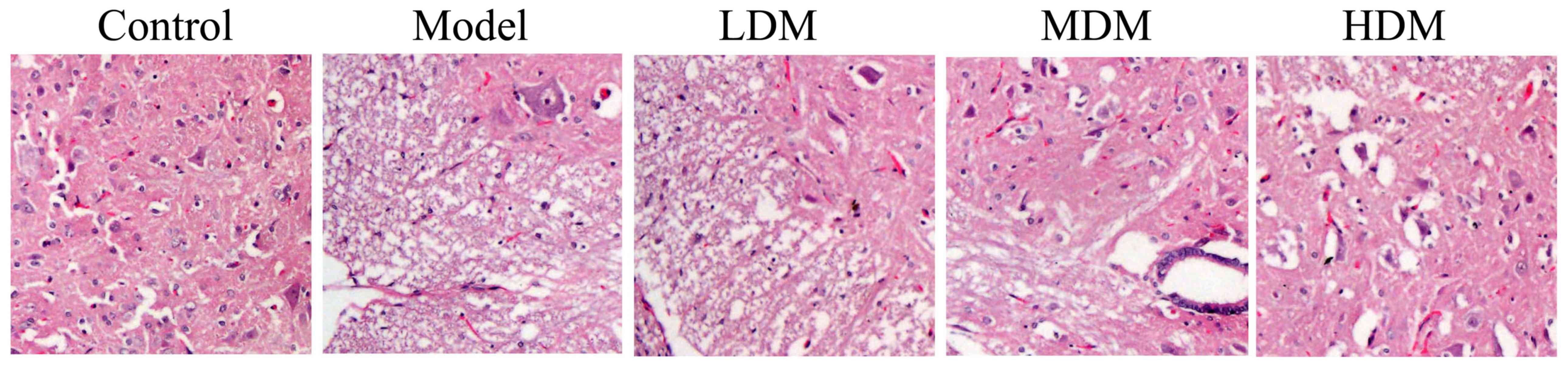
After treatment for 8 weeks, the extent of knee
osteoarthritis in the PTKO model group was higher compared with
that of the control group, as observed by hematoxylin and eosin
staining (Fig. 1). Low dose myrtol
treatment did not have a significant effect on knee osteoarthritis
in PTKO mice (Fig. 1). However,
treatment with medium or high dose of myrtol significantly reduced
the knee osteoarthritis in PTKO mice (Fig. 1; P=0.0034 and P=0.0012,
respectively).
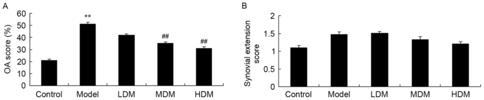
Myrtol improves knee tissue score in
PTKO model
The osteoarthritis and synovial extension scores of
each knee tissue sample were graded in order to examine the effect
of myrtol treatment on PTKO. The osteoarthritis score of the PTKO
model group was significantly higher compared with that of the
control group (P=0.0016), and low dose myrtol treatment had no
significant effect on this score in PTKO mice (Fig. 2A). However, treatment with medium or
high dose of myrtol significantly inhibited the osteoarthritis
score in PTKO mice (Fig. 2A;
P=0.0066 and P=0.0049, respectively). By contrast, the synovial
extension score was not significantly different in the PTKO and
myrtol-treated groups in comparison with the control (Fig. 2B).
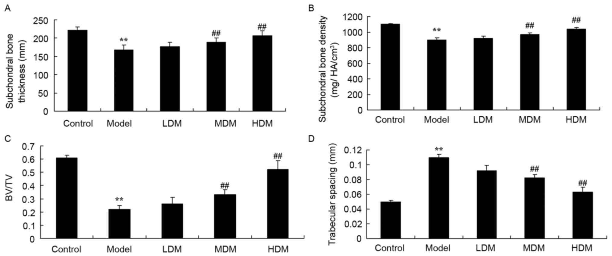
Myrtol improves the results of
micro-CT analyses in the PTKO model
The subchondral bone thickness (Fig. 3A; P=0.0059), subchondral bone density
(Fig. 3B; P=0.0073) and BV/TV ratio
(Fig. 3C; P=0.0023) in the tissues
of the PTKO model group were significantly lower when compared with
those of the control group. As shown in Fig. 3D, trabecular bone spacing in the PTKO
model group was higher in comparison with that of the control group
(P=0.0042). Low myrtol treatment did not markedly affect these four
parameters in PTKO mice (Fig. 3).
However, treatment with medium and high dose myrtol significantly
reversed the subchondral bone thickness (P=0.0077 and P=0.0062,
respectively), subchondral bone density (P=0.0095 and P=0.0089,
respectively), BV/TV (P=0.0081 and P=0.0058, respectively) and
trabecular bone spacing (P=0.0047 and P=0.0031, respectively.) in
PTKO mice (Fig. 3A-D).
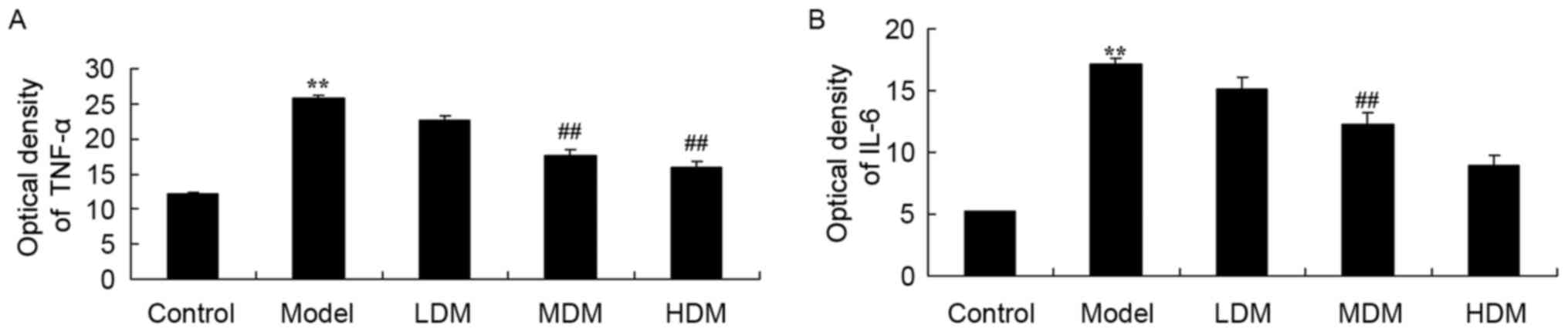
Myrtol improves the activities of
TNF-α and IL-6 in the PTKO model
To explore the effect of myrtol on the levels of
inflammation factors in the PTKO model, the activities of TNF-α and
IL-6 were measured using ELISA kits. As shown in Fig. 4, the activities of TNF-α (P=0.0025)
and IL-6 (P=0.0013) in the PTKO model group were significantly
increased when compared with those of the control group. Treatment
with medium or high dose myrtol significantly reduced the TNF-α
(Fig. 4A; P=0.0054 and P=0.0048) and
IL-6 (Fig. 4B; P=0.0066 and
P=0.0032) activities in the PTKO mice. By contrast, TNF-α and IL-6
activities in the low dose myrtol treatment group were not
evidently altered compared with the model group (Fig. 4).
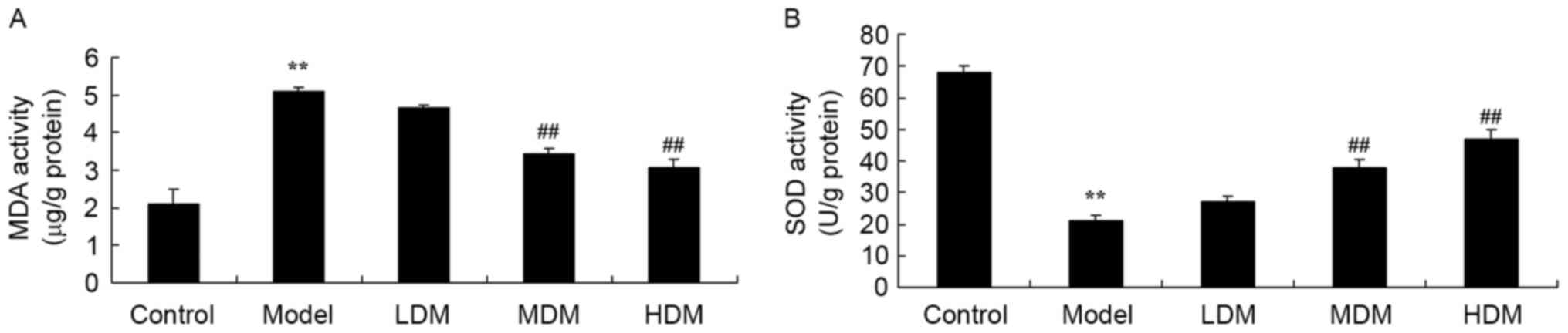
Myrtol improves the levels of MDA and
SOD in the PTKO model
To further explore the effect of myrtol on oxidative
stress in the PTKO mouse model, the activities of MDA and SOD were
measured using ELISA kits. As shown in Fig. 5, the activity of MDA (P=0.0031) was
increased and the activity of SOD (P=0.0027) was reduced in the
PTKO model group in comparison with those of the control group.
Treatment with medium or high dose myrtol significantly reversed
the PTKO-altered MDA (P=0.0068 and P=0.0055, respectively) and SOD
(P=0.0042 and P=0.0031, respectively) activities, while low dose
treatment had no significant effect in PTKO mice (Fig. 5).
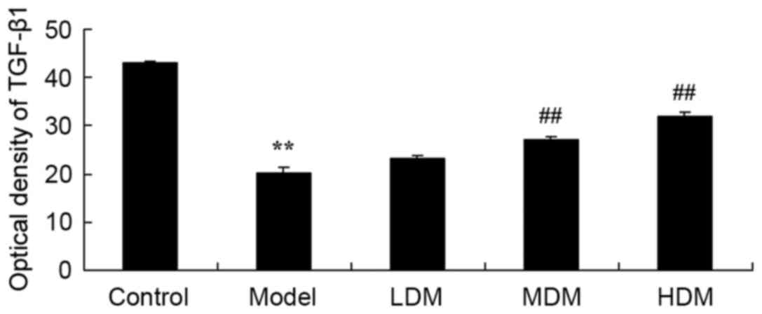
Myrtol improves the level of TGF-β1 in
the PTKO model
To examine the mechanism underlying the action of
myrtol treatment in PTKO, TGF-β1 level was detected using an ELISA
kit. As shown in Fig. 6, there was a
significant increase in TGF-β1 level of the PTKO model group,
compared with the control group (P=0.0039). In the medium and high
myrtol treatment groups, the TGF-β1 activation was significantly
inhibited compared with the PTKO model group (Fig. 6; P=0.0066 and P=0.0021,
respectively).
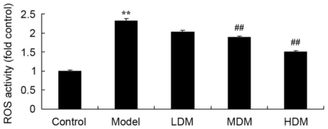
Myrtol improves the level of ROS in
the PTKO model
The effect of myrtol on PTKO-induced ROS generation
was determined using an ELISA kit, in order to analyze the
underlying mechanism of myrtol on PTKO. As shown in Fig. 7, the ROS level in the PTKO model
group was markedly increased compared with that of the control
group (P=0.0018). Treatment with medium or high dose myrtol
significantly reduced the PTKO-induced ROS level in PTKO mice
(Fig. 7; P=0.0083 and P=0.0041,
respectively).
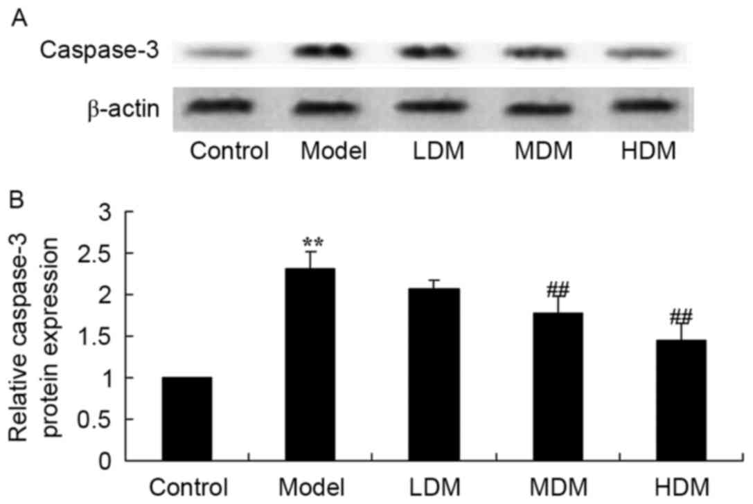
Myrtol improves the level of caspase-3
protein in the PTKO model
To examine the mechanism of myrtol on the apoptosis
of PTKO, caspase-3 protein was measured using western blot
analysis. As shown in Fig. 8, the
level of caspase-3 protein in PTKO model mice was higher than that
of the control group (P=0.0025). Medium or high dose myrtol
significantly suppressed the PTKO-induced caspase-3 protein in PTKO
mice (Fig. 8; P=0.0076 and P=0.0046,
respectively).
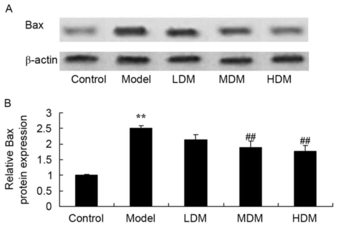
Myrtol improves the level of Bax
protein in the PTKO model
To further examine the mechanism of myrtol on
apoptosis in PTKO mice, Bax protein expression was measured using
western blot analysis. As shown in Fig.
9, the level of Bax protein in the PTKO model was significantly
increased compared with that of the control group (P=0.0031).
Medium or high dose myrtol treatment significantly suppressed the
PTKO-induced Bax protein in the PTKO mice (Fig. 9; P=0.0082 and P=0.0059,
respectively).
Discussion
PTKO is a frequently-occurring disease that is
characterized of soft tissue injury around the joint region, which
can lead to articular cartilage injury and eventually involve the
entire joint, resulting in articular cartilage degeneration,
fibrosis and fracture, and anabrosis (16). Knee pain, stiffness and muscular
movement dysfunction are the main clinical manifestations of PTKO
(17). Among them, myodynamia
reduction of quadriceps femoris muscles is a characteristic
manifestation of PTKO muscle dysfunction (18). In the present study, myrtol treatment
significantly improved knee osteoarthritis, inhibited the
osteoarthritis score, and reversed the PTKO-induced changes in
subchondral bone thickness, subchondral bone density, BV/TV and
trabecular bone spacing. These findings are in agreement with a
previous study demonstrating that myrtol ameliorated cartilage
lesions by downregulating the expression of TNF-α, IL-6 and Bax in
an osteoarthritis rat model (19).
Activation of IL-1β induces damage and synovial
inflammation and is an important cytokine causing articular
cartilage damage (20). Activation
of IL-1β promotes proliferation and differentiation of synovial
cells, and disintegration of the cartilage matrix and local immune
complex. Free collagen and other decomposition products stimulate
the synthesis of IL-1β, which forms a vicious circle (20). Furthermore, TNF-α can synthesize PGE2
and collagenase in decomposition lesions, lead to the absorption
and destruction of the bone and cartilage (11), as well as promote hyperplasia of
fibroblasts. Although IL-6 cannot stimulate synovial cells,
chondrocytes synthesize PGE2 and collagenase. However, IL-1β and
TNF-α can induce the production of IL-6. Consequently, IL-6, as an
inflammatory factor, is considered to be an amplification factor of
IL-1β and TNF-α. In the current study, myrtol treatment
significantly reduced the TNF-α and IL-6 activities in PTKO mice.
Similarly, Beuscher et al (12) revealed that myrtol standardized
inhibited inflammatory (TNF-α and IL-6) and allergic mediators.
In the cartilage of PTKO mice, expression quantity
has been upregulated to a certain degree, which may initiate the
self-rehabilitation of cartilage successively after the cartilage
damage. Thus, upregulation of TGF-β1 gene and protein promotes
downstream signal transduction and regulates the proliferation and
differentiation of cartilage (6).
Under the effects of specific inflammatory mediators and tumor
necrosis factors, the self-rehabilitation ability of cartilage has
been restricted, so that the cartilage cannot be fully repaired
(6). In the present study, myrtol
treatment significantly inhibited the activation of TGF-β1 level in
PTKO mice. Zhao et al (13)
also observed that myrtol standardized inhibited TGF-β1 expression,
and suppressed the levels of TNF-α, IL-1β and IL-6 in
radiation-induced lung injury.
The damage of PTKO articular cartilage and bone
tissue are closely associated with abnormalities of the cell
apoptosis process. Over-proliferation of synovial cells is
originated from comparative deficiency of synovial cell apoptosis
(21). In recent years, research has
detected that the second messenger ROS is associated with PTKO
damage (22). The oxidative stress
caused by ROS is able to damage mitochondrial respiratory chain.
Damaged mitochondria can lead to cell senescence and death.
However, ROS generation has an important effect on the
over-proliferation of PTKO and synovial membrane (23). Previous studies have suggested that
ROS can regulate cell proliferation, differentiation, programmed
cell death and aging. A small increase in ROS level promotes cell
proliferation (22,24), while a moderate increase can induce
cell apoptosis, and high ROS levels directly cause necrocytosis.
High concentration of ROS can directly or indirectly damage the
mitochondrial membrane structure, induce voltage reduction of
mitochondrial membrane, and lead to mitochondrial bulging (21). Mitochondrial bulging indicates that
the changes of mitochondrial membrane structure will result in loss
of the ATP synthesis ability (24).
In the present study, myrtol treatment significantly reduced the
PTKO-induced ROS level, and reversed the PTKO-altered MDA and SOD
activities in a mouse model. Similarly, Rantzsch et al
(25) reported that myrtol had
effective antioxidative properties in patients with chronic
obstructive pulmonary disease.
Cell apoptosis occurs in various physiological and
pathological processes, and is a cell death program with strict
regulation of energy dependence (26). In particular, Bax is the ‘molecular
switch’ initiating cell apoptosis, and serves a key function on
determines cell apoptosis (27). In
addition, caspase is not only an important molecule in apoptosis,
but also serves as a key protease of Bax (28). Activation of the caspase
decomposition specificity substrate may lead to cell apoptosis
(29). The present study found that
myrtol significantly suppressed PTKO-induced caspase-3 and Bax
protein expression in PTKO mice. A study by Ying et al
(19) also demonstrated that myrtol
ameliorates cartilage lesions by downregulating the expression of
Bax in an osteoarthritis rat model.
In conclusion, myrtol treatment improved knee
osteoarthritis, inhibited the osteoarthritis score, and reversed
the PTKO-induced changes in subchondral bone thickness, subchondral
bone density, BV/TV and trabecular bone spacing in a mouse model of
PTKO. Myrtol also inhibited inflammation and oxidative stress,
reduced ROS and TGF-β1 levels, and suppressed caspase-3 and Bax
protein expression in PTKO mice. These results indicated that
myrtol improves PTKO and may be a promising drug for clinical
therapy.
References
|
1
|
Swärd P, Fridén T, Boegård T, Kostogiannis
I, Neuman P and Roos H: Association between varus alignment and
post-traumatic osteoarthritis after anterior cruciate ligament
injury. Knee Surg Sports Traumatol Arthrosc. 21:2040–2047. 2013.
View Article : Google Scholar : PubMed/NCBI
|
|
2
|
Wolsko PM, Eisenberg DM, Simon LS, Davis
RB, Walleczek J, Mayo-Smith M, Kaptchuk TJ and Phillips RS:
Double-blind placebo-controlled trial of static magnets for the
treatment of osteoarthritis of the knee: Results of a pilot study.
Altern Ther Health Med. 10:36–43. 2004.PubMed/NCBI
|
|
3
|
Giannini S, Buda R, Ruffilli A, Pagliazzi
G, Ensini A, Grigolo B, Desando G and Vannini F: Failures in
bipolar fresh osteochondral allograft for the treatment of
end-stage knee osteoarthritis. Knee Surg Sports Traumatol Arthrosc.
23:2081–2089. 2015. View Article : Google Scholar : PubMed/NCBI
|
|
4
|
Jiang Q, Qiu YT, Chen MJ, Zhang ZY and
Yang C: Synovial TGF-β1 and MMP-3 levels and their correlation with
the progression of temporomandibular joint osteoarthritis combined
with disc displacement: A preliminary study. Biomed Rep. 1:218–222.
2013. View Article : Google Scholar : PubMed/NCBI
|
|
5
|
Wilbers RH, Westerhof LB, van Raaij DR,
van Adrichem M, Prakasa AD, Lozano-Torres JL, Bakker J, Smant G and
Schots A: Co-expression of the protease furin in Nicotiana
benthamiana leads to efficient processing of latent transforming
growth factor-β1 into a biologically active protein. Plant
Biotechnol J. 14:1695–1704. 2016. View Article : Google Scholar : PubMed/NCBI
|
|
6
|
Fahlgren A, Andersson B and Messner K:
TGF-beta1 as a prognostic factor in the process of early
osteoarthrosis in the rabbit knee. Osteoarthritis Cartilage.
9:195–202. 2001. View Article : Google Scholar : PubMed/NCBI
|
|
7
|
Vonwil D, Trüssel A, Haupt O, Gobaa S,
Barbero A, Shastri VP and Martin I: Substrate elasticity modulates
TGF beta stimulated re-differentiation of expanded human articular
chondrocytes. Drug Deliv Transl Res. 2:351–362. 2012. View Article : Google Scholar : PubMed/NCBI
|
|
8
|
Heard BJ, Solbak NM, Achari Y, Chung M,
Hart DA, Shrive NG and Frank CB: Changes of early post-traumatic
osteoarthritis in an ovine model of simulated ACL reconstruction
are associated with transient acute post-injury synovial
inflammation and tissue catabolism. Osteoarthritis Cartilage.
21:1942–1949. 2013. View Article : Google Scholar : PubMed/NCBI
|
|
9
|
Lewis JS, Hembree WC, Furman BD, Tippets
L, Cattel D, Huebner JL, Little D, DeFrate LE, Kraus VB, Guilak F
and Olson SA: Acute joint pathology and synovial inflammation is
associated with increased intra-articular fracture severity in the
mouse knee. Osteoarthritis Cartilage. 19:864–873. 2011. View Article : Google Scholar : PubMed/NCBI
|
|
10
|
Bradley EW, Carpio LR, McGee-Lawrence ME,
Becerra C Castillejo, Amanatullah DF, Ta LE, Otero M, Goldring MB,
Kakar S and Westendorf JJ: Phlpp1 facilitates post-traumatic
osteoarthritis and is induced by inflammation and promoter
demethylation in human osteoarthritis. Osteoarthritis Cartilage.
24:1021–1028. 2016. View Article : Google Scholar : PubMed/NCBI
|
|
11
|
Huebner KD, Shrive NG and Frank CB:
Dexamethasone inhibits inflammation and cartilage damage in a new
model of post-traumatic osteoarthritis. J Orthop Res. 32:566–572.
2014. View Article : Google Scholar : PubMed/NCBI
|
|
12
|
Beuscher N, Kietzmann M, Bien E and
Champeroux P: Interference of myrtol standardized with inflammatory
and allergic mediators. Arzneimittelforschung. 48:985–989.
1998.PubMed/NCBI
|
|
13
|
Zhao DY, Qu HJ, Guo JM, Zhao HN, Yang YY,
Zhang P, Cao K, Lei X, Cui JG, Liu C, et al: Protective effects of
Myrtol standardized against radiation-induced lung injury. Cell
Physiol Biochem. 38:619–634. 2016. View Article : Google Scholar : PubMed/NCBI
|
|
14
|
Christiansen BA, Anderson MJ, Lee CA,
Williams JC, Yik JH and Haudenschild DR: Musculoskeletal changes
following non-invasive knee injury using a novel mouse model of
post-traumatic osteoarthritis. Osteoarthritis Cartilage.
20:773–782. 2012. View Article : Google Scholar : PubMed/NCBI
|
|
15
|
Furman BD, Strand J, Hembree WC, Ward BD,
Guilak F and Olson SA: Joint degeneration following closed
intraarticular fracture in the mouse knee: A model of posttraumatic
arthritis. J Orthop Res. 25:578–592. 2007. View Article : Google Scholar : PubMed/NCBI
|
|
16
|
Bégué T, Mebtouche N and Levante S:
One-stage procedure for total knee arthroplasty in post-traumatic
osteoarthritis of the knee with wound defect. Usefulness of
navigation and flap surgery. Knee. 19:948–950. 2012. View Article : Google Scholar : PubMed/NCBI
|
|
17
|
Nordenvall R, Bahmanyar S, Adami J,
Mattila VM and Felländer-Tsai L: Cruciate ligament reconstruction
and risk of knee osteoarthritis: The association between cruciate
ligament injury and post-traumatic osteoarthritis. a population
based nationwide study in Sweden, 1987–2009. PLoS One.
9:e1046812014. View Article : Google Scholar : PubMed/NCBI
|
|
18
|
Bala A, Penrose CT, Seyler TM, Mather RC
3rd, Wellman SS and Bolognesi MP: Outcomes after total knee
arthroplasty for post-traumatic arthritis. Knee. 22:630–639. 2015.
View Article : Google Scholar : PubMed/NCBI
|
|
19
|
Ying B, Maimaiti AK, Song D and Zhu S:
Myrtol ameliorates cartilage lesions in an osteoarthritis rat
model. Int J Clin Exp Pathol. 8:1435–1442. 2015.PubMed/NCBI
|
|
20
|
Satkunananthan PB, Anderson MJ, De Jesus
NM, Haudenschild DR, Ripplinger CM and Christiansen BA: In vivo
fluorescence reflectance imaging of protease activity in a mouse
model of post-traumatic osteoarthritis. Osteoarthritis Cartilage.
22:1461–1469. 2014. View Article : Google Scholar : PubMed/NCBI
|
|
21
|
Jiang L, Li L, Geng C, Gong D, Jiang L,
Ishikawa N, Kajima K and Zhong L: Monosodium iodoacetate induces
apoptosis via the mitochondrial pathway involving ROS production
and caspase activation in rat chondrocytes in vitro. J Orthop Res.
31:364–369. 2013. View Article : Google Scholar : PubMed/NCBI
|
|
22
|
Courties A, Gualillo O, Berenbaum F and
Sellam J: Metabolic stress-induced joint inflammation and
osteoarthritis. Osteoarthritis Cartilage. 23:1955–1965. 2015.
View Article : Google Scholar : PubMed/NCBI
|
|
23
|
Ziskoven C, Jäger M, Zilkens C, Bloch W,
Brixius K and Krauspe R: Oxidative stress in secondary
osteoarthritis: From cartilage destruction to clinical
presentation? Orthop Rev (Pavia). 2:e232010. View Article : Google Scholar : PubMed/NCBI
|
|
24
|
Xie L, Lin AS, Kundu K, Levenston ME,
Murthy N and Guldberg RE: Quantitative imaging of cartilage and
bone morphology, reactive oxygen species, and vascularization in a
rodent model of osteoarthritis. Arthritis Rheum. 64:1899–1908.
2012. View Article : Google Scholar : PubMed/NCBI
|
|
25
|
Rantzsch U, Vacca G, Dück R and Gillissen
A: Anti-inflammatory effects of Myrtol standardized and other
essential oils on alveolar macrophages from patients with chronic
obstructive pulmonary disease. Eur J Med Res. 14 Suppl 4:S205–S209.
2009. View Article : Google Scholar
|
|
26
|
Bar-Yehuda S, Rath-Wolfson L, Del Valle L,
Ochaion A, Cohen S, Patoka R, Zozulya G, Barer F, Atar E,
Piña-Oviedo S, et al: Induction of an antiinflammatory effect and
prevention of cartilage damage in rat knee osteoarthritis by CF101
treatment. Arthritis Rheum. 60:3061–3071. 2009. View Article : Google Scholar : PubMed/NCBI
|
|
27
|
Li H, Lei M, Yu C, Lv Y, Song Y and Yang
L: Mechano growth factor-E regulates apoptosis and inflammatory
responses in fibroblast-like synoviocytes of knee osteoarthritis.
Int Orthop. 39:2503–2509. 2015. View Article : Google Scholar : PubMed/NCBI
|
|
28
|
Lee CH, Wen ZH, Chang YC, Huang SY, Tang
CC, Chen WF, Hsieh SP, Hsieh CS and Jean YH: Intra-articular
magnesium sulfate (MgSO4) reduces experimental osteoarthritis and
nociception: Association with attenuation of N-methyl-D-aspartate
(NMDA) receptor subunit 1 phosphorylation and apoptosis in rat
chondrocytes. Osteoarthritis Cartilage. 17:1485–1493. 2009.
View Article : Google Scholar : PubMed/NCBI
|
|
29
|
Bouderlique T, Vuppalapati KK, Newton PT,
Li L, Barenius B and Chagin AS: Targeted deletion of Atg5 in
chondrocytes promotes age-related osteoarthritis. Ann -Rheum Dis.
75:627–631. 2016. View Article : Google Scholar : PubMed/NCBI
|