Introduction
Coronary atherosclerosis is a serious vascular
disease, which may lead to coronary artery stenosis that is a
common cause of angina and myocardial infarction (1). Long-term myocardial ischemia and
hypoxia can result in heart enlargement, decrease of compliance and
myocardial contraction dysfunction, leading to chronic heart
failure (2). Coronary
atherosclerotic plaque, particularly the vulnerable plaque of the
coronary artery with a thrombus formation tendency, is one of the
most important reasons for coronary artery stenosis (3). Coronary atherosclerosis has a greater
impact on the quality of life of patients, due to its long clinical
treatment, easy relapse and poor prognosis. Therefore, studies on
coronary atherosclerosis at the molecular level are important for
the clinical treatment of coronary atherosclerosis.
MicroRNAs (miRNAs or miRs) are a class of
non-encoding RNA molecules with 18–22 nucleotides in eukaryotes,
which regulate the expression of their target proteins at the mRNA
level (4–6). Altered expression of various miRNA
molecules and a variety of proteins in coronary atherosclerosis
suggests that miRNAs serve important roles in the regulation of
proteins that are associated with coronary atherosclerosis
(7,8). Studies have demonstrated that
atherosclerosis is a type of artery intimal inflammation with a
series of basic features of inflammation (9,10).
Furthermore, cyclooxygenase (COX)-2 is a key inflammatory factor in
the human body. Activation of the COX-2 signaling pathway causes
and aggravates the pathogenesis of atherosclerosis (11). Atherosclerosis can be considered
chronic inflammation. COX-2 is an important rate-limiting enzyme in
the synthesis of prostaglandin and thromboxane from arachidonic
acid and is highly expressed at inflammatory sites (12). It has been demonstrated that COX-2 is
expressed in atherosclerotic lesions and is regulated by a variety
of inflammatory cytokines, including IL-1, IFN-γ and TNF-α
(12). The metabolites of COX-2 also
promote inflammation, indicating that COX-2 is involved in the
pathological process of atherosclerosis occurrence and development
(13,14). However, the mechanism of COX-2
regulation on coronary atherosclerotic plaques remains unclear.
In the present study, the expression levels of COX-2
mRNA and protein in the plaque tissues, peripheral blood
mononuclear cells (PBMCs) and serum of coronary atherosclerosis
patients were examined. In addition, the association between
miR-381 and COX-2 was also investigated.
Materials and methods
Patients
A total of 36 patients with coronary atherosclerosis
who received coronary endarterectomy at Linyi People's Hospital and
Junan Hospital of Traditional Chinese Medicine (Linyi, China)
between January 2013 and June 2016 were enrolled into the present
study, including 19 males and 17 females (age range, 38–72 years;
median age, 59 years). In addition, 39 healthy subjects were
enrolled into the control group, including 18 males and 21 females
(age range, 39–75 years; median age, 61 years). All patients with
coronary atherosclerosis were diagnosed by electrocardiography,
echocardiography and coronary angiography. None of the patients had
any complications or infections in the liver or kidney, or
immune-associated diseases. Subjects in the control group did not
suffer from coronary atherosclerosis. The age, gender, history of
diabetes and history of hypertension of all patients were recorded.
Gensini scoring scale (15) was used
to evaluate the degree of coronary atherosclerosis. For severity
scoring, stenosis with 0-<25, 25–50, 50–75, 75–90, 90–99 and
>90% luminal diameter reduction scored 1, 2, 3, 8, 16 and 32
points, respectively. Segment multiplying factors for the left main
trunk, proximal left anterior descending branch, middle left
anterior descending branch, D1 diagonal branch, D2 diagonal
branches, proximal left circumflex branch, distal left circumflex
branch, posterior descending branch, posterior lateral branch and
the proximal, middle and distal right coronary artery was 5, 2.5,
1.5, 1, 0.5, 2.5, 1, 1, 0.5 and 1, respectively. The Gensini score
was calculated by multiplying severity scores with their respective
segment multiplying factors. All procedures were approved by the
Ethics Committee of Linyi People's Hospital (Linyi, China). Written
informed consents were obtained from all patients or their
families.
Samples
Plaque tissues and adjacent artery intimal tissues
(serving as the controls) were obtained from all patients by
coronary endarterectomy and stored in liquid nitrogen until further
use. In addition, peripheral blood was collected from all patients
and healthy subjects (control) for the separation of serum by
centrifugation at a speed of 400 × g at 4°C for 10 min. In order to
obtain PBMCs, a mixture of the heparin anticoagulated venous blood
and equal amount of serum-free Iscove's modified Dulbecco's medium
(v/v, 1:1; P12440053; Thermo Fisher Scientific, Inc., Waltham, MA,
USA) was added to the lymphocyte separation medium prior to
centrifugation at a speed of 400 × g for 30 min at room
temperature. Following centrifugation, the middle layer was
aspirated and mixed with 5 volumes of Hank's solution (H1020;
Beijing Solarbio Science & Technology Co., Ltd., Beijing,
China) prior to further centrifugation at a speed of 300 × g for 10
min at 4°C. Subsequent to two washes with PBS, the cells were
counted and diluted to a density of 1×10⁶ cells/ml.
Finally, 3×10⁶ cells were seeded onto a round culture
plate with a bottom area of 9 cm2, followed by
incubation at 37°C and 5% CO2 for 1–2 h. The cells that
attached on the bottom (PBMCs) were collected by discarding the
medium.
Reverse transcription-quantitative
polymerase chain reaction (RT-qPCR)
Serum (1 ml) was mixed with 1 ml TRIzol reagent
(cat. no. 10606ES60; Yeasen Biotechnology Co., Ltd., Shanghai,
China) for lysis. Next, total RNA was extracted using miRNeasy
Serum/Plasma kit (cat. no. JL217184; Guangzhou Jianlun Biological
Technology Co., Ltd., Guangzhou, China). The purity of RNA was then
determined by measuring the absorbance at 260/280 nm using
ultraviolet spectrophotometry (Nanodrop ND-1000; Thermo Fisher
Scientific, Inc.). Subsequently, cDNA was obtained by reverse
transcription using TIANScript II cDNA First Strand Synthesis kit
(cat. no. KR107; Tiangen Biotech Co., Ltd., Beijing, China) from 1
µg RNA and stored at −20°C.
SuperReal PreMix (SYBR-Green) kit (Tiangen Biotech
Co., Ltd.) was used to detect the mRNA expression of COX-2, using
β-actin as an internal reference. The reaction system (total
volume, 20 µl) was composed of 10 µl qPCR Mix, 0.5 µl upstream
primer (COX-2 mRNA, 5′-CAGCCATACAGCAAATCCTTG-3′; β-actin,
5′-CACCAGGGCGTGATGGT-3′), 0.5 µl downstream primer (COX-2 mRNA,
5′-CAAATGTGATCTGGATGTCAAC-3′; β-actin,
5′-CTCAAACATGATCTGGGTCAT-3′), 1 µl cDNA and 8 µl ddH2O.
qPCR was conducted under the following condition: Initial
denaturation at 95°C for 30 sec; 40 cycles of denaturation at 95°C
for 5 sec and annealing at 60°C for 34 sec (iQ5 Reverse
transcription PCR Detection System; Bio-Rad Laboratories, Inc.,
Hercules, CA, USA). The 2−ΔΔCq method (16) was used to calculate the relative
expression of COX-2 mRNA against β-actin. Each sample was tested in
triplicate.
The expression of miR-381 was determined by miRcute
miRNA qPCR detection kit (cat. no. FP401; Tiangen Biotech Co.,
Ltd.), using U6 as an internal reference. The reaction system
(total volume, 25 µl) contained 12.5 µl SYBR Premix Ex Taq, 1 µl
upstream primer (miR-381, 5′-ACACTCCAGCTGGGTATACAAGGGCAAGCT-3′; U6,
5′-CTCGCTTCGGCAGCACA-3′), 1 µl downstream primer (miR-381,
5′-TGGTGTCGTGGAGTCG-3′; U6, 5′-AACGCTTCACGAATTTGCGT-3′), 2 µl
template and 8.5 µl ddH2O. The reaction protocol was as
follows: Initial denaturation at 95°C for 5 min; 40 cycles of
denaturation at 95°C for 15 sec, annealing at 60°C for 30 sec and
elongation at 72°C for 30 sec (iQ5; Bio-Rad Laboratories, Inc.).
The 2−ΔΔCq method was used to calculate the relative
expression of miR-381 against U6. Each sample was tested in
triplicate.
Western blotting
Tissues and cells were collected and mixed with 100
µl precooled radioimmunoprecipitation assay lysis buffer (P0013B;
Beyotime Institute of Biotechnology, Shanghai, China) containing 1
mM phenylmethylsulfonyl fluoride for lysis for 15 min at 4°C. Next,
the mixture was centrifuged at 12,000 × g at 4°C for 5 min. The
supernatant was used to determine the protein concentration with a
bicinchoninic acid protein concentration determination kit (cat.
no. RTP7102; Real-Times Biotechnology Co., Ltd., Beijing, China).
Protein samples were then mixed with sodium dodecyl sulfate (SDS)
loading buffer prior to denaturation in a boiling water bath for 5
min. Subsequently, the samples (20 µg) were subjected to 10%
SDS-polyacrylamide gel electrophoresis. The resolved proteins were
transferred to polyvinylidene difluoride membranes on ice (100 V; 2
h) and blocked with 5% skimmed milk at room temperature for 1 h.
The membranes were then incubated with rabbit anti-human COX-2
polyclonal primary antibody (1:1,000; cat. no. ab15191; Abcam,
Cambridge, UK) and rabbit anti-human β-actin primary antibody
(1:5,000; cat. no. ab129348; Abcam) at 4°C overnight. Following
washing three times for 5 min each, with phosphate-buffered saline
with Tween-20 (TBST), the membranes were incubated with polyclonal
goat anti-rabbit horseradish peroxidase-conjugated secondary
antibody (1:3,000; cat. no. ab6721; Abcam) for 1 h at room
temperature, followed by washing with TBST for three times for 15
min each. Subsequently, the membrane was developed with an enhanced
chemiluminescence detection kit (cat. no. ab65623; Abcam) for
imaging. Image lab version 3.0 software (Bio-Rad Laboratories,
Inc.) was used to acquire and analyze imaging signals. The relative
expression of COX-2 protein was calculated against that of
β-actin.
Enzyme-linked immunosorbent assay
(ELISA)
Blood samples were centrifuged at a speed of 1,000 ×
g at 4°C for 10 min and serum was tested using a COX-2 ELISA kit
(cat. no. KB1567; Shanghai Kemin Biotech Co., Ltd., Shanghai,
China). Standards (50 µl; provided by the kit), serum samples (10
µl sample liquid and 40 µl diluent) and blanks were added into the
predefined wells of a 96-well microplate. In the standard and serum
wells, horseradish peroxidase-labelled conjugates (100 µl) were
added prior to sealing the plates for incubation at 37°C for 1 h.
Subsequent to five washes using wash liquid (part of the kit),
substrates A (50 µl) and B (50 µl) were added into each well. After
incubation at 37°C for 15 min, stop solution (50 µl) was added into
each well and the absorbance was measured at 450 nm within 15
min.
Dual-luciferase reporter assay
Bioinformatics prediction is a powerful tool used to
study the functions of miRNAs. In order to understand the
regulatory mechanism of COX-2 in coronary atherosclerosis, the
online software miRanda (www.microma.org/rnicroma/home.do), TargetScan
(www.targetscan.org), PITA (genie.weizmann.ac.il/pubs/mir07/mir07_data.html),
RNAhybrid (bibiserv.techfak.uni-bielefeld.de/rnahybrid) and
PicTar (pictar.mdc-berlin.de) were used to
predict miRNA molecules that may regulate COX-2. The analysis
identified that miR-381 was able to potentially regulate COX-2.
The cells were divided into negative control (NC),
wild-type (WT) and mutant groups. According to the bioinformatics
results, WT and mutant seed regions of miR-381 in the
3′-untranslated region (UTR) of the COX-2 gene were chemically
synthesized in vitro, added with SpeI and HindIII
restriction sites for the insertion of plasmids and then cloned
into pMIR-REPORT luciferase reporter plasmids (Ambion; Thermo
Fisher Scientific, Inc.). Plasmids (0.8 µg) with WT or mutant
3′-UTR DNA sequences were co-transfected with agomiR-381 (100 nM;
Sangon Biotech Co., Ltd., Shanghai, China) into 293T cells [ATCC,
Manassas, VA, USA; cultured in 24-well plates containing Dulbecco's
modified Eagle's medium (Thermo Fisher Scientific, Inc.) and 10%
fetal bovine serum (Sangon Biotech Co. Ltd.) at 37°C and 5%
CO2]. Following cultivation for 24 h, the cells were
lysed using a dual-luciferase reporter assay kit (Promega
Corporation, Madison, WI, USA) according to the manufacturer's
protocol and fluorescence intensity was measured using a GloMax
20/20 luminometer (Promega Corporation). Using Renilla
fluorescence activity as an internal reference, the fluorescence
values of each group of cells were measured.
Statistical analysis
The results were analyzed using SPSS version 18.0
statistical software (SPSS, Inc., Chicago, IL, USA). The data are
expressed as the mean ± standard deviation. Data were tested for
normality and multigroup measurement data were analyzed using
one-way analysis of variance. In case of homogeneity of variance,
the least significant difference and Student-Newman-Keuls methods
were used, whereas in case of heterogeneity of variance, Tamhane's
T2 or Dunnett's T3 method was used. P<0.05 indicated
statistically significant differences.
Results
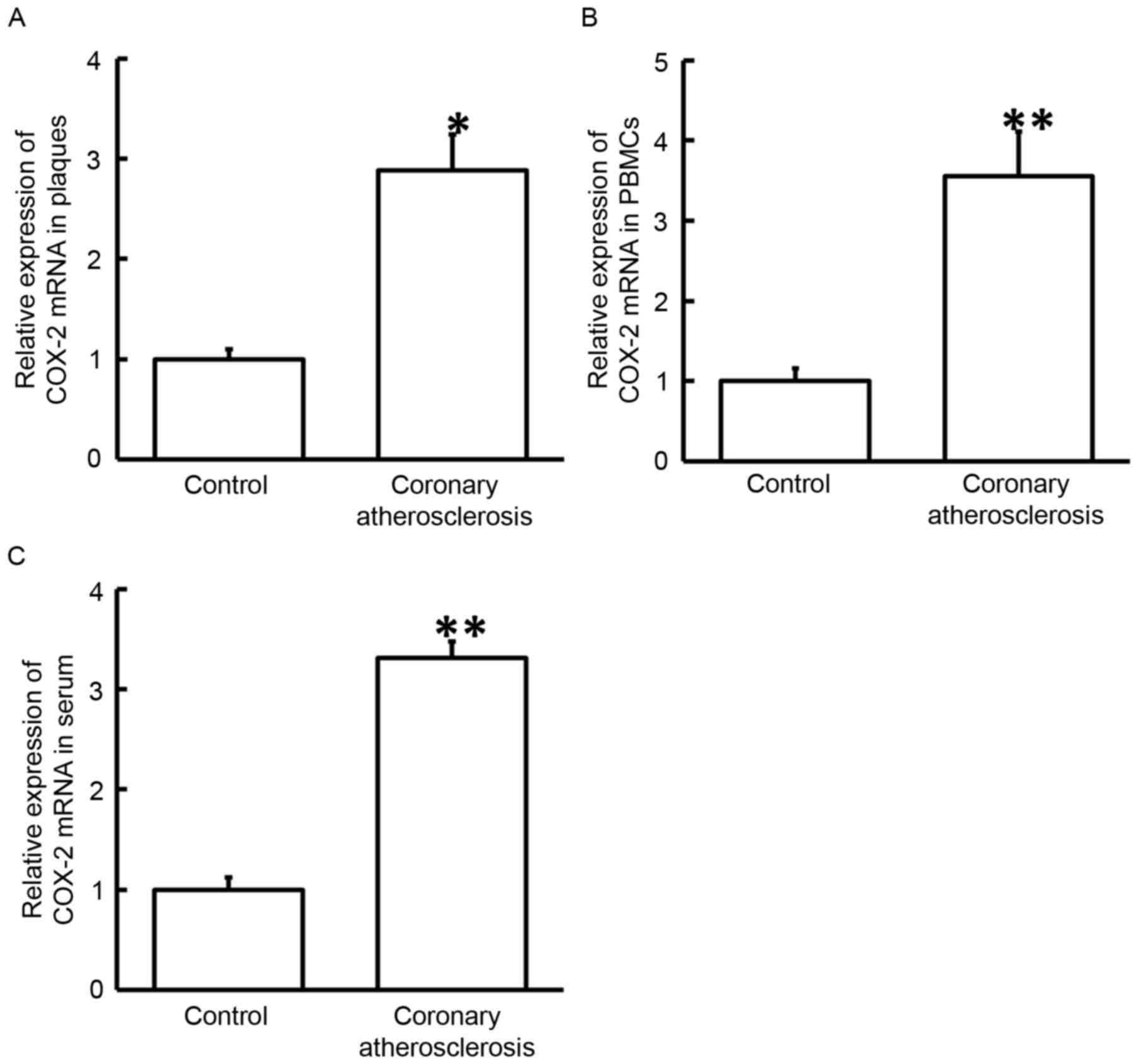
COX-2 mRNA expression is upregulated
in the plaque tissues, PBMCs and serum of patients with coronary
atherosclerosis
To measure the expression of COX-2 mRNA, RT-qPCR was
employed. The data demonstrated that the levels of COX-2 mRNA in
the plaques, PBMCs and serum of patients with coronary
atherosclerosis were significantly higher when compared with those
in normal adjacent tissues or healthy subjects (P<0.05; Fig. 1). These results suggested that
upregulation of COX-2 mRNA in plaque tissues, PBMCs and serum was
associated with the occurrence of coronary atherosclerosis.
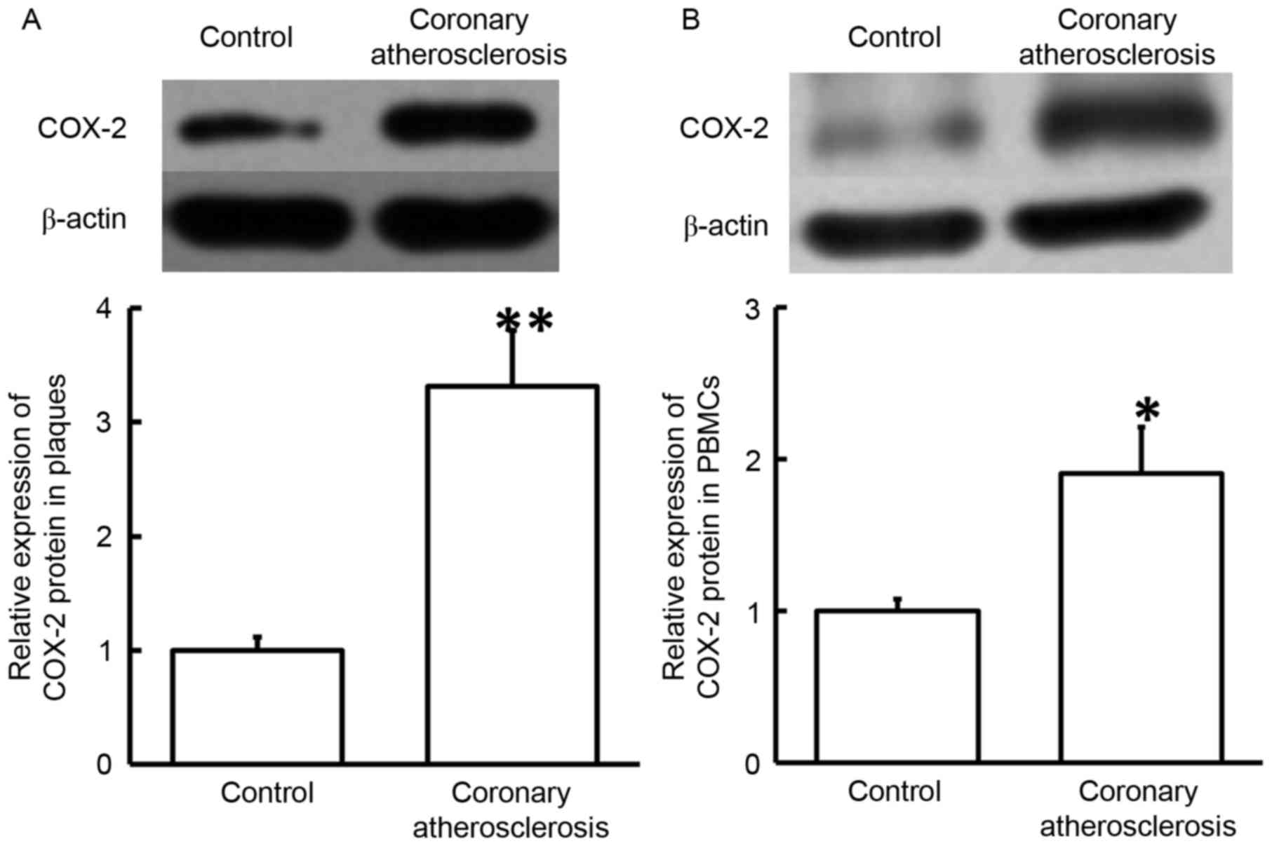
Increased expression of COX-2 protein
in plaque tissues and PBMCs suggests its regulatory role in
coronary atherosclerosis
To determine COX-2 protein expression in plaque
tissues and PBMCs, western blotting was used. The data revealed
that COX-2 protein levels in plaque tissues and PBMCs from patients
with coronary atherosclerosis were significantly elevated compared
with those in the corresponding control groups (P<0.05; Fig. 2). The results indicated that
increased expression of COX-2 protein in the plaque tissues and
PBMCs may serve a regulatory role in coronary atherosclerosis.
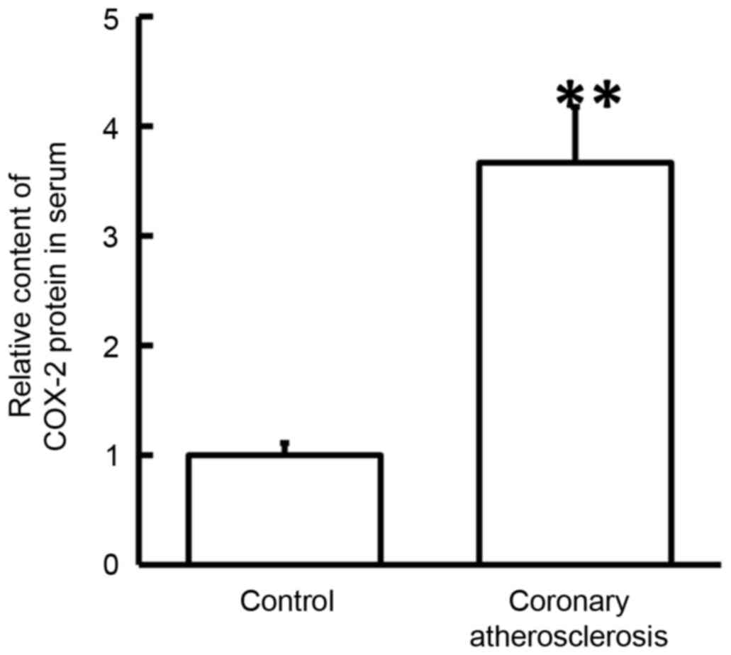
Secretion of COX-2 protein into the
blood by PBMCs is promoted in coronary atherosclerosis
To examine the contents of COX-2 protein in the
serum, ELISA was conducted. The data indicated that the serum level
of COX-2 protein in patients with coronary atherosclerosis was
significantly higher in comparison with that in healthy subjects
(P<0.05; Fig. 3). These findings
suggested that the secretion of COX-2 protein into the blood by
PBMCs was promoted in coronary atherosclerosis.
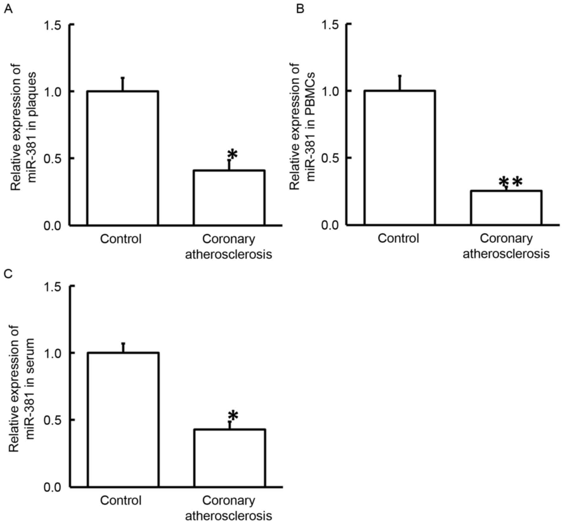
miR-381 may serve a regulatory role in
the pathogenesis of coronary atherosclerosis
To study the expression of miR-381, RT-qPCR was
performed. The results revealed that the levels of miR-381 in
plaque tissues, PBMCs and serum obtained from patients with
coronary atherosclerosis were significantly reduced as compared
with the corresponding control groups (P<0.05; Fig. 4). These results indicate that miR-381
may serve a regulatory role in the pathogenesis of coronary
atherosclerosis.
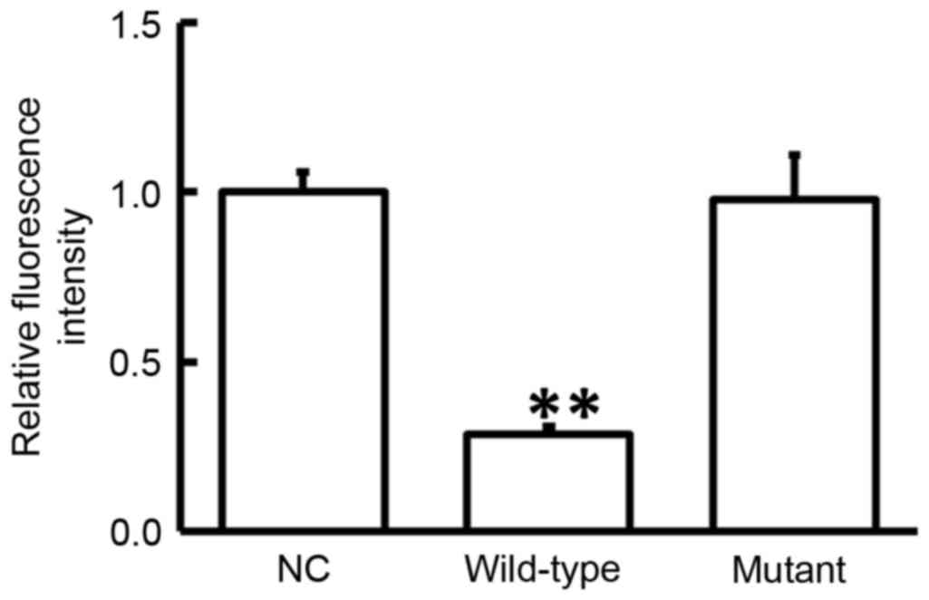
miR-381 regulates the expression of
COX-2 by directly binding with the 3′-UTR of COX-2
Analyses performed using miRanda, TargetScan, PITA,
RNAhybrid and PicTar demonstrated that miR-381 may target COX-2
(data not shown). To test whether miR-381 directly targets COX-2,
dual-luciferase reporter assay was performed. The data demonstrated
that transfection with agomiR-381 and pMIR-REPORT in the WT group
resulted in a significantly reduced fluorescence intensity compared
with the negative control (P<0.05). By contrast, fluorescence
intensity in the mutant group was not significantly different from
that in the negative control (P>0.05; Fig. 5). These observations suggest that
miR-381 regulated the expression of COX-2 by directly binding with
the 3′-UTR of COX-2.
Discussion
During the occurrence and development of chronic
inflammation, COX-2 serves an important role. COX-2 is induced by
external stimuli (physical, chemical or biological factors) and
participates in inflammatory reactions by catalyzing the synthesis
of prostaglandins (17). Due to the
importance of COX-2 in inflammation, high expression of COX-2 in
tissues has become a significant marker of inflammation (18). In addition to antipyretic and
analgesic effects, COX-2 inhibitors can also serve an auxiliary
role in antitumor therapy (19). In
previous studies, the association between COX-2 and atherosclerosis
has been examined. It has been reported that the genetic variation
of COX-2 is associated with cardiovascular diseases (20). Another study observed that
endotoxin-treated porcine coronary artery contraction induced by
bradykinin was achieved via the COX-2 pathway (21). Furthermore, COX-2 is considered to
exert certain therapeutic effects in the process of atherosclerosis
by selectively inhibiting celecoxib (22). Consistently, the results of the
present study revealed that the expression of COX-2 was elevated in
the plaque tissues, PBMCs and serum of coronary atherosclerosis
patients, suggesting that the onset of this disease is similar to
that of vascular intimal inflammation, in which PBMCs and
lymphocytes are activated. These cells secrete a great amount of
COX-2 to initiate abundant antigen immune responses.
Previous studies have suggested that miRNA molecules
are important in the occurrence and development of various diseases
(23,24) and may slice the mRNA of COX-2 and
inhibit its translation (25). It
has been reported that miR-381 is downregulated in colon cancer
tissues, leading to upregulated liver receptor homolog-1, as well
as the proliferation and invasion of colon cancer cells (26). In addition, miR-381, which exists in
the negative feedback loop of p53/pituitary tumor-transforming 1,
inhibits the growth of hypophysoma (27). miR-381 is also closely associated
with multidrug resistance protein 1 gene and serves important roles
in multidrug resistance (28). Along
with miR-424, miR-381 can target WEE1 gene and inhibit the activity
of cyclin-dependent kinase 2 in renal carcinoma cells (29). Furthermore, miR-381 is also closely
associated with lung adenocarcinoma (30). These previous studies suggested that
miR-381 expression may be closely correlated with various human
diseases.
According to bioinformatics prediction conducted in
the present study, miR-381 was predicted to be a potential upstream
regulator of COX-2. The data of the present study suggested that
miR-381 expression was downregulated and COX-2 was upregulated in
the plaques and PBMCs of coronary atherosclerosis patients. This
suggests that the body may decrease miR-381 levels to negatively
regulate its slicing effect on COX-2, while enhanced expression of
COX-2 then produces immune responses. Similar results were observed
in the serum of patients, indicating that increased COX-2 in PBMCs
can also be released into the blood. Therefore, the levels of
miR-381 and COX-2 in the serum may reflect the degrees of
inflammation responses and tissue injuries in coronary
atherosclerosis. Notably, the current study data of dual-luciferase
reporter assay revealed that miR-381 directly binds with the 3′-UTR
of COX-2 mRNA and regulates the expression of COX-2.
In conclusion, the present study demonstrated that
enhanced levels of COX-2 expression in patients with coronary
atherosclerosis were associated with the downregulation of miR-381
expression. Thus, miR-381 may regulate the occurrence and immune
responses of coronary atherosclerosis via COX-2.
Acknowledgements
The authors would like to thank the doctors and
nurses at the Linyi People's Hospital (Linyi, China) who assisted
in the plasma specimen and clinical data collection.
Funding
No funding was received.
Availability of data and materials
The analyzed data sets generated during the present
study are available from the corresponding author on reasonable
request.
Authors' contributions
KS and LL conceived and designed the study. LL and
GS performed the experiments and collected the data. KS and YW
wrote the paper and interpretated the data. KS and LL reviewed and
edited the manuscript. All authors read and approved the
manuscript.
Ethics approval and consent to
participate
All procedures were approved by the Ethics Committee
of Linyi People's Hospital (Linyi, China). Written informed
consents were obtained from all patients or their families.
Consent for publication
Not applicable.
Competing interests
The authors declare that they have no competing
interests.
References
|
1
|
Alexy T, Pais E, Wenby RB, Mack WJ, Hodis
HN, Kono N, Wang J, Baskurt OK, Fisher TC and Meiselman HJ:
Abnormal blood rheology and chronic low grade inflammation:
Possible risk factors for accelerated atherosclerosis and coronary
artery disease in Lewis negative subjects. Atherosclerosis.
239:248–251. 2015. View Article : Google Scholar : PubMed/NCBI
|
|
2
|
Kucharska-Newton AM, Heiss G, Ni H,
Stearns SC, Puccinelli-Ortega N, Wruck LM and Chambless L:
Identification of heart failure events in medicare claims: The
atherosclerosis risk in communities (ARIC) study. J Card Fail.
22:48–55. 2016. View Article : Google Scholar : PubMed/NCBI
|
|
3
|
Naghavi M, Libby P, Falk E, Casscells SW,
Litovsky S, Rumberger J, Badimon JJ, Stefanadis C, Moreno P,
Pasterkamp G, et al: From vulnerable plaque to vulnerable patient:
A call for new definitions and risk assessment strategies: Part I.
Circulation. 108:1664–1672. 2003. View Article : Google Scholar : PubMed/NCBI
|
|
4
|
Jiang XI, Luo Y, Zhao S, Chen Q, Jiang C,
Dai Y, Chen Y and Cao Z: Clinical significance and expression of
microRNA in diabetic patients with erectile dysfunction. Exp Ther
Med. 10:213–218. 2015. View Article : Google Scholar : PubMed/NCBI
|
|
5
|
Jia W, Wu Y, Zhang Q, Gao GE, Zhang C and
Xiang Y: Expression profile of circulating microRNAs as a promising
fingerprint for cervical cancer diagnosis and monitoring. Mol Clin
Oncol. 3:851–858. 2015. View Article : Google Scholar : PubMed/NCBI
|
|
6
|
Graziano A, Lo Monte G, Piva I, Caserta D,
Karner M, Engl B and Marci R: Diagnostic findings in adenomyosis: A
pictorial review on the major concerns. Eur Rev Med Pharmacol Sci.
19:1146–1154. 2015.PubMed/NCBI
|
|
7
|
Tang Y, Zhang YC, Chen Y, Xiang Y, Shen CX
and Li YG: The role of miR-19b in the inhibition of endothelial
cell apoptosis and its relationship with coronary artery disease.
Sci Rep. 5:151322015. View Article : Google Scholar : PubMed/NCBI
|
|
8
|
Ouimet M, Ediriweera HN, Gundra UM, Sheedy
FJ, Ramkhelawon B, Hutchison SB, Rinehold K, van Solingen C,
Fullerton MD, Cecchini K, et al: MicroRNA-33-dependent regulation
of macrophage metabolism directs immune cell polarization in
atherosclerosis. J Clin Invest. 125:4334–4348. 2015. View Article : Google Scholar : PubMed/NCBI
|
|
9
|
Ross R: The pathogenesis of
atherosclerosis-an update. N Engl J Med. 314:488–500. 1986.
View Article : Google Scholar : PubMed/NCBI
|
|
10
|
Ross R: Atherosclerosis-an inflammatory
disease. N Engl J Med. 340:115–126. 1999. View Article : Google Scholar : PubMed/NCBI
|
|
11
|
Oguz N, Kirca M, Cetin A and Yesilkaya A:
Effect of uric acid on inflammatory COX-2 and ROS pathways in
vascular smooth muscle cells. J Recept Signal Transduct Res.
37:500–505. 2017. View Article : Google Scholar : PubMed/NCBI
|
|
12
|
Rumzhum NN and Ammit AJ: Cyclooxygenase 2:
Its regulation, role and impact in airway inflammation. Clin Exp
Allergy. 46:397–410. 2016. View Article : Google Scholar : PubMed/NCBI
|
|
13
|
Schonbeck U, Sukhova GK, Graber P, Coulter
S and Libby P: Augmented expression of cyclooxygenase-2 in human
atherosclerotic lesions. Am J Pathol. 155:1281–1291. 1999.
View Article : Google Scholar : PubMed/NCBI
|
|
14
|
Baker CS, Hall RJ, Evans TJ, Pomerance A,
Maclouf J, Creminon C, Yacoub MH and Polak JM: Cyclooxygenase-2 is
widely expressed in atherosclerotic lesions affecting native and
transplanted human coronary arteries and colocalizes with inducible
nitric oxide synthase and nitrotyrosine particularly in
macrophages. Arterioscler Thromb Vasc Biol. 19:646–655. 1999.
View Article : Google Scholar : PubMed/NCBI
|
|
15
|
Dursunoglu N, Dursunoglu D, Yildiz AI,
Uludag B, Alaçam ZN and Sarıçopur A: Severity of coronary
atherosclerosis in patients with COPD. Clin Respir J. 11:751–756.
2017. View Article : Google Scholar : PubMed/NCBI
|
|
16
|
Livak KJ and Schmittgen TD: Analysis of
relative gene expression data using real-time quantitative PCR and
the 2(-Delta Delta C(T)) method. Methods. 25:402–408. 2001.
View Article : Google Scholar : PubMed/NCBI
|
|
17
|
Alhouayek M and Muccioli GG: COX-2-derived
endocannabinoid metabolites as novel inflammatory mediators. Trends
Pharmacol Sci. 35:284–292. 2014. View Article : Google Scholar : PubMed/NCBI
|
|
18
|
Motino O, Agra N, Contreras Brea R,
Domínguez-Moreno M, García-Monzón C, Vargas-Castrillón J, Carnovale
CE, Boscá L, Casado M, Mayoral R, et al: Cyclooxygenase-2
expression in hepatocytes attenuates non-alcoholic steatohepatitis
and liver fibrosis in mice. Biochim Biophys Acta. 1862:1710–1723.
2016. View Article : Google Scholar : PubMed/NCBI
|
|
19
|
Kim HJ, Yim GW, Nam EJ and Kim YT:
Synergistic effect of COX-2 inhibitor on paclitaxel-induced
apoptosis in the human ovarian cancer cell line OVCAR-3. Cancer Res
Treat. 46:81–92. 2014. View Article : Google Scholar : PubMed/NCBI
|
|
20
|
Ross S, Eikelboom J, Anand SS, Eriksson N,
Gerstein HC, Mehta S, Connolly SJ, Rose L, Ridker PM, Wallentin L,
et al: Association of cyclooxygenase-2 genetic variant with
cardiovascular disease. Eur Heart J. 35:2242–2248a. 2014.
View Article : Google Scholar : PubMed/NCBI
|
|
21
|
More AS, Kim HM, Zhao R, Khang G,
Hildebrandt T, Bernlöhr C, Doods H, Lee D, Lee SH, Vanhoutte PM and
Wu D: COX-2 mediated induction of endothelium-independent
contraction to bradykinin in endotoxin-treated porcine coronary
artery. J Cardiovasc Pharmacol. 64:209–217. 2014. View Article : Google Scholar : PubMed/NCBI
|
|
22
|
Papageorgiou N, Zacharia E, Briasoulis A,
Charakida M and Tousoulis D: Celecoxib for the treatment of
atherosclerosis. Expert Opin Investig Drugs. 25:619–633. 2016.
View Article : Google Scholar : PubMed/NCBI
|
|
23
|
Lewis BP, Burge CB and Bartel DP:
Conserved seed pairing, often flanked by adenosines, indicates that
thousands of human genes are microRNA targets. Cell. 120:15–20.
2005. View Article : Google Scholar : PubMed/NCBI
|
|
24
|
Chen K and Rajewsky N: The evolution of
gene regulation by transcription factors and microRNAs. Nat Rev
Genet. 8:93–103. 2007. View
Article : Google Scholar : PubMed/NCBI
|
|
25
|
Liu D, Wang D, Xu Z, Gao J, Liu M, Liu Y,
Jiang M and Zheng D: Dysregulated expression of miR-101b and
miR-26b lead to age-associated increase in LPS-induced COX-2
expression in murine macrophage. Age (Dordr). 37:972015. View Article : Google Scholar : PubMed/NCBI
|
|
26
|
Liang Y, Zhao Q, Fan L, Zhang Z, Tan B,
Liu Y and Li Y: Down-regulation of MicroRNA-381 promotes cell
proliferation and invasion in colon cancer through up-regulation of
LRH-1. Biomed Pharmacother. 75:137–141. 2015. View Article : Google Scholar : PubMed/NCBI
|
|
27
|
Liang HQ, Wang RJ, Diao CF, Li JW, Su JL
and Zhang S: The PTTG1-targeting miRNAs miR-329, miR-300, miR-381
and miR-655 inhibit pituitary tumor cell tumorigenesis and are
involved in a p53/PTTG1 regulation feedback loop. Oncotarget.
6:29413–29427. 2015. View Article : Google Scholar : PubMed/NCBI
|
|
28
|
Xu Y, Ohms SJ, Li Z, Wang Q, Gong G, Hu Y,
Mao Z, Shannon MF and Fan JY: Changes in the expression of miR-381
and miR-495 are inversely associated with the expression of the
MDR1 gene and development of multi-drug resistance. PLoS One.
8:e820622013. View Article : Google Scholar : PubMed/NCBI
|
|
29
|
Chen B, Duan L, Yin G, Tan J and Jiang X:
Simultaneously expressed miR-424 and miR-381 synergistically
suppress the proliferation and survival of renal cancer cells-Cdc2
activity is up-regulated by targeting WEE1. Clinics (Sao Paulo).
68:825–833. 2013. View Article : Google Scholar : PubMed/NCBI
|
|
30
|
Rothschild SI, Tschan MP, Jaggi R, Fey MF,
Gugger M and Gautschi O: MicroRNA-381 represses ID1 and is
deregulated in lung adenocarcinoma. J Thorac Oncol. 7:1069–1077.
2012. View Article : Google Scholar : PubMed/NCBI
|