Introduction
Endometrial carcinoma (EC) is one of the most common
malignant tumors in the female reproductive system. It often occurs
in perimenopausal women, mainly those aged 50 years. Recent studies
have shown that its incidence rate has an increasing trend
(1). Special AT-rich sequence
binding protein 1 (SATB1) is a binding protein that can bind to
AT-rich bases and regulate the downstream gene expression (2). Previous studies showed that SATB1 plays
an important role in the differentiation of T cells; at the same
time, SATB1 is highly expressed in a variety of tumors and can
regulate the expression of genes and promote the cell
proliferation, invasion and metastasis (3). Studies have shown that SATB1 is
expressed abnormally in gastric cancer, esophageal cancer (4), liver cancer (5) and other malignant tumors, which is
closely related to the tumor growth, development and prognosis, but
there has been no research on the expression of SATB1 in tissues of
EC patients. E-cadherin (E-cad) is a key gene in
epithelial-mesenchymal transition (EMT). Studies have shown that
the downregulation of E-cad in tissues promotes the invasion and
metastasis of malignant tumors. Therefore, the expressions of SATB1
and E-cad in 104 cases of EC tissues and 104 cases of
para-carcinoma tissues were detected using the immunohistochemical
method in this study, the relationship of their expression with
clinicopathological features of EC patients was analyzed, and then
the influence on the occurrence and metastasis of EC were
investigated.
Materials and methods
General materials
One hundred and four cases of carcinoma tissues of
patients pathologically diagnosed as EC in Affiliated Hospital of
Jining Medical University (Jining, China) from from August 2015 to
August 2016 were selected. All patients underwent panhysterectomy,
bilateral adnexectomy and pelvic lymph node dissection. Patients
did not receive chemotherapy, radiotherapy and biological targeted
therapy, before operation. Patients were aged from 31 to 67 years
with an average of 47.56±15.49 years. According to the degree of
pathological differentiation, there were 38 cases of high
differentiation, 34 cases of moderate differentiation and 32 cases
of low differentiation. According to the staging criteria of the
International Federation of Gynecology and Obstetrics (IFGO) in
2009, there were 26 cases of stage I, 31 cases of stage II, 29
cases of stage III and 18 cases of stage IV. In terms of
pathological type, there were 59 cases of endometrioid
adenocarcinoma, 23 cases of clear cell carcinoma and 22 cases of
papillary serous carcinoma and 104 para-carcinoma tissues of EC
patients during the same period were selected. The patients were
aged from 29 to 70 years with an average of 48.39±16.21 years. The
study was approved by the Ethics Committee of the Affiliated
Hospital of Jining Medical University and written informed consents
were signed by the patients and/or guardians.
Materials and methods
Major reagents
Rabbit anti-human SATB1 monoclonal antibody (cat.
no. ab92307) and rabbit anti-human E-cad monoclonal antibody (cat.
no. 1ab40772) were purchased from Abcam (Cambridge, MA, USA);
immunohistochemical SP kit, diaminobenzidine (DAB) developing kit
and hematoxylin were purchased from Beijing Zhongshan Golden Bridge
Biological Technology Co., Ltd. (Beijing, China).
Immunohistochemistry
The tissue paraffin block of EC patients was
continuously sliced into 4 µm-thick sections and heated at 65°C for
2 h, followed by dewaxing and hydration using the
immunohistochemical SP method in strict accordance with the
instructions of kit. Primary antibody SATB1 (1:500) or E-cad
(1:400) was added at 4°C overnight, and then goat anti-rabbit
secondary polyclonal antibody (dilution 1:2,000; cat. no. ab150077;
Abcam Cambridge, MA, USA) was used, instead of the primary
antibody, as the negative control in the experiment, and the known
positive sections were used as positive controls.
Determination of results
The positive expression of both SATB1 and E-cad
showed the brown yellow or yellow particles. The positive
expression of SATB1 was located in the nucleus, while that of E-cad
was located in the cytoplasm or membrane. Six visual fields were
randomly selected under the microscope (×400), and the results were
determined according to the percentage of positive cells and the
staining depth. i) According to the cell staining depth: Negative,
0 point; pale yellow, 1 point; brown yellow, 2 points; dark brown,
3 points. ii) According to the percentage of positive cells in
total cells: 0–30%, 1 point; 30–70%, 2 points; 70–100%, 3 points.
The product of both scores greater than or equal to 3 points
indicated positive expression; otherwise, it indicated negative
expression (6).
Statistical analysis
SPSS 22.0 (version X; IBM, Armonk, NY, USA) software
was used for statistical analysis of data, and Chi-square test was
used for enumeration data. P<0.05 suggested that the difference
was statistically significant.
Results
Expression of SATB1 in tissues of EC
patients
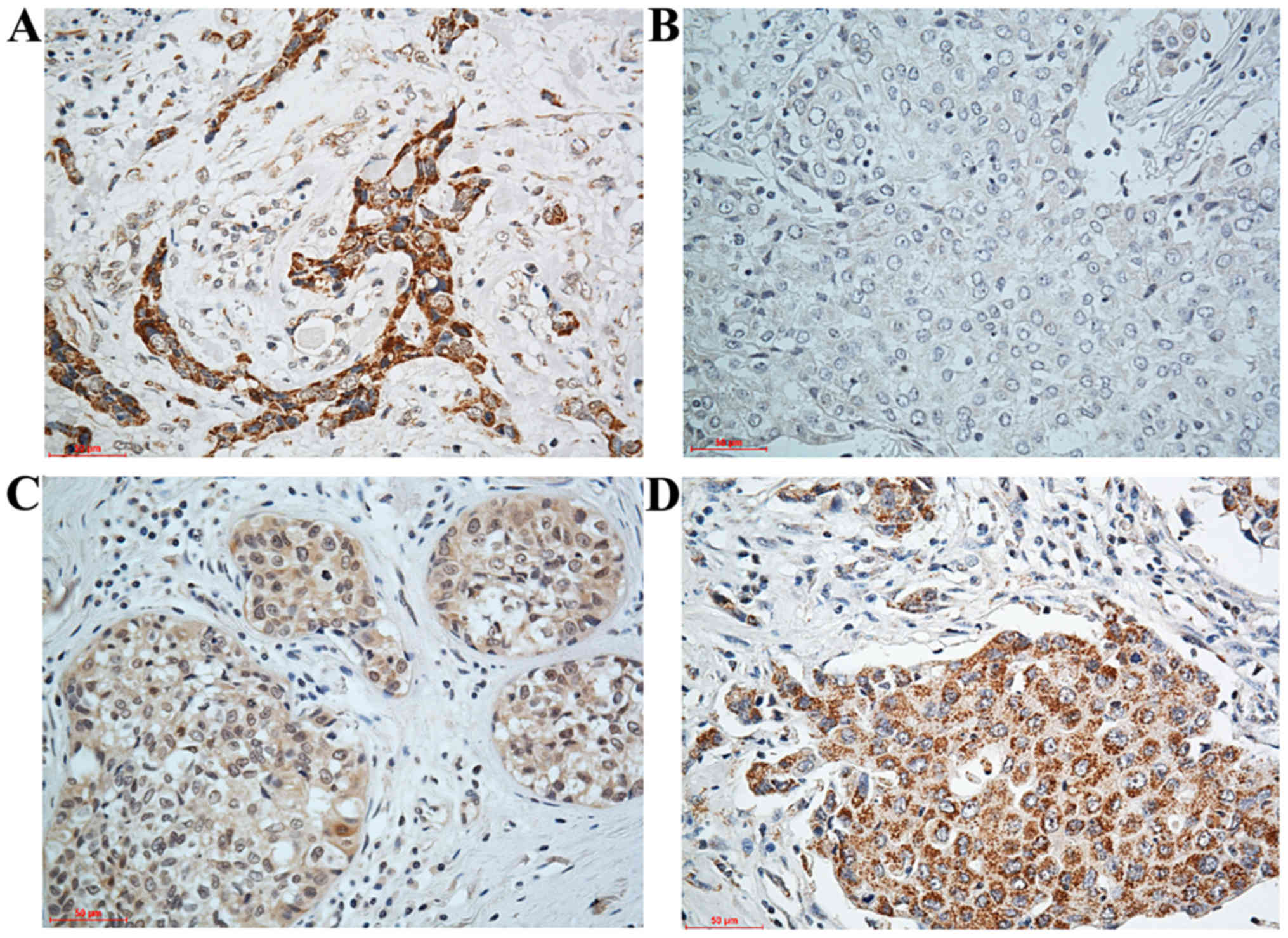
Microscopic observation revealed that the positive
expression of SATB1 was located in the nucleus, showing dark brown
or yellow particles. In this study, immunohistochemical results
showed that SATB1 was positively expressed in 71 out of 104 cases
of EC tissues (the expression rate was 68.27%) and in 25 out of 104
cases of para-carcinoma tissues (the expression rate was 24.03%).
The expression of SATB1 in EC tissues was significantly higher than
that in para-carcinoma tissues (χ2=27.849, P<0.05).
Besides, microscopic observation also revealed that the positive
expression of E-cad was located in the cytoplasm and membrane,
showing dark brown or yellow particles. E-cad was positively
expressed in 60 out of 104 cases of carcinoma tissues (the
expression rate was 57.6%) and 95 out of 104 cases of
para-carcinoma tissues (the positive expression rate was 91.3%)
(χ2=7.516, P<0.05) (Fig.
1 and Table I).
 | Table I.Expression of SATB1 and E-cad in
breast cancer tissues. |
Table I.
Expression of SATB1 and E-cad in
breast cancer tissues.
|
|
| SATB1 | E-cad |
|---|
|
|
|
|
|
|---|
| Group | Case (n) | − | + | Positive rate
(%) | − | + | Positive rate
(%) |
|---|
| Breast cancer
tissue | 104 | 33 | 71 | 68.27 | 44 | 60 | 57.6 |
| Para-carcinoma
tissue | 104 | 79 | 25 | 24.03 | 9 | 95 | 91.3 |
Relationship of SATB1 and E-cad
expression in tissues of EC patients with clinicopathological
features
The expression of SATB1 and E-cad in EC tissues were
not associated with the menopausal status and age of patients
(P>0.05), but correlated with the histological grade of EC,
depth of tumor invasion, lymph node metastasis and TNM staging
(P<0.05) (Table II).
| Table II.Relationship of the expression of
SATB1 and E-cad with the clinicopathological features of EC. |
Table II.
Relationship of the expression of
SATB1 and E-cad with the clinicopathological features of EC.
|
|
| SATB1 positive
expression | E-cad positive
expression |
|---|
|
|
|
|
|
|---|
| Pathological
feature | Total case | n | Percentage | χ2 | P-value | n | Percentage | χ2 | P-value |
|---|
| Menopausal
status |
| Yes | 64 | 46 | 71.88 | 0.142 | >0.05 | 49 | 76.56 | 0.172 | >0.05 |
| No | 40 | 26 | 65.00 |
|
| 30 | 75.00 |
|
|
| Age (years) |
| ≥50 | 80 | 56 | 70.00 | 0.473 | >0.05 | 44 | 55.00 | 0.382 | >0.05 |
|
<50 | 24 | 16 | 66.67 |
|
| 13 | 66.67 |
|
|
| Histological
grade |
| I | 42 | 30 | 71.43 | 13.287 | <0.05 | 34 | 80.95 | 15.382 | <0.05 |
| II | 30 | 22 | 73.33 |
|
| 21 | 70.00 |
|
|
| III | 32 | 19 | 59.38 |
|
| 11 | 34.38 |
|
|
| Depth of
invasion |
|
Shallow | 38 | 24 | 63.16 | 5.692 | <0.05 | 32 | 84.21 | 7.482 | <0.05 |
| Deep | 66 | 47 | 71.21 |
|
| 49 | 74.24 |
|
|
| Lymph node
metastasis |
| No | 58 | 38 | 65.52 | 8.472 | <0.05 | 31 | 53.45 | 10.372 | <0.05 |
| Yes | 46 | 33 | 71.74 |
|
| 35 | 76.09 |
|
|
| TNM staging |
| I,
II | 42 | 28 | 66.67 | 6.489 | <0.05 | 30 | 71.43 | 7.386 | <0.05 |
| III,
IV | 62 | 45 | 72.58 |
|
| 51 | 82.26 |
|
|
Discussion
SATB1 is a binding protein of tissue-specific
expression in matrix attachment region, located in 3p23 region of
chromosome 3 and a total of 763 amino acids in length (7,8). SATB1
contains an AT-rich MAR sequence with a high base pairing region.
It has been found that SATB1 can promote the stable binding of
SATB1 and MAR sequence through anchoring the chromosome ring, thus
participating in the structural remodeling, methylation and histone
acetylation of chromosomes (9,10). SATB1
is basically not expressed in normal tissues and abnormally
expressed in a variety of malignant tumor tissues. Alvarez et
al (11) established an animal
model with SATB1 gene knockout, and randomly observed the
expression of 597 genes. The results showed that 10 genes were
upregulated, while 1 gene was downregulated. The analysis of
expression profile of SATB1 gene knockout found that SATB1
regulates the expression of genes with poor prognosis, such as
genes that control cell cycle, signal transduction pathways and
apoptosis. Through upregulating the expression of CDK4, SATB1 can
induce cell proliferation, promote the cell cycle, inhibit the
Fas-related protein-mediated apoptosis pathway and inhibit cell
apoptosis (12). Han et al
(13) found that the mRNA and
protein in SATB1 are highly expressed in breast cancer cell lines,
and showed that SATB1 is closely related to the prognosis of
patients combined with 1,318 cases of tissue samples of breast
cancer patients, and the survival of patients with high expression
of SATB1 is short. At the same time, it was found that the SATB1
can upregulate the expression of genes related to invasion and
metastasis of breast cancer cells, and inhibit the expression of
tumor suppressor genes, thus promoting the tumor growth and
metastasis. Zheng et al (14)
reported that SATB1 is highly expressed in invasive breast cancer
tissues. Zhang et al (15)
found that SATB1 is not expressed in para-breast carcinoma tissues,
the positive expression rate in breast cancer tissues is 67.9%, and
the expression difference between the para-carcinoma tissues and
carcinoma tissues was statistically significant. In addition, it
was also found that the expression of SATB1 is closely related to
the histological grade and clinicopathological staging of breast
cancer patients. The higher the clinicopathological staging is the
poorer the differentiation of tumor cells is, the higher the
positive expression rate of SATB1 in tissues will be, suggesting
that SATB1 is involved in the proliferation and metastasis of
breast cancer. The COX regression model revealed that SATB1 can
also be used as an independent factor for the prognosis of breast
cancer patients. However, there are different results in studies
during the same period. It was found (15) that the expression of SATB1 is not
correlated with the prognosis of patients. The gene chip analysis
showed that there is no significant relationship between the
survival time of breast cancer patients and the expression of
SATB1; and the expression of SATB1 does not significantly affect
the proliferation and metastasis of breast cancer cells, so it is
thought that SATB1 is not involved in the proliferation and
metastasis of breast cancer, and it will not affect the prognosis
of patients. In this study, the expressions of SATB1 in 104 cases
of EC tissues and 104 cases of para-carcinoma tissues were detected
using the immunohistochemical method, and the correlation of
expression with clinicopathological features of EC patients was
also analyzed. The research results revealed that the positive
expression rate of SATB1 in EC tissues was 68.27%, and its
expression is related to the histological grade, depth of tumor
invasion, lymph node metastasis and TNM staging, and the
differences were statistically significant (P<0.05). The results
suggested that SATB1 may be associated with the occurrence of EC,
but its specific mechanism remains unclear and needs further
study.
E-cad is a key protein in EMT, whose main function
is to maintain the normal cellular morphology, and it also plays an
important role in continuing the tissue integrity and reducing the
cell dispersion (16). The
expression of E-cad is different in cytoplasm and membrane of
various normal epithelial cells in the body; at the same time, the
downregulation of E-cad expression in cells will promote the
invasion and metastasis of tumor cells (17,18). The
results of this study showed that E-cad was positively expressed in
60 out of 104 cases of carcinoma tissues (the expression rate was
57.6%) and 95 out of 104 cases of para-carcinoma tissues (the
positive expression rate was 91.3%) (χ2=7.516,
P<0.05), which was consistent with the research of scholars
world-wide.
In conclusion, the above results suggested that the
downregulation of E-cad expression leads to the tumor invasion and
metastasis, which is consistent with the results reported by Yu
et al (19). In conclusion,
SATB1 and E-cad are involved in the occurrence and development of
EC, which are of great significance to the potential therapeutic
target and prognosis estimation of EC.
Acknowledgements
Not applicable.
Funding
No funding was received.
Availability of data and materials
The datasets used and/or analyzed during the present
study are available from the corresponding author on reasonable
request.
Authors' contributions
YF wrote the manuscript. YF and XW performed
immunohistochemistry and made substantial contributions to analysis
and interpretation of data. QW conceived and designed the study,
and gave final approval of the version to be published. All authors
read and approved the final manuscript.
Ethics approval and consent to
participate
The study was approved by the Ethics Committee of
Affiliated Hospital of Jining Medical University (Jining, China).
Signed written informed consents were obtained from the
patients.
Consent for publication
Not applicable.
Competing interests
The authors declare that they have no competing
interests.
References
|
1
|
van der Steen MJ, de Waal YR, Westermann
A, Tops B, Leenders W and Ottevanger PB: An impressive response to
pazopanib in a patient with metastatic endometrial carcinoma. Neth
J Med. 74:410–413. 2016.PubMed/NCBI
|
|
2
|
Wen J, Huang S, Rogers H, Dickinson LA,
Kohwi-Shigematsu T and Noguchi CT: SATB1 family protein expressed
during early erythroid differentiation modifies globin gene
expression. Blood. 105:3330–3339. 2005. View Article : Google Scholar : PubMed/NCBI
|
|
3
|
Yuan CL, Li L, Zhou X, Liz H and Han L:
Expression of SATB1 and HER2 in gastric cancer and its clinical
significance. Eur Rev Med Pharmacol Sci. 20:2256–2264.
2016.PubMed/NCBI
|
|
4
|
Song G, Liu K, Yang X, Mu B, Yang J, He L,
Hu X, Li Q, Zhao Y, Cai X, et al: SATB1 plays an oncogenic role in
esophageal cancer by upregulation of FN1 and PDGFRB. Oncotarget.
8:17771–17784. 2017.PubMed/NCBI
|
|
5
|
Tu W, Luo M, Wang Z, Yan W, Xia Y, Deng H,
He J, Han P and Tian D: Upregulation of SATB1 promotes tumor growth
and metastasis in liver cancer. Liver Int. 32:1064–1078. 2012.
View Article : Google Scholar : PubMed/NCBI
|
|
6
|
Zhang Y, Tian X, Ji H, Guan X, Xu W, Dong
B, Zhao M, Wei M, Ye C, Sun Y, et al: Expression of SATB1 promotes
the growth and metastasis of colorectal cancer. PLoS One.
9:e1004132014. View Article : Google Scholar : PubMed/NCBI
|
|
7
|
Fang XF, Hou ZB, Dai XZ, Chen C, Ge J,
Shen H, Li XF, Yu LK and Yuan Y: Special AT-rich sequence-binding
protein 1 promotes cell growth and metastasis in colorectal cancer.
World J Gastroenterol. 19:2331–2339. 2013. View Article : Google Scholar : PubMed/NCBI
|
|
8
|
Zhou LY, Liu F, Tong J, Chen QQ and Zhang
FW: Expression of special AT-rich sequence-binding protein mRNA and
its clinicopathological significance in non-small cell lung cancer.
Nan Fang Yi Ke Da Xue Xue Bao. 29:534–537. 2009.(In Chinese).
PubMed/NCBI
|
|
9
|
Zhao XL and Wang P: Expression of SATB1
and BRMS1 in ovarian serous adenocarcinoma and its relationship
with clinieopathological features. Sichuan Da Xue Xue Bao Yi Xue
Ban. 42(82–85): 1052011.(In Chinese).
|
|
10
|
Cheng C, Lu X, Wang G, Zheng L, Shu X, Zhu
S, Liu K, Wu K and Tong Q: Expression of SATB1 and heparanase in
gastric cancer and its relationship to clinicopathologic features.
APMIS. 118:855–863. 2010. View Article : Google Scholar : PubMed/NCBI
|
|
11
|
Alvarez JD, Yasui DH, Niida H, Joh T, Loh
DY and Kohwi-Shigematsu T: The MAR-binding protein SATB1
orchestrates temporal and spatial expression of multiple genes
during T-cell development. Genes Dev. 14:521–535. 2000.PubMed/NCBI
|
|
12
|
Heubner M, Kimmig R, Aktas B, Siffert W
and Frey UH: The haplotype of three polymorphisms in the SATB1
promoter region impacts survival in breast cancer patients. Oncol
Lett. 7:2007–2012. 2014. View Article : Google Scholar : PubMed/NCBI
|
|
13
|
Han HJ, Russo J, Kohwi Y and
Kohwi-Shigematsu T: SATB1 reprogrammes gene expression to promote
breast tumour growth and metastasis. Nature. 452:187–193. 2008.
View Article : Google Scholar : PubMed/NCBI
|
|
14
|
Zheng M, Xing W, Liu Y, Li M and Zhou H:
Tetramerization of SATB1 is essential for regulating of gene
expression. Mol Cell Biochem. 430:171–178. 2017. View Article : Google Scholar : PubMed/NCBI
|
|
15
|
Zhang S, Gao X, Ma Y, Jiang J, Dai Z, Yin
X, Min W, Hui W and Wang B: Expression and significance of SATB1 in
the development of breast cancer. Genet Mol Res. 14:3309–3317.
2015. View Article : Google Scholar : PubMed/NCBI
|
|
16
|
Jang SM, Sim J, Han H, Ahn HI, Kim H, Yi
K, Jun YJ, Rehman A, Chung MS, Jang K, et al: Clinicopathological
significance of CADM4 expression in invasive ductal carcinoma of
the breast. J Clin Pathol. 66:681–686. 2013. View Article : Google Scholar : PubMed/NCBI
|
|
17
|
Kim S, Lee J, Jeon M, Nam SJ and Lee JE:
Elevated TGF-β1 and -β2 expression accelerates the epithelial to
mesenchymal transition in triple-negative breast cancer cells.
Cytokine. 75:151–158. 2015. View Article : Google Scholar : PubMed/NCBI
|
|
18
|
Kim S, Lee J, Oh SJ, Nam SJ and Lee JE:
Differential effect of EGFR inhibitors on tamoxifen-resistant
breast cancer cells. Oncol Rep. 34:1613–1619. 2015. View Article : Google Scholar : PubMed/NCBI
|
|
19
|
Yu Z, Sun M, Jin F, Xiao Q, He M, Wu H,
Ren J, Zhao L, Zhao H, Yao W, et al: Combined expression of ezrin
and E-cadherin is associated with lymph node metastasis and poor
prognosis in breast cancer. Oncol Rep. 34:165–174. 2015. View Article : Google Scholar : PubMed/NCBI
|