Introduction
Recurrent convulsion during developmental stage
endangers the lives of children, affecting children's intellectual
development and physical health (1).
Convulsion has paroxysmal, recurrent and persistent characteristics
and may be accompanied by abnormalities of corresponding cognitive,
psychological and social behavior that may continue into adulthood,
which incurs a heavy psychological and economic burden on the
children, their family and society (2,3).
Apoptosis is the main form of brain damage after recurrent
convulsion (4). Among apoptotic
death receptors, endoplasmic reticulum signals and mitochondrial
signaling pathways, the mitochondrial pathway is an important
pathway of apoptosis involved in central nervous system diseases
(5). Therefore, it is always a
hotspot to further study the pathogenesis of convulsion in order to
explore a relatively safe and effective therapeutic approach.
Materials and methods
Animals
Thirty-six Sprague-Dawley male rats
(2.5–3-month-old; weighing 280±350 g) were obtained from Beijing
Vital River Laboratory Animal Technology Co., Ltd. (Beijing,
China). The rats were kept in cages with controlled temperature and
light cycles (24°C and 12/12 light cycles). The humidity was 40%
and free access to water and food.
Behavioral characteristics of
flurothyl-induced recurrent convulsion in rats during developmental
stage
The rats in the recurrent-seizure group were dripped
with flurothylin in the experimental cabin. After 40–60 sec, onset
of convulsion occurred, and rats were manifested by dysphoria, head
nodding, jumping around, accompanied with scream, followed by skin
mucous cyanosis, rigidity of limbs and spasm, namely generalized
tonic-clonic seizure and other seizure behaviors at grade III or
above as per Racine scoring criteria (Racine grade III: facial
clonus, including spasm-like blink, beard moving, rhythmic
mastication, rhythmic nod, forelimb clonus). Each attack lasted 4–5
min, followed by intermittence for 2 to 3 min, and consciousness in
the intermission of convulsion was not recovered. The rats in the
control group (NS group) had normal activity without convulsive
attack.
Main reagents
Bicinchoninic acid (BCA) Protein Assay kit (Beyotime
Institute of Biotechnology, Shanghai, China); TRIzol Total RNA
Extraction kit and reverse transcription-polymerase chain reaction
(RT-PCR) kit (both from Tiangen Biotech Co., Ltd., Beijing, China);
terminal deoxynucleotidyltransferase (TdT)-mediated dUTP nick
end-labeling (TUNEL) kit (Roche, Basel, Switzerland);
anti-glyceraldehyde 3-phosphate dehydrogenase (GAPDH),
anti-caspase-3 and cytochrome c (Cyt c) monoclonal
antibodies and secondary antibodies (all from Cell Signaling
Technology, Inc., Boston, MA, USA) were used in the present
study.
Preparation of paraffin-embedded
tissues
Paraffin wax sample was prepared by routine
dehydration, paraffin dipping and embedding, followed by coronal
serial section with a thickness of 5 µm. Three sets of adjacent
slices containing hippocampal tissue were selected, and slides were
coated with poly-lysine for anti-stripping. The slides were removed
and placed in an incubator at 62°C overnight, followed by
hematoxylin and eosin (H&E) staining and in situ cell
apoptosis detection.
H&E staining
Paraffin section was treated by routine dewaxing and
hydration, followed by rinsing with phosphate-buffered saline (PBS)
for 3 min, for a total of three times. The sample was stained with
hematoxylin for 5 min, faded with 1% HCl and rinsed using
double-distilled water for 5 min, for a total of six times. The
sample was blued with lithium carbonate saturated solution for 1 to
2 min and rinsed in double-distilled water for 5 min, three times,
followed by color separation using 80% alcohol and rinsing with
double-distilled water for 5 min, for a total of three times.
Subsequently, the sample was stained with eosin for 5 min and
rinsed in double-distilled water for 5 min, for a total of three
times. Finally, dehydration, hyalinization and mounting using resin
were conducted.
Detection of apoptosis
According to the instructions, paraffin sections
were treated by routine dewaxing and hydration, followed by rinsing
with PBS for 3 min, for a total of three times. Then it was
incubated with 1% H2O2 at room temperature
for 20 min to inhibit endogenous peroxidase, followed by rinsing
with PBS for 3 min, three times and digestion with 20 µg/ml
protease K at 37°C for 20 min. Subsequently, the sample was rinsed
with PBS for 3 min, a total of three times and incubated in TdT
buffer solution for 10 min. The sample was soaked in mixture (100
µl solution containing 1 µl TdT and 1 µl Biotin-11-dUTP mixture) at
37°C for 90 min and rinsed by PBS for 3 min, a total of three
times. Tris-buffered solution was added at room temperature for 15
min to terminate the reaction. The sample was added with
anti-avidin-horseradish peroxidase (HRP) immediate solution drip by
drip at 37°C for 1 h and rinsed with PBS for 3 min, three times,
followed by coloration with diaminobenzidine (DAB) for 20 to 30
min, after which it was washed using running water to terminate the
reaction. At higher magnification (10×40-fold), three visual fields
in the hippocampal CA1 region were randomly observed, and the mean
was calculated as the apoptotic number in this animal.
Determination of hippocampus neuronal
mitochondrial membrane potential (ΔΨm) after convulsion
Single-cell suspension of hippocampus was prepared
with the concentration of cells as 1×10⁹/l. The JC-1
working fluid was added into cell suspension to make the final
concentration was 5 mg/l. Then it was incubated at room temperature
for 30 min in the dark, and the cell count was 1×10⁴.
The mitochondrial ΔΨm was measured by using an excitation
wavelength of 488 nm.
RT-PCR analysis
Tissues of different groups were extracted according
to operation procedures of RNAiso Plus kit (Tiangen Biotech Co.,
Ltd., Beijing, China), and the purity and content of the extracted
RNA sample were calculated. The samples were packed separately and
stored at −80°C for experimental use. The reverse transcription
reaction liquid was prepared according to the scale on the
instructions of PrimeScript® RT reagent kit (Tiangen
Biotech Co., Ltd.) with gDNA Eraser and added with the
corresponding RNA sample, followed by reverse transcription to
obtain cDNA. The level of mRNA was detected according to the
protocol of SYBR® Premix Ex Taq™ II (Tli RNaseH Plus)
kit (Tiangen Biotech Co., Ltd.). The corresponding RNA primer
sequences are shown in Table I.
 | Table I.Quantitative PCR analysis of primer
sequences of related genes. |
Table I.
Quantitative PCR analysis of primer
sequences of related genes.
| Gene name | Forward primer
(5′-3′) | Reverse primer
(5′-3′) |
|---|
| Mfn2 |
ATGTCCCTGCTCTTTTCTCGA |
CTATCTGCTGGGCTGCAGGTA |
| Drp1 |
ATGGAGGGGCTGATCCCGGTC |
TCACCAAAGATGAGTGTGTCG |
| Caspase-3 |
GAGGCCGACTTCCTGTATGC |
TGACCCGTCCCTTGAATTTC |
| Cyt c |
GGTGATGTTGAAAAAGGCAAGAA |
TGCTTGCCTCCTTTTTCCA |
| GAPDH |
GCACCGTCAAGGCTGAGAAC |
TGGTGAAGACGCCAGTGGA |
Western blot analysis
Operations were conducted in accordance with the
instructions of the Total Protein Extraction kit (Beyotime
Institute of Biotechnology, Shanghai, China). The sample was added
with lysate and centrifuged at 12,000 × g and at 4°C for 10 min.
The supernatant was collected, namely total protein. The
concentration of protein was detected by BCA Protein Assay kit
(Beyotime Institute of Biotechnology), and samples were packed
separately and stored at −80°C for reservation. The total protein
extraction fluid was mixed well with 2X loading buffer (100+4 µl
β-mercaptoethanol) according to the proportion of 1:1, and then
placed into a boiling water bath for 5 min, cooled and stored in a
refrigerator at 4°C for preservation. The appreciable proportion of
SDS-PAGE separation gel was prepared according the molecular weight
of the target protein, followed by solidification for approximately
1 h, and then 5% SDS-PAGE concentrated gel was prepared, followed
by solidification for approximately 30 min. The electrophoretic
buffer was added, and the denatured protein sample was added into
the loading sample well. The total protein content of each well was
the same according to the concentration loading of protein. The
sample was treated via electrophoresis under a constant pressure of
220 V, until bromophenol blue was evident at the bottom of gel, and
electrophoresis was stopped. The gel was cut off according to the
molecular weight of target protein, and placed into transfer
buffer.
A layer of polyvinylidene difluoride (PVDF) membrane
and six layers of filter paper were cut according to the size of
the gel. Firstly, PVDF membrane was soaked in carbinol for 10 sec,
and then PVDF membrane and filter paper were placed into the
transfer buffer. Then it was placed in the transfer apparatus
according to positive pole - three-layer filter paper - PVDF
membrane - gel - three-layer filter paper - negative pole, with
attention to edge alignment to prevent blistering. Electrophoresis
was performed under constant pressure of 110 V for 2 h. PVDF
membrane with protein was placed in 5% skimmed milk powder and
sealed on the table concentrator at room temperature for 3 h. The
sealed membrane was rinsed using Tween/Tris-buffered salt solution
(TTBS) for 5 min and placed into rabbit anti-rat GAPDH, Mfn2, Drp1,
caspase-3, Cyt c primary monoclonal antibody (1:800; cat.
nos. 5014, 9482, 8570, 9665, 4272; Cell Signaling Technology, Inc.,
Boston, MA, USA) with corresponding proportion, followed by
incubation at 4°C overnight. The membrane was rinsed with TTBS
three times, each for 10 min, followed by placement into the
corresponding goat anti-rabbit secondary polyclonal antibody
(1:1,500; cat. no. 7074; Cell Signaling Technology, Inc.). Then it
was incubated on the table concentrator at room temperature for 3 h
and rinsed with TTBS three times, each for 10 min. The gel imager
was started up to preheat for 30 min. The reagents of A and B in
the enhanced chemiluminescence (ECL) reagent box were mixed
uniformly as the equal volume, followed by dropping on the PVDF
membrane for full contact and developing avoiding light for 1 min.
By using filter paper, the excess liquid around the membrane was
sucked, and the membrane was placed in the gel imager. The image
was taken using dynamic integral model, and the results were
observed. Image analysis was completed by Lab Works 4.6
professional image analysis software.
Statistical analysis
Experimental data were expressed as mean ± standard
deviation (mean ± SD). Experimental results were statistically
analyzed by Statistical Product and Service Solutions (SPSS) 17.0
(SPSS Inc., Chicago, IL, USA) software. The t-test was adopted for
comparison of mean between the two groups. One-way analysis of
variance (ANOVA) was utilized for comparison of mean among multiple
samples and the post hoc test was Dunnett's test. The t-test was
used for pairwise comparison. P<0.05 was considered to indicate
a statistically significant difference.
Results
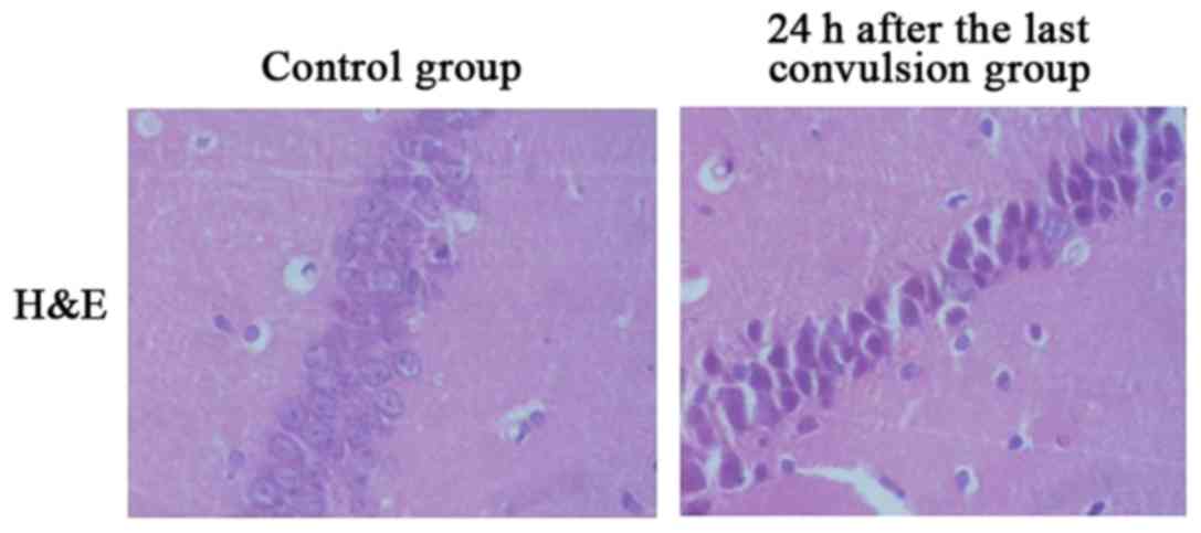
H&E staining
As shown in Fig. 1,
in the control group, pyramidal cells in CA1 region of hippocampus
were manifested by tight arrangement, distinct gradations, clear
margin, transparent cytoplasm, elliptical or round cells, uniform
distribution of chromatin in the nucleus and clear nucleoli. After
recurrent convulsion, pyramidal cells in CA1 region of hippocampus
were manifested by gradually scattered arrangement, cellular
swelling and some cell fracture, followed by the characteristic
apoptosis changes such as cells becoming smaller, margination and
condensation of chromatin, karyopyknosis and hyperchromasia and
extracellularhalos. The peak of changes was at 24 h.
Dynamic changes of mitochondrial ΔΨm
in rat neurons after sustained convulsion
As shown in Table
II, mitochondrial ΔΨm in hippocampal neurons of experimental
rats were significantly reduced at 1.5, 3 and 12 h after
convulsion, which reached a trough at 12 h, and was rapidly
elevated from 24 h [p<0.05, p<0.01 (n=6)].
| Table II.Dynamic changes of ΔΨm in rat neurons
after sustained convulsion. |
Table II.
Dynamic changes of ΔΨm in rat neurons
after sustained convulsion.
| Group | Mitochondrial
ΔΨm |
|---|
| Control | 8.68±0.97 |
| 0 h | 8.62±1.08 |
| 1.5 h | 7.47±0.89 |
| 3 h | 7.32±1.02 |
| 12 h | 6.82±0.99 |
| 24 h | 7.28±1.03 |
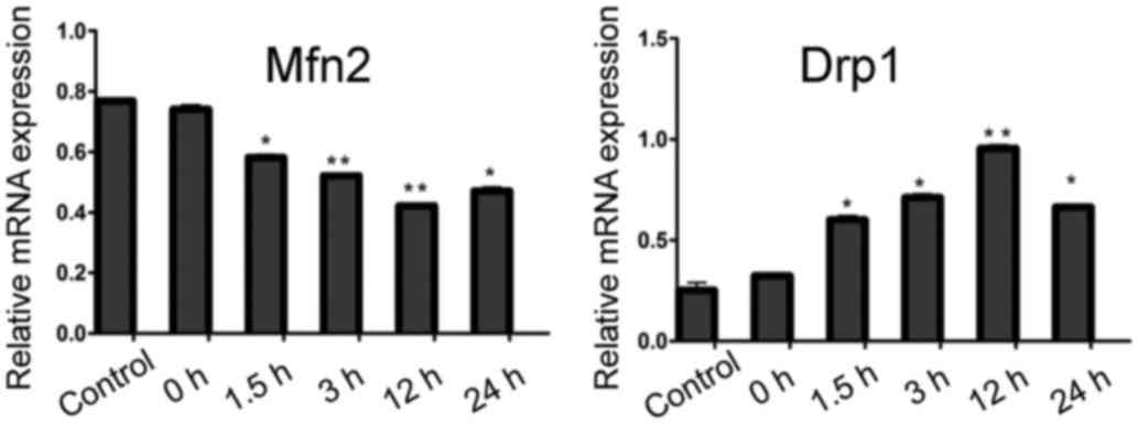
RT-PCR results of mitochondrial fusion
and division-related genes, Mfn2 and Drpl
The expression of Mfn2 mRNA in hippocampus tissue
was significantly lower than that in the control group at 1.5, 3,
12 and 24 h after recurrent convulsion (p<0.05), whereas that of
Drp1 mRNA in hippocampus tissue was distinctly higher than that in
the control group at 1.5, 3, 12 and 24 h after recurrent convulsion
(p<0.05) (Fig. 2).
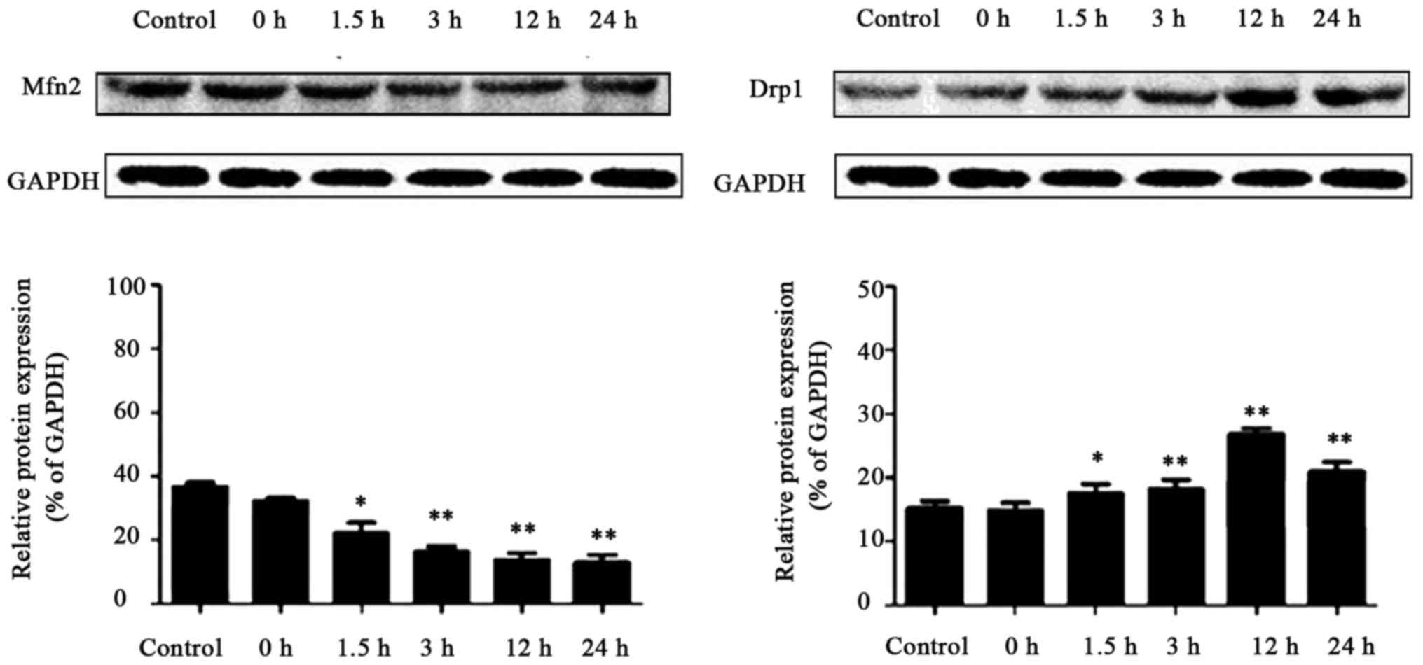
Expression of Mfn2 and Drpl proteins
in each group
As shown in Fig. 3,
the results of western blot analysis revealed that Mfn2 protein
expression in hippocampus tissue was obviously lower than that in
the control group at 1.5, 3, 12 and 24 h after recurrent
convulsion. By contrast, the expression of Drp1 protein in
hippocampus tissue was significantly higher than that in the
control group at 1.5, 3, 12 and 24 h after recurrent
convulsion.
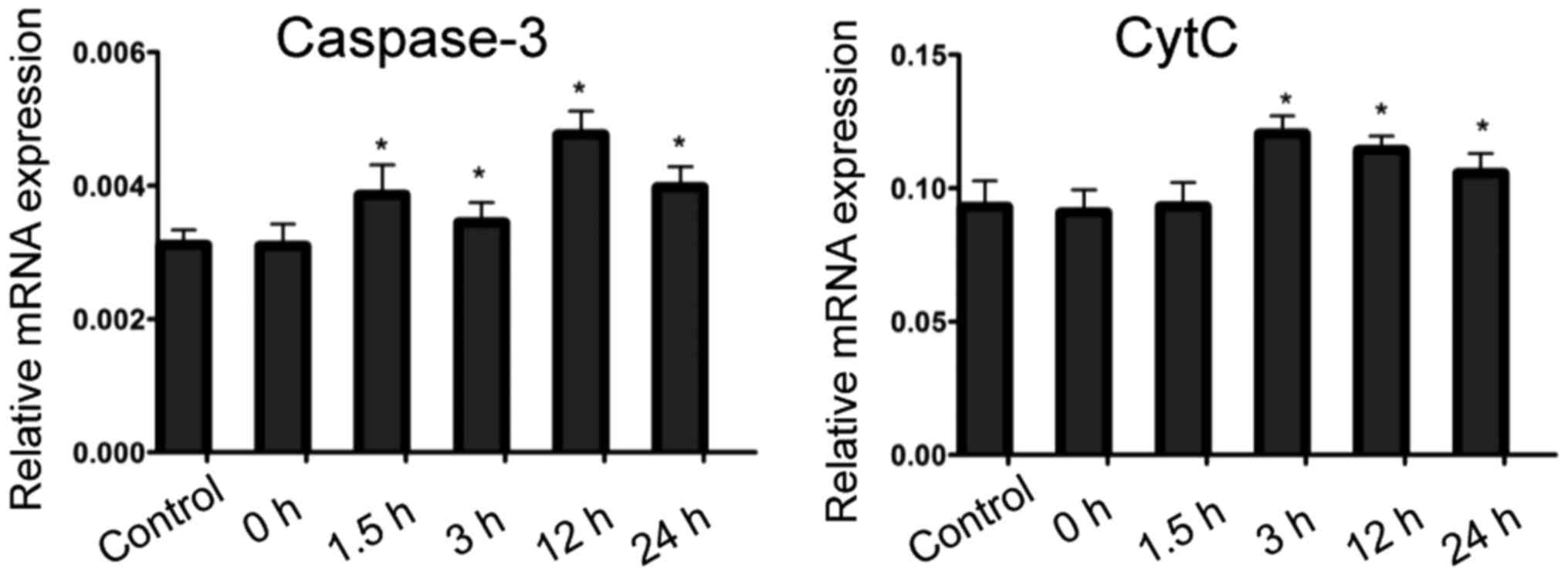
RT-PCR results of apoptosis-related
factors
Compared with that in the control group, caspase-3
expression in the hippocampus of rats in convulsion group began to
rise at 1.5 h after the last convulsion, and lasted 24 h after
convulsion. The expression of Cyt c in hippocampus of rats in
convulsion group was increased at 3 h after the last convulsion,
and lasted 24 h after convulsion (Fig.
4).
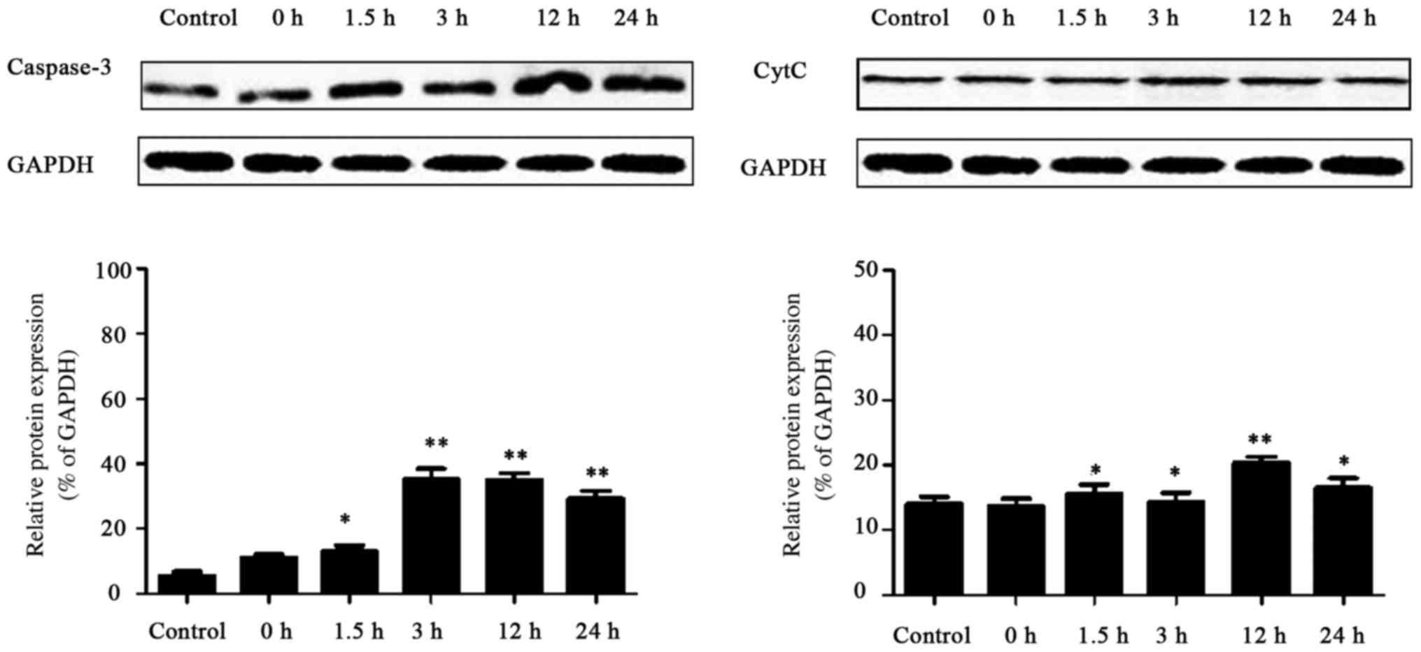
Expression of caspase-3 and Cyt c
proteins in each group
As shown in Fig. 5,
the results of western blot analysis revealed that compared with
that in the control group, caspase-3 expression in the hippocampus
of rats in the convulsion group began to rise at 1.5 h after the
last convulsion, and lasted 24 h after convulsion. The expression
of Cyt c in hippocampus of rats in convulsion group was
increased at 3 h after the last convulsion, and lasted 24 h after
convulsion. The results indicated that the expression of
apoptosis-related genes, caspase-3 and Cyt c, were increased
after the recurrent convulsion during the developmental stage,
suggesting that apoptosis occurs in the early stage after recurrent
convulsion.
Correlation analysis of changes in
mitochondrial function and neuronal apoptosis
As shown in Table
III, the changes of mitochondrial membrane potential were at
different time-points, and apoptotic rates were also distinctly
changed at the same time. It was evident that the change in
mitochondrial function was closely related to neuronal
apoptosis.
| Table III.Correlation between changes in
mitochondrial function and neuronal apoptosis. |
Table III.
Correlation between changes in
mitochondrial function and neuronal apoptosis.
| Group | Mitochondrial
membrane potential | Apoptosis rate
(%) |
|---|
| Control | 8.68±0.97 | 1±2 |
| 0 h | 8.62±1.08 | 2±1 |
| 1.5 h | 7.47±0.89 | 18±5 |
| 3 h | 7.32±1.02 | 22±3 |
| 12 h | 6.82±0.99 | 32±4 |
| 24 h | 7.28±1.03 | 21±5 |
Discussion
The occurrence of convulsion is very complex, and
although a great deal of research has been conducted, the mechanism
involved remains unclear. Previous studies in China and foreign
countries have revealed that apoptosis and necrosis are the main
pathogenesis of neuronal damage after convulsion (6,7).
However, recent studies have found that mitochondrial function
changes and apoptosis play important roles in the occurrence and
development of convulsion (8–10).
Neurons are non-differentiated cells, which cannot remove the
damaged misfolded proteins and organelles through cell division;
thus, they are especially sensitive to apoptosis and closely
associated with many chronic nervous system diseases (11).
Previous findings have indicated that apoptosis is
programmed cell death induced by a variety of pathological and
physiological stimuli, and whether or not apoptosis occurs in
multiple cells, including neurons, is affected by many links in the
upstream (12). Therein, the
reduction of mitochondrial ΔΨm and release of apoptosis-related
factors are considered to be important links of apoptosis (13). ΔΨm is the potential difference
between the two sides of biofilm (14). The reduction of ΔΨm causes the
opening of the mitochondrial permeability transition pore, increase
of nuclear permeability, release of ion and some proteins in
mitochondrial matrix and disappearance of ion gradient across the
inner membrane, thus forming a vicious cycle and resulting in a
further decline in ΔΨm until complete disintegration (15–17).
After the mitochondrial permeability transition pore opening and
the rupture of outer mitochondrial membrane, mitochondrial
interstitial apoptosis-promoting factor C is released to cytoplasm
(18). Under the action of
apoptosis-promoting gene, the assembly that promotes aspartic acid
specific cysteine protease complex is formed, incurring apoptosis
inevitably (19,20). Thus, the reduction of mitochondrial
permeability transition pore and the release of apoptotic factors
are considered the early events of apoptosis.
In this study, flurothyl was used to induce
recurrent convulsion in rats for seven consecutive days during
developmental stage at eight days after birth (the 8th day). At 0,
1.5, 3, 12 and 24 h after the last convulsion at 14th day, the
dynamic changes of mitochondrial fusion and division-related genes,
Mfn2 and Drp1, in the hippocampus after recurrent
convulsion and expressions of apoptosis-related genes
(caspase-3, Cyt c) in rat hippocampus were detected by
RT-PCR and western blot analysis. The changes in mitochondrial ΔΨm
in hippocampal neurons were detected by flow cytometer, and
expressions of apoptosis-related factors (caspase-3, Cyt c) were
determined by western blot analysis eventually. The study aimed to
investigate the changes in mitochondrial function and the role of
apoptosis in brain injury after recurrent convulsion during
developmental stage, so as to provide a new intervention target for
clinical treatment of convulsion.
Acknowledgements
Not applicable.
Funding
No funding was received.
Availability of data and materials
The datasets used and/or analyzed during the present
study are available from the corresponding author on reasonable
request.
Authors' contributions
YL and JC were responsible for collection and
preparation of paraffin-embedded tissues. MJ and ZL performed and
analyzed H&E staining. TT helped with PCR. LL and HN
contributed to western blot analysis. All authors read and approved
the final manuscript.
Ethics approval and consent to
participate
The study was approved by the Ethics Committee of
the Children's Hospital of Soochow University (Jiangsu, China).
Consent for publication
Not applicable.
Competing interests
There were no competing interests to declare.
References
|
1
|
Shinnar S, Pellock JM, Moshé SL, Maytal J,
O'Dell C, Driscoll SM, Alemany M, Newstein D and DeLorenzo RJ: In
whom does status epilepticus occur: Age-related differences in
children. Epilepsia. 38:907–914. 1997. View Article : Google Scholar : PubMed/NCBI
|
|
2
|
Huang L, Cilio MR, Silveira DC, McCabe BK,
Sogawa Y, Stafstrom CE and Holmes GL: Long-term effects of neonatal
seizures: A behavioral, electrophysiological, and histological
study. Brain Res Dev Brain Res. 118:99–107. 1999. View Article : Google Scholar : PubMed/NCBI
|
|
3
|
Chin RF, Neville BG, Peckham C, Wade A,
Bedford H and Scott RC: Treatment of community-onset, childhood
convulsive status epilepticus: A prospective, population-based
study. Lancet Neurol. 7:696–703. 2008. View Article : Google Scholar : PubMed/NCBI
|
|
4
|
Roy H, Lippé S, Lussier F, Sauerwein HC,
Lortie A, Lacroix J and Lassonde M: Developmental outcome after a
single episode of status epilepticus. Epilepsy Behav. 21:430–436.
2011. View Article : Google Scholar : PubMed/NCBI
|
|
5
|
Sheppard E and Lippé S: Cognitive outcome
of status epilepticus in children. Epilepsy Res Treat.
2012:9841242012.PubMed/NCBI
|
|
6
|
Liu Z, Yang Y, Silveira DC, Sarkisian MR,
Tandon P, Huang LT, Stafstrom CE and Holmes GL: Consequences of
recurrent seizures during early brain development. Neuroscience.
92:1443–1454. 1999. View Article : Google Scholar : PubMed/NCBI
|
|
7
|
Gómez-Gonzalo M, Losi G, Chiavegato A,
Zonta M, Cammarota M, Brondi M, Vetri F, Uva L, Pozzan T, de Curtis
M, et al: An excitatory loop with astrocytes contributes to drive
neurons to seizure threshold. PLoS Biol. 8:e10003522010. View Article : Google Scholar : PubMed/NCBI
|
|
8
|
Wang H, Yan WJ, Zhang JL, Zhang FY, Gao C,
Wang YJ, Law Bond W and Tao L: Adiponectin partially rescues high
glucose/high fat-induced impairment of mitochondrial biogenesis and
function in a PGC-1α dependent manner. Eur Rev Med Pharmacol Sci.
21:590–599. 2017.PubMed/NCBI
|
|
9
|
Ouyang YB, Lu Y, Yue S and Giffard RG:
miR-181 targets multiple Bcl-2 family members and influences
apoptosis and mitochondrial function in astrocytes. Mitochondrion.
12:213–219. 2012. View Article : Google Scholar : PubMed/NCBI
|
|
10
|
January CT, Wann LS, Alpert JS, Calkins H,
Cigarroa JE, Cleveland JC Jr, Conti JB, Ellinor PT, Ezekowitz MD,
Field ME, et al: American College of Cardiology/American Heart
Association Task Force on Practice Guidelines: 2014 AHA/ACC/HRS
guideline for the management of patients with atrial fibrillation:
A report of the American College of Cardiology/American Heart
Association Task Force on Practice Guidelines and the Heart Rhythm
Society. J Am Coll Cardiol. 64:e1–e76. 2014. View Article : Google Scholar : PubMed/NCBI
|
|
11
|
Arends MJ and Wyllie AH: Apoptosis:
Mechanisms and roles in pathology. Int Rev Exp Pathol. 32:223–254.
1991. View Article : Google Scholar : PubMed/NCBI
|
|
12
|
Schwartzman RA and Cidlowski JA:
Apoptosis: The biochemistry and molecular biology of programmed
cell death. Endocr Rev. 14:133–151. 1993. View Article : Google Scholar : PubMed/NCBI
|
|
13
|
Savill J and Fadok V: Corpse clearance
defines the meaning of cell death. Nature. 407:784–788. 2000.
View Article : Google Scholar : PubMed/NCBI
|
|
14
|
Falasca L, Bergamini A, Serafino A,
Balabaud C and Dini L: Human Kupffer cell recognition and
phagocytosis of apoptotic peripheral blood lymphocytes. Exp Cell
Res. 224:152–162. 1996. View Article : Google Scholar : PubMed/NCBI
|
|
15
|
Fadok VA, Voelker DR, Campbell PA, Cohen
JJ, Bratton DL and Henson PM: Exposure of phosphatidylserine on the
surface of apoptotic lymphocytes triggers specific recognition and
removal by macrophages. J Immunol. 148:2207–2216. 1992.PubMed/NCBI
|
|
16
|
Yin XM: Signal transduction mediated by
Bid, a pro-death Bcl-2 family proteins, connects the death receptor
and mitochondria apoptosis pathways. Cell Res. 10:161–167. 2000.
View Article : Google Scholar : PubMed/NCBI
|
|
17
|
Duchen MR: Mitochondria and calcium: From
cell signalling to cell death. J Physiol. 529:57–68. 2000.
View Article : Google Scholar : PubMed/NCBI
|
|
18
|
Zou H, Li Y, Liu X and Wang X: An
APAF-1.cytochrome c multimeric complex is a functional
apoptosome that activates procaspase-9. J Biol Chem.
274:11549–11556. 1999. View Article : Google Scholar : PubMed/NCBI
|
|
19
|
Acehan D, Jiang X, Morgan DG, Heuser JE,
Wang X and Akey CW: Three-dimensional structure of the apoptosome:
Implications for assembly, procaspase-9 binding, and activation.
Mol Cell. 9:423–432. 2002. View Article : Google Scholar : PubMed/NCBI
|
|
20
|
Desagher S and Martinou JC: Mitochondria
as the central control point of apoptosis. Trends Cell Biol.
10:369–377. 2000. View Article : Google Scholar : PubMed/NCBI
|