Introduction
Paraquat is a widely used non-selective herbicide,
commonly used in agriculture (1).
However, paraquat is also one of the herbicides mostly used for
suicide. Paraquat has caused 20 deaths per million people
worldwide, and the mortality rate is 60–70% (2,3).
Paraquat poisoning toxicity involves multiple organs such as the
digestive tract, skin and respiratory tract, which can lead to
multiple organ dysfunction syndrome and renal failure (4). The main target organ of paraquat
poisoning is the lung. In the later stage, the pulmonary alveolar
and interstitial pulmonary fibrosis gradually occurred in patients.
The reason of death is mostly due to respiratory failure caused by
acute lung injury or acute respiratory distress syndrome in
patients (5). Therefore, early
diagnosis and early treatment are critical in patients with
pulmonary fibrosis caused by paraquat poisoning. A large number of
studies have found that curcumin is a phenolic pigment extracted
from the underground rhizome of turmeric. It has antibacterial and
anti-fibrotic pharmacological effects, and has become a major
target of study in the medical field (6,7).
Currently, the condition and prognosis of patients
with paraquat poisoning are mostly determined by blood and urinary
paraquat content. However, there is no agreed evaluation standard
in the medical community for the time being, so the search for
reliable prognostic factors will be helpful for clinical guidance
in treatment (8). The blood gas
index can directly reflect the air function of the lungs in the
body, and is used to measure the gas and acid-base balance in the
blood of the body. It is currently used as an indicator to observe
whether the patient has symptoms such as hypoxemia, respiratory
failure and coma (9). The Mothers
against decapentaplegic homolog 4 (Smad4) is a transforming growth
factor-β1 (TGF-β1) that transmits signals to the nucleus through
specific receptors on the cell membrane (10). However, Smad ubiquitination
regulatory factor2 (Smurf2) mediates the Smad signaling pathway and
plays a pivotal role in the regulation of TGF-β1 signaling activity
(11,12). Interferon-γ (IFN-γ) is a Th-1 type
cytokine that inhibits the proliferation of fibroblasts and
promotes the degradation of fibroblasts, and has anti-pulmonary
fibrosis (13). Interleukin-4 (IL-4)
is a Th-2 type cytokine that promotes the proliferation of
fibroblasts and also fibrosis through anti-apoptotic effects
(14).
Currently, there are only a few clinical studies on
the arterial blood gas index and the expression of Smad4, Smurf2,
IL-4 and IFN-γ in rats with pulmonary fibrosis induced by paraquat
poisoning. Therefore, this study observed the rats with pulmonary
fibrosis induced by paraquat poison, compared the changes of artery
blood gas index and Smad4, Smurf2, IL-4 and IFN-γ expression in
rats with pulmonary fibrosis induced by curcumin.
Materials and methods
Animal data
A total of 54 clean 6-week-old Wistar rats weighing
224.24±4.36 g were purchased from the Experimental Center of Gansu
University of traditional Chinese Medicine (production license no.
SCXK 2012–0002; Gansu, China). The rats were raised at the
temperature 24.00±2.00°C, humidity 50.00±5.00%, 12 h day and night
alternative, in normal single cages and free feeding and drinking
water environment. This experiment was approved by the Ethics
Committee of Gansu Provincial Hospital (Lanzhou, China).
Modeling
After feeding the rats for one week, 36 rats were
randomly selected to model the lung fibrosis of paraquat poisoning.
Paraquat (20%) was diluted with distilled water 20 times to 0.2%
paraquat (Beijing Bai Ao Si Biotechnology Co., Ltd., Beijing,
China). The solution was configured with a concentration of 10
mg/ml, and the rats were given a single oral gavage at 20 mg/kg.
There was no convulsion, death or vomiting during the intragastric
administration. The pulmonary fibrosis was judged according to the
rat lung diffusion function. The pulmonary fibrosis was determined
according to the function of lung diffusion in rats. After 1 h of
gavage, 18 rats were randomly selected to administer curcumin
(Shanghai Baoman Bio-technology Co., Ltd., Shanghai, China)
suspension of 200 mg/kg through intraperitoneal injection and
classified as the curcumin group. The remaining 18 rats were given
a saline injection of 10 ml/kg intraperitoneally and classified as
the paraquat group. A further 18 rats were reared normally and were
not processed, and classified as the control group.
Detection method
Rats in each group were randomly selected to obtain
0.5 ml of venous blood on days 1 and 5 after modeling. After
leaving the blood for 30 min, the serum was separated by
centrifugation at 3,000 × g for 15 min at 4°C, stored at −20°C by
using an enzyme-linked immunosorbent assay (ELISA) method. The
following kits were used: rat SMAD4 ELISA kit (item no. H-EL-Smad4;
Shanghai Zeye Biotechnology Co., Ltd., Shanghai, China), rat Smurf2
ELISA kit (article no. wu-El082ra-s96; Shanghai Wuyi Biotechnology
Co., Ltd., Shanghai, China), rat IL-4 ELISA kit (article no.
RA20088; Wuhan Hualianke Biotechnology Co., Ltd., Wuhan, China),
rat INF-γ ELISA kit (article no. 865.010.048; Beijing Borede
Biotechnology Co., Ltd., Beijing, China). The expression levels of
Smad4, Smurf2, IL-4 and INF-γ were measured in accordance with the
operating instructions. Then, 0.5 ml of abdominal aorta blood was
drawn for the determination of pH, arterial partial pressure of
oxygen (PaO2) and arterial partial pressure of carbon
dioxide (PaCO2) using a Thunder ABL80 blood gas analyzer
(Nanjing Li Ai Trading Co., Ltd., Nanjing, China). After the blood
was drawn, the rats were sacrificed by cervical dislocation.
Satistical analysis
The data were analyzed and processed by SPSS19.6
statistical software (Beijing Sitron Weida Information Technology
Co., Ltd., Beijing, China). The results of the experiments were
expressed as the mean ± standard deviation. Multivariate time
comparison was performed by repeated measures of ANOVA and the LSD
post hoc test. A comparison between the two pairs was performed
using a paired t-test. P<0.05 was considered to indicate a
statistically significant difference.
Results
Modeling results
In the 54 modeled rats, 53 were successfully
modeled, and the modeling success rate was 98.15%. There were 18
rats in the control group, 18 in the curcumin group, and 17 in the
paraquat group.
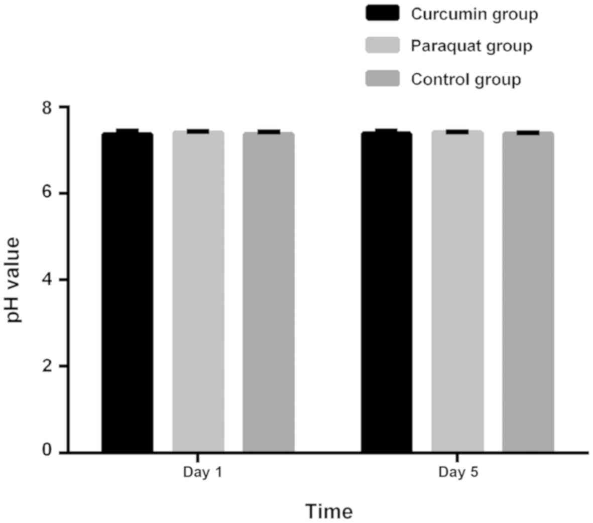
Comparison of pH values of arterial
whole blood in three groups of rats
The pH values of artery blood on days 1 and 5 in the
curcumin group were 7.37±0.08 and 7.39±0.06, respectively. The pH
values of artery blood on days 1 and 5 in the paraquat group were
7.40±0.04 and 7.41±0.02, respectively. The pH values of artery
blood on days 1 and 5 in the control group were 7.37±0.06 and
7.38±0.03, respectively. There was no significant difference in the
pH value of artery blood between days 1 and 5 in the curcumin
group, the paraquat group or the control group (P>0.05; Fig. 1). On days 1 and 5, the artery blood
pH of the three groups was measured. There was no significant
difference and it was not statistically significant
(P>0.05).
Comparison of PaO2 in
artery blood of rats between the three groups
The PaO2 of arterial blood on days 1 and
5 in the curcumin group was 83.75±13.65 and 67.39±15.57 mmHg,
respectively. The PaO2 of artery blood on days 1 and 5
in the paraquat group was 76.37±13.19 and 50.40±16.53 mmHg, the
artery blood PaO2 of the control group on days 1 and 5
was 89.65±16.43 and 87.38±12.34 mmHg, respectively (Fig. 2).
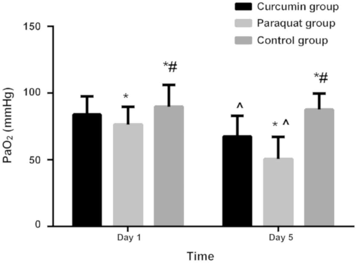 | Figure 2.Comparison of PaO2 in
artery blood of rats between the three groups. The PaO2
of artery blood on days 1 and 5 in the curcumin group was
83.75±13.65 and 67.39±15.57 mmHg, respectively. The PaO2
of artery blood on days 1 and 5 in the paraquat group was
76.37±13.19 and 50.40±16.53 mmHg, respectively, the artery blood
PaO2 of the control group on days 1 and 5 were
89.65±16.43 and 87.38±12.34 mmHg, respectively. PaO2 in
artery blood of rats in the curcumin group on day 1 was
significantly higher than that on day 5, which was statistically
significant (P<0.05). PaO2 in artery blood of rats in
the paraquat group was significantly higher on day 1 than that on
day 5, which was statistically significant (P<0.001). There was
no significant difference in PaO2 of artery blood on
days 1 and 5 in the control group, which was not statistically
significant (P>0.05). On days 1 and 5, PaO2 in the
artery blood of rats between the three groups was statistically
significant (P<0.001). The artery blood PaO2 of the
control group was higher than that in the curcumin group and
paraquat group, which were statistically significant (P<0.05).
The PaO2 of artery blood in the curcumin group was
higher than that in the paraquat group, which was statistically
significant (P<0.05). *P<0.05, when compared with the
curcumin group; #P<0.05, when compared with the
paraquat group and ^P<0.05, when compared to day
1. |
The artery blood PaO2 of rats in the
curcumin group was significantly higher on day 1 than day 5, which
was statistically significant (t=3.352, P=0.002). The artery blood
PaO2 of rats in the paraquat group was significantly
higher on day 1 than that on day 5, which was statistically
significant (t=5.063, P<0.001). There was no significant
difference in PaO2 of artery blood on days 1 and 5 in
the control group, which was not statistically significant
(P>0.05). On days 1 and 5, the artery blood PaO2 of
rats in the three groups was statistically significant (F=3.666,
P=0.033; F=27.080, P<0.001) (data not shown). The artery blood
PaO2 in the control group was higher than that in the
curcumin and paraquat groups, and were statistically significant
(P<0.05). The PaO2 of the artery blood in the
curcumin group was higher than that in the paraquat group
(P<0.05; Fig. 2).
Comparison of PaCO2 values
in artery blood of rats in the three groups
The PaCO2 of artery blood on days 1 and 5
in the curcumin group was 40.41±5.16 and 53.73±5.72 mmHg,
respectively. The PaCO2 of artery blood on days 1 and 5
in the paraquat group was 43.56±6.39 and 69.67±6.53 mmHg,
respectively. PaCO2 of artery blood on days 1 and 5 in
the control group were 35.48±5.92 and 36.54±5.61 mmHg, respectively
(Fig. 3). The artery blood
PaCO2 of rats in the curcumin group was significantly
lower on day 1 than that on day 5, which was statistically
significant (t=4.230, P<0.001). The artery blood
PaCO2 of rats in the paraquat group was significantly
lower on day 1 than that on day 5, which was statistically
significant (t=4.014, P<0.001). There was no significant
difference in PaCO2 between the control group on days 1
and 5, which was not statistically significant (P>0.05). On days
1 and 5, the artery blood PaCO2 of rats in the three
groups was statistically significant (F=8.366, P<0.001;
F=26.030, P<0.001) (data not shown). The arterial blood
PaCO2 of the control group was lower than that in the
curcumin and paraquat groups, which was statistically significant
(P<0.05). The PaCO2 of artery blood in the curcumin
group was lower than that in the paraquat group (P<0.05;
Fig. 3).
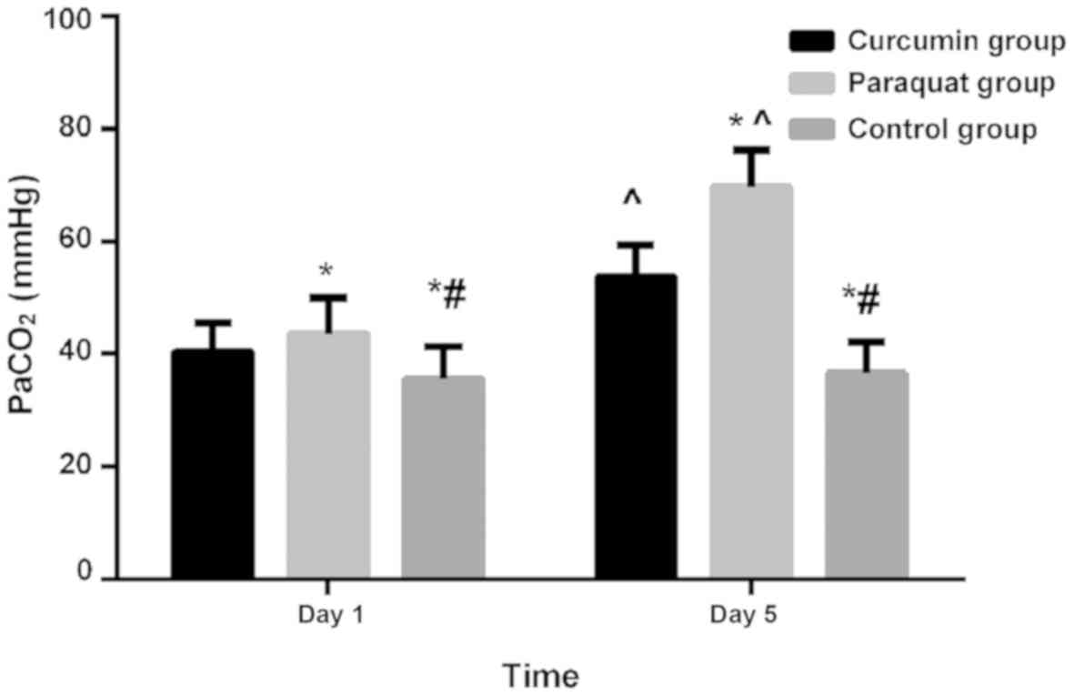 | Figure 3.Comparison of artery blood
PaCO2 values of rats between the three groups. The
PaCO2 of artery whole blood on days 1 and 5 in the
curcumin group were 40.41±5.16 mmHg and 53.73±5.72 mmHg,
respectively. The PaCO2 of artery blood on days 1 and 5
in the paraquat group was 43.56±6.39 mmHg and 69.67±6.53 mmHg,
respectively. The PaCO2 of artery blood days 1 and 5 in
the control group was 35.48±5.92 mmHg and 36.54±5.61 mmHg,
respectively. PaCO2 in artery blood of rats in the
curcumin group was significantly lower on day 1 than that on day 5,
which was statistically significant (P<0.001). PaCO2
in artery blood of rats in the paraquat group was significantly
lower on day 1 than that on day 5, which was statistically
significant (P<0.001); There was no significant difference in
PaCO2 on days 1 and 5 in the control group, which was
not statistically significant (P>0.05). On days 1 and 5, the
artery blood PaCO2 of rats between the three groups was
statistically significant (P<0.001); The artery blood
PaCO2 of the control group was lower than that in the
curcumin group and paraquat group, which were statistically
significant (P<0.05). The PaCO2 of artery blood in
the curcumin group was lower than that in the paraquat group, which
was statistically significant (P<0.05). *P<0.05, when
compared with the curcumin group; #P<0.05, when
compared with the paraquat group and ^P<0.05,
compared to day 1. |
Comparison of serum Smad4 values in
rats between the three groups
The serum Smad4 of the curcumin group was lower on
day 1 than that on day 5, which was statistically significant
(t=2.260, P=0.030). The serum Smad4 of the paraquat group was
significantly lower on day 1 than that on day 5, which was
statistically significant (t=5.223, P<0.001); There was no
significant difference in serum Smad4 between days 1 and 5 in the
control (P>0.05). On days 1 and 5, the serum Smad4 of rats in
the three group was statistically significant (F=28.420,
P<0.001; F=92.180, P<0.001). The serum Smad4 of rats in the
control group was significantly lower than that in the curcumin and
paraquat group (P<0.05). The serum Smad4 in the curcumin group
was lower than that in the paraquat group (P<0.05; Table I).
 | Table I.Comparison of serum Smad4 values in
rats between the three groups (pg/ml). |
Table I.
Comparison of serum Smad4 values in
rats between the three groups (pg/ml).
| Groups | Day 1 | Day 5 | t | P-value |
|---|
| Curcumin group
(n=18) | 58.45±12.36 |
67.35±11.24 | 2.260 |
0.030 |
| Paraquat group
(n=17) |
75.27±13.73a |
100.92±14.88a | 5.223 | <0.001 |
| Control group
(n=18) |
44.73±9.62a,b |
45.19±10.15a,b | 0.140 |
0.890 |
| F | 28.420 | 92.180 |
|
|
| P-value | <0.001 | <0.001 |
|
|
Comparison of serum Smurf2 values of
rats in the three groups
The serum Smurf2 of the curcumin group was lower on
day 1 than that on day 5, which was statistically significant
(t=5.188, P<0.001). The serum Smurf2 of the paraquat group was
significantly lower on day 1 than that on day 5, which was
statistically significant (t=4.786, P<0.001); There was no
significant difference in serum Smurf2 between the control group on
days 1 and 5 (P>0.05). On days 1 and 5, the serum Smurf2 of rats
between the three groups was statistically significant (F=66.480,
P<0.001; F=135.000, P<0.001). Serum Smurf2 in the control
group was significantly lower than that in the curcumin and
paraquat groups (P<0.05). Serum Smurf2 in the curcumin group was
lower than that in the paraquat group (P<0.05; Table II).
| Table II.Comparison of serum Smurf2 values in
rats between the three groups (pg/ml). |
Table II.
Comparison of serum Smurf2 values in
rats between the three groups (pg/ml).
| Groups | Day 1 | Day 5 | t | P-value |
|---|
| Curcumin group
(n=18) | 53.41±8.72 | 68.83±9.11 | 5.188 | <0.001 |
| Paraquat group
(n=17) |
64.55±9.28a |
81.56±11.34a | 4.786 | <0.001 |
| Control group
(n=18) |
31.62±7.84a,b |
30.77±8.05a,b | 0.321 |
0.750 |
| F | 66.480 | 135.000 |
|
|
| P-value | <0.001 |
<0.001 |
|
|
Comparison of serum IL-4 in rats in
the three groups
Serum IL-4 in the curcumin group was lower on day 1
than that on day 5, which was statistically significant (t=4.749,
P<0.001). The serum IL-4 level in the paraquat group was
significantly lower on day 1 than that on day 5, which was
statistically significant (t=6.248, P<0.001). There was no
significant difference in serum IL-4 between days 1 and 5 in the
control group, which was not statistically significant (P>0.05).
On days 1 and 5, serum IL-4 of rats in the three groups was
statistically significant (F=202.200, P<0.001; F=330.400,
P<0.001). Serum IL-4 in the control group was significantly
lower than that in the curcumin group and the paraquat group
(P<0.05). Serum IL-4 was lower in the curcumin group than that
in the paraquat group, which was statistically significant
(P<0.05; Table III).
| Table III.Comparison of serum IL-4 values of
rats between the three groups (ng/l). |
Table III.
Comparison of serum IL-4 values of
rats between the three groups (ng/l).
| Groups | Day 1 | Day 5 | t | P-value |
|---|
| Curcumin group
(n=18) | 262.73±9.81 | 278.54±10.16 | 4.749 | <0.001 |
| Paraquat group
(n=17) |
284.49±10.72a |
308.48±11.65a | 6.248 | <0.001 |
| Control group
(n=18) |
221.52±7.58a,b |
222.69±8.13a,b | 0.447 | 0.658 |
| F | 202.200 | 330.400 |
|
|
| P-value | <0.001 | <0.001 |
|
|
Comparison of serum INF-γ in rats
between the three groups
Serum INF-γ in the curcumin group was significantly
higher on day 1 than that on day 5, which was statistically
significant (t=5.959, P<0.001). The serum INF-γ in the paraquat
group was significantly higher on day 1 than that on day 5, which
was statistically significant (t=4.118, P<0.001); there was no
significant difference in serum INF-γ between days 1 and 5 in the
control group, which was not statistically significant (P>0.05).
On days 1 and 5, serum INF-γ of rats in the three groups was
statistically significant (F=9.138, P<0.001; F=26.030,
P<0.001). The serum INF-γ of the control group was significantly
higher than that in the curcumin group and the paraquat group
(P<0.05). The serum INF-γ of the curcumin group was higher than
that in the paraquat group, which was statistically significant
(P<0.05; Table IV).
| Table IV.Comparison of serum INF-γ values of
rats between the three groups (ng/l). |
Table IV.
Comparison of serum INF-γ values of
rats between the three groups (ng/l).
| Groups | Day 1 | Day 5 | t | P-value |
|---|
| Curcumin group
(n=18) | 528.91±18.42 | 491.66±19.08 | 5.959 | <0.001 |
| Paraquat group
(n=17) |
473.15±19.86a |
445.95±18.63a | 4.118 | <0.001 |
| Control group
(n=18) |
580.62±20.75a,b |
577.36±18.28a,b | 0.500 | 0.620 |
| F | 9.138 | 26.030 |
|
|
| P-value | <0.001 | <0.001 |
|
|
Discussion
Pulmonary fibrosis caused by paraquat is a chronic
progressive inflammatory disease that can cause diffuse alveolitis
and alveolar structural disorders. However, due to the lack of
effective treatment methods, the mortality rate remains high and
seriously endangers the life and health of patients (15). In recent years, it has been reported
that curcumin can inhibit the transformation of epithelial to
mesenchymal, and has anti-angiogenic, pro-apoptotic and
immunomodulatory effects, which can inhibit the occurrence and
development of tumors and make curcumin a focus of research
(16,17). Curcumin has the characteristics of
large safe concentration of drug, small adverse reaction and low
price, and has been widely used in the treatment of many diseases,
such as colorectal cancer, lung cancer and inflammatory diseases
(18–20). Therefore, this study investigated the
effects of curcumin on a rat model of pulmonary fibrosis induced by
paraquat.
In this study, we found that the PaO2 in
the artery blood of the curcumin group was higher than that in the
paraquat group, the PaCO2 was lower than that in the
paraquat group. The PaO2 and PaCO2 levels can
be used to determine whether a patient has symptoms such as
CO2 retention and hypoxia, and can reflect the degree or
type of respiratory disease of the patient (21). Due to the massive replacement of
alveolar fibrosis in the absence of gas exchange function in
pulmonary fibrosis, normal lung tissue structure degeneration and
partial lung function loss, CO2 retention may occur in
the blood, which may lead to poor breathing, hypoxia, and can even
lead to death of the patient (22).
According to studies reported by Lelli et al (23), curcumin has a variety of pro-growth
effects, which may play a role in the treatment of lung diseases
such as lung cancer, as well as the regulation of transcription
factors, cytokines, adhesion molecules and enzymes that play a key
role in inflammation and cancer. Therefore, rats with pulmonary
fibrosis caused by paraquat poisoning had lower PaO2 and
higher PaCO2 than normal rats. When paraquat-poisoned
rats were treated with curcumin, their blood gas indexes were
normal compared with those in the paraquat group. Recent studies
have found that curcumin has an obvious anti-pulmonary fibrosis
effect, and its conclusions can be used to support our research
(24). In recent years, it has been
reported that TGF-β plays a key role in the development and
progression of pulmonary fibrosis, and TGF-β1 causes pulmonary
fibrosis by promoting the proliferation, invasion and activation of
lung fibroblasts (25). Smurfs plays
a pivotal role in the regulation of TGF-β1 signaling by selectively
mediating the degradation of key components in the Smad signaling
pathway, and its dysfunction leads to abnormal TGF-β1/Smads
signaling and to a series of pathophysiological changes. It has
been reported that Smad4 and Smurf2 can promote the development of
pulmonary fibroblasts in pulmonary fibrosis (26,27).
Furthermore, they have a key role in pulmonary fibrosis, which is
in agreement with our research results. The report of Yallapu et
al (28), indicates that
curcumin inhibits the synthesis of extracellular matrix and
inhibits collagen synthesis through Smad-mediated signaling
pathway, thereby reducing cardiac repair and improving cardiac
function after ischemia-reperfusion. It was found that curcumin can
reduce myocardial ischemia and fibrosis as well as significantly
downregulated the expression of TGF-β1 and Smad in
ischemia-reperfusion myocardium (28). Furthermore, curcumin, not only
reduced myocardial ischemia and fibrosis, but significantly
downregulated the expression of TGF-β1 and Smad in
ischemia-reperfusion myocardium (28). Those findings may support our
research results. When the stress response is repaired after the
lung is damaged, IFN-γ can inhibit the activation and proliferation
of fibroblasts and promote the synthesis of collagen (29). The expression of TGF-β1 and Smad in
ischemia-reperfusion myocardium were also significantly
downregulated, which may support our findings. When lung damage
occurs and stress response is repaired, IFN-γ can inhibit the
activation and proliferation of fibroblasts and promote collagen
synthesis (29). IL-4 can induce
fibroblast proliferation and promote exogenous collagen to cause
fibrosis, which accumulates in the lungs and causes lung damage
through oxidation and inflammation processes (30). When the body is normal, IFN-γ and
IL-4 are in a state of equilibrium, which plays a key regulatory
role in the body's immune response. However, fibrosis occurs when
IL-4 is transferred. According to the in vitro studies of
LITERAT (31), curcumin can inhibit
the production of alveolar inflammatory cytokines IL-β1 and IL-8.
In addition, the nasal administration of curcumin can inhibit
airway inflammation and pulmonary fibrosis (32), which further validates our
experiment.
Further aspects remain to be explored, including the
mechanism influence of curcumin on paraquat poisoning. Of note,
differences between animal models and human body should be
considered. Consequently, experiments regarding the effect of
curcumin on paraquat poisoning in human are to be conducted in
future studies.
In summary, curcumin can effectively improve
pulmonary fibrosis in rats after treatment of paraquat poisoning.
In addition, it is expected to be an effective drug for treating
paraquat and providing effective reference and guidance for
clinical treatment.
Acknowledgements
Not applicable.
Funding
This study was supported by Lanzhou Health Science
and Technology Development Project (no. LZWSKY2014-2-18).
Availability of data and materials
The datasets used and/or analyzed during the present
study are available from the corresponding author on reasonable
request.
Authors' contributions
HC wrote the manuscript. HC and XF were responsible
for construction of the lung fibrosis model. HC and RY performed
ELISA. RY and YT helped with determination of pH, PaO2
and PaCO2. All authors read and approved the final
manuscript.
Ethics approval and consent to
participate
The study was approved by the Ethics Committee of
Gansu Provincial Hospital (Lanzhou, China).
Patient consent for publication
Not applicable.
Competing interests
The authors declare that they have no competing
interests.
References
|
1
|
Rasooli R, Rajaian H, Pardakhty A and
Mandegary A: Preference of aerosolized pirfenidone to oral intake:
an experimental model of pulmonary fibrosis by paraquat. J Aerosol
Med Pulm Drug Deliv. 31:25–32. 2018. View Article : Google Scholar : PubMed/NCBI
|
|
2
|
Kim HJ, Kim HK, Lee H, Bae JS, Kown JT,
Gil HW and Hong SY: Toxicokinetics of paraquat in Korean patients
with acute poisoning. Korean J Physiol Pharmacol. 20:35–39. 2016.
View Article : Google Scholar : PubMed/NCBI
|
|
3
|
Bromilow RH: Paraquat and sustainable
agriculture. Pest Manag Sci. 60:340–349. 2004. View Article : Google Scholar : PubMed/NCBI
|
|
4
|
Wen C, Wang Z, Zhang M, Wang S, Geng P,
Sun F, Chen M, Lin G, Hu L, Ma J, et al: Metabolic changes in rat
urine after acute paraquat poisoning and discriminated by support
vector machine. Biomed Chromatogr. 30:75–80. 2016. View Article : Google Scholar : PubMed/NCBI
|
|
5
|
Khalighi Z, Rahmani A, Cheraghi J, Ahmadi
MR, Soleimannejad K, Asadollahi R and Asadollahi K: Perfluorocarbon
attenuates inflammatory cytokines, oxidative stress and
histopathologic changes in paraquat-induced acute lung injury in
rats. Environ Toxicol Pharmacol. 42:9–15. 2016. View Article : Google Scholar : PubMed/NCBI
|
|
6
|
Gunes H, Gulen D, Mutlu R, Gumus A, Tas T
and Topkaya AE: Antibacterial effects of curcumin: An in vitro
minimum inhibitory concentration study. Toxicol Ind Health.
32:246–250. 2016. View Article : Google Scholar : PubMed/NCBI
|
|
7
|
Xiao J, Sheng X, Zhang X, Guo M and Ji X:
Curcumin protects against myocardial infarction-induced cardiac
fibrosis via SIRT1 activation in vivo and in vitro. Drug Des Devel
Ther. 10:1267–1277. 2016.PubMed/NCBI
|
|
8
|
Wang HR, Pan J, Shang AD and Lu YQ:
Time-dependent haemoperfusion after acute paraquat poisoning. Sci
Rep. 7:22392017. View Article : Google Scholar : PubMed/NCBI
|
|
9
|
Huang R and Li M: Protective effect of
Astragaloside IV against sepsis-induced acute lung injury in rats.
Saudi Pharm J. 24:341–347. 2016. View Article : Google Scholar : PubMed/NCBI
|
|
10
|
Zandvoort A, Postma DS, Jonker MR,
Noordhoek JA, Vos JT and Timens W: Smad gene expression in
pulmonary fibroblasts: indications for defective ECM repair in
COPD. Respir Res. 16:832008. View Article : Google Scholar
|
|
11
|
Cai Y, Zhou CH, Fu D and Shen XZ:
Overexpression of Smad ubiquitin regulatory factor 2 suppresses
transforming growth factor-β mediated liver fibrosis. J Dig Dis.
13:327–334. 2012. View Article : Google Scholar : PubMed/NCBI
|
|
12
|
Nakano A, Koinuma D, Miyazawa K, Uchida T,
Saitoh M, Kawabata M, Hanai J, Akiyama H, Abe M, Miyazono K, et al:
Pin1 down-regulates transforming growth factor-beta (TGF-beta)
signaling by inducing degradation of Smad proteins. J Biol Chem.
284:6109–6115. 2009. View Article : Google Scholar : PubMed/NCBI
|
|
13
|
Ziesche R and Block LH: Mechanisms of
antifibrotic action of interferon gamma-1b in pulmonary fibrosis.
Wien Klin Wochenschr. 112:785–790. 2000.PubMed/NCBI
|
|
14
|
Kodera T, McGaha TL, Phelps R, Paul WE and
Bona CA: Disrupting the IL-4 gene rescues mice homozygous for the
tight-skin mutation from embryonic death and diminishes TGF-beta
production by fibroblasts. Proc Natl Acad Sci USA. 99:3800–3805.
2002. View Article : Google Scholar : PubMed/NCBI
|
|
15
|
Javad-Mousavi SA, Hemmati AA, Mehrzadi S,
Hosseinzadeh A, Houshmand G, Rashidi Nooshabadi MR, Mehrabani M and
Goudarzi M: Protective effect of Berberis vulgaris fruit extract
against Paraquat-induced pulmonary fibrosis in rats. Biomed
Pharmacother. 81:329–336. 2016. View Article : Google Scholar : PubMed/NCBI
|
|
16
|
Mirzaei H, Naseri G, Rezaee R, Mohammadi
M, Banikazemi Z, Mirzaei HR, Salehi H, Peyvandi M, Pawelek JM and
Sahebkar A: Curcumin: A new candidate for melanoma therapy? Int J
Cancer. 139:1683–1695. 2016. View Article : Google Scholar : PubMed/NCBI
|
|
17
|
Mirzaei H, Khoi MJ, Azizi M and Goodarzi
M: Can curcumin and its analogs be a new treatment option in cancer
therapy? Cancer Gene Ther. 23:4102016. View Article : Google Scholar : PubMed/NCBI
|
|
18
|
Mudduluru G, George-William JN, Muppala S,
Asangani IA, Kumarswamy R, Nelson LD and Allgayer H: Curcumin
regulates miR-21 expression and inhibits invasion and metastasis in
colorectal cancer. Biosci Rep. 31:185–197. 2011. View Article : Google Scholar : PubMed/NCBI
|
|
19
|
Chin KY: The spice for joint inflammation:
Anti-inflammatory role of curcumin in treating osteoarthritis. Drug
Des Devel Ther. 10:3029–3042. 2016. View Article : Google Scholar : PubMed/NCBI
|
|
20
|
Ting CY, Wang HE, Yu CC, Liu HC, Liu YC
and Chiang IT: Curcumin triggers DNA damage and inhibits expression
of DNA repair proteins in human lung cancer cells. Anticancer Res.
35:3867–3873. 2015.PubMed/NCBI
|
|
21
|
Spindelboeck W, Gemes G, Strasser C,
Toescher K, Kores B, Metnitz P, Haas J and Prause G: Arterial blood
gases during and their dynamic changes after cardiopulmonary
resuscitation: A prospective clinical study. Resuscitation.
106:24–29. 2016. View Article : Google Scholar : PubMed/NCBI
|
|
22
|
Campo G, Pavasini R, Malagù M, Mascetti S,
Biscaglia S, Ceconi C, Papi A and Contoli M: Chronic obstructive
pulmonary disease and ischemic heart disease comorbidity: Overview
of mechanisms and clinical management. Cardiovasc Drugs Ther.
29:147–157. 2015. View Article : Google Scholar : PubMed/NCBI
|
|
23
|
Lelli D, Sahebkar A, Johnston TP and
Pedone C: Curcumin use in pulmonary diseases: State of the art and
future perspectives. Pharmacol Res. 115:133–148. 2017. View Article : Google Scholar : PubMed/NCBI
|
|
24
|
Punithavathi D, Venkatesan N and Babu M:
Protective effects of curcumin against amiodarone-induced pulmonary
fibrosis in rats. Br J Pharmacol. 139:1342–1350. 2003. View Article : Google Scholar : PubMed/NCBI
|
|
25
|
Richter K and Kietzmann T: Reactive oxygen
species and fibrosis: Further evidence of a significant liaison.
Cell Tissue Res. 365:591–605. 2016. View Article : Google Scholar : PubMed/NCBI
|
|
26
|
Li YJ, Azuma A, Usuki J, Abe S, Matsuda K,
Sunazuka T, Shimizu T, Hirata Y, Inagaki H, Kawada T, et al: EM703
improves bleomycin-induced pulmonary fibrosis in mice by the
inhibition of TGF-beta signaling in lung fibroblasts. Respir Res.
7:162006. View Article : Google Scholar : PubMed/NCBI
|
|
27
|
Bellaye PS, Wettstein G, Burgy O, Besnard
V, Joannes A, Colas J, Causse S, Marchal-Somme J, Fabre A, Crestani
B, et al: The small heat-shock protein αB-crystallin is essential
for the nuclear localization of Smad4: Impact on pulmonary
fibrosis. J Pathol. 232:458–472. 2014. View Article : Google Scholar : PubMed/NCBI
|
|
28
|
Yallapu MM, Jaggi M and Chauhan SC:
Curcumin nanoformulations: A future nanomedicine for cancer. Drug
Discov Today. 17:71–80. 2012. View Article : Google Scholar : PubMed/NCBI
|
|
29
|
Midgley AC, Morris G, Phillips AO and
Steadman R: 17β-estradiol ameliorates age-associated loss of
fibroblast function by attenuating IFN-γ/STAT1-dependent miR-7
upregulation. Aging Cell. 15:531–541. 2016. View Article : Google Scholar : PubMed/NCBI
|
|
30
|
Amirshahrokhi K and Khalili AR: Carvedilol
attenuates paraquat-induced lung injury by inhibition of
proinflammatory cytokines, chemokine MCP-1, NF-κB activation and
oxidative stress mediators. Cytokine. 88:144–153. 2016. View Article : Google Scholar : PubMed/NCBI
|
|
31
|
Literat A, Su F, Norwicki M, Durand M,
Ramanathan R, Jones CA, Minoo P and Kwong KY: Regulation of
pro-inflammatory cytokine expression by curcumin in hyaline
membrane disease (HMD). Life Sci. 70:253–267. 2001. View Article : Google Scholar : PubMed/NCBI
|
|
32
|
Chauhan PS, Dash D and Singh R: Intranasal
curcumin inhibits pulmonary fibrosis by modulating matrix
metalloproteinase-9 (MMP-9) in ovalbumin-induced chronic asthma.
Inflammation. 40:248–258. 2017. View Article : Google Scholar : PubMed/NCBI
|