Introduction
Bacillary angiomatosis is an angioproliferative
cutaneous and extracutaneous disease, caused by infection with the
intracellular Gram-negative coccobacillus Bartonella species
(Bartonella (B) henselae and Bartonella (B)
quintana) (1). The
physiopathology of this neoproliferative process is based on the
production of angiogenetic molecules, such as vascular endothelial
growth factor (VEGF) and IL-8 (1-3).
Cats are the most important reservoir for B.
henselae and the transmission between cats is realised through
a flea vector (Ctenocephalides felis) (4). In immunocompetent patients, the
infection with this bacterium can lead to the ‘cat scratch disease’
(5). In these situations, almost 90%
of patients admit a recent contact with a cat (a scratch or a bite)
(4). In both immunodeficient and
immunocompetent patients, B. henselae can produce bacillary
angiomatosis, but in this case, fleas may play a major role in
transmitting the infection to humans, considering the fact that
only 20% of patients or less admit exposure to cats (4,6). In
order to sustain this hypothesis, B. henselae DNA was
detected in fleas (7-9).
B. quintana has a human reservoir and among
humans it can be transmitted by body lice (Pediculus
humanus) and fleas as well, especially in the context of
poverty, lack of hygienic conditions and homelessness (5,10).
Moreover, it was the causative organism of trench fever among
militaries in World War I (11).
Case report
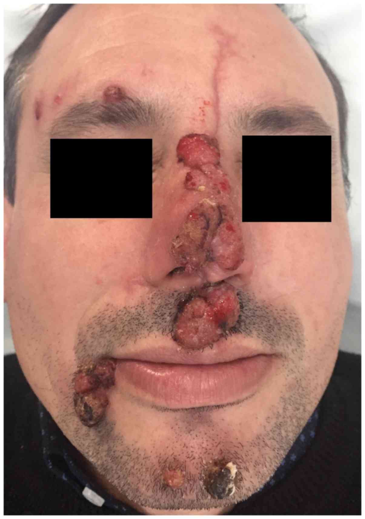
We present the case of a 43-year-old patient, who
had an impressive facial eruption evolving for three weeks,
associated with cervical lymphadenopathy. Consent for publication
was obtained from the patient. Clinical examination revealed
multiple angiomatous erythematous papules and nodules on the nasal
pyramid, philtrum region, right supraorbital area, mental
protuberance and right commissure of the lips, either isolated or
grouped, some of them covered by hematic and honey-like yellowish
crusts (Fig. 1).
The lesions appeared one week after a severe local
trauma produced by a bicycle accident. In the place where the wound
on the nose was sutured, the patient noticed red macules that
evolved very quickly into papules and nodules, followed by the
appearance of other lesions on the face. The oral cavity was normal
and he did not have any other complains. He denied any contact with
cats or travel in other countries. The clinical aspect of the
eruption pointed towards the diagnosis of bacillary
angiomatosis.
Results
The complete blood count, blood chemistry, liver and
renal tests, urine analysis and erythrocyte sedimentation rate were
within normal values and pulmonary radiography and abdominal
ultrasound did not show any anomalies. HIV serology assessed by
ELISA was negative, as well as the screening tests for hepatitis B
and C. A bacterial culture performed from the lesion showed
Staphylococcus aureus colonization.
Histopathological examination revealed in the dermis
a diffuse proliferation of small caliber vascular structures, some
of them dilated, lined by edematous endothelial cells. The
edematous stroma was associated with a polymorphous inflammatory
infiltrate (mostly neutrophils, but also lymphocytes, plasma cells
and histiocytes) dispersed throughout the lesion. Moreover,
derma-epidermal ulceration was present, with a necrotic detritus
with fibrin, leucocytes and small microbial colonies, and positive
Giemsa and Warthin Starry stains. The histopathological findings
were compatible with pyogenic granuloma/bacillary angiomatosis or
verruga peruana. Verruga peruana was excluded because
the patient denied traveling in South America. In vivo
imaging techniques do not provide much support for the diagnosis,
although progress has been made in recent years in some
inflammatory skin diseases (12).
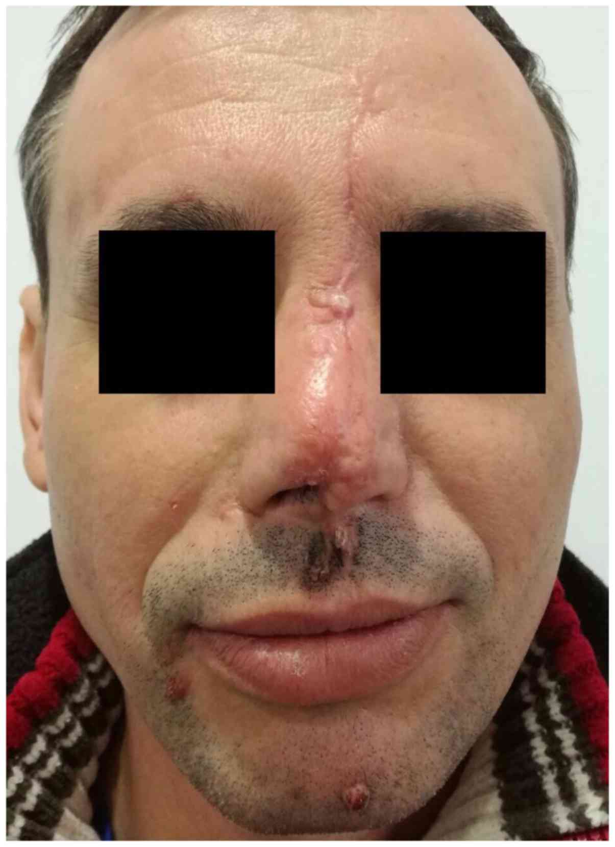
The patient was prescribed clarithromycin 500 mg
twice daily for six weeks. After the first ten days of treatment, a
gradual reduction in size of the lesions was observed. After one
month, there were only two remnant papules on the face that were
shave biopsied (Fig. 2). Considering
the clinical aspect of the lesions, the histopathological findings
and the rapid remission after initiation of the antibiotic
treatment (which is not characteristic for pyogenic granuloma) a
final diagnosis of bacillary angiomatosis was made. In the year of
follow-up, there was no recurrence.
Discussion
Bacillary angiomatosis was first described several
decades before in HIV-positive patients (13) as an opportunistic infection, but it
may also occur in the context of other conditions that may impair
the immune status, such as chronic lymphocytic leukemia (11,14,15),
chemotherapy (16), systemic
corticotherapy (17), solid organ
transplantation (18-20),
hepatitis B infection (3).
Nevertheless, immunocompetent patients can be affected by this
cutaneous disorder (6,17,21-24).
It can also appear in pediatric patients (20,24,25).
The presented patient was HIV, HBV (hepatitis B
virus) and HCV (hepatitis C virus) negative, other causes of
immunosuppression were absent and he was not taking any drugs.
Only a case with lesions on the face resembling
those of the presented patient was found in the literature, that
appeared on the site of a recent burn (6), as well as lesions induced by recent
trauma (3). Moreover, a healthy
child, without any sign of immunodeficiency, developed bacillary
angiomatosis on the face after a traumatic wound, as in the case of
the presented patient (24). In
these cases, the infection could be transmitted by sandflies, ticks
or unknown vehicle (3).
Clinically, the lesions may mimic the aspect of
pyogenic granulomas, Kaposi's sarcoma or verruga peruana,
described as bright red, purple or violaceous vascular papules and
nodules, with sharp demarcation and a chronic evolution over days
to months (7,26,27). The
surface may be friable, with tendency to bleed or it may be covered
by serous and hematic crust (7).
More rarely, the clinical picture may be represented by
subcutaneous nodules or indurated plaques (26-29).
In HIV patients bacillary angiomatosis presents as a large,
subcutaneous tumoral mass (5,30). A
case of bacillary angiomatosis in an immunocompetent patient,
presenting erythematous nodules on the legs which became ulcerated,
has been described (27). The
eruption usually involves the face, the neck, the trunk and the
limbs, but other regions can be affected as well, including the
scalp (22). Bacillary angiomatosis
can also affect the mucous membranes (tarsal and oral mucosa)
(31).
Clinical examination can reveal one single papule or
nodule, but hundreds of lesions can also be present (32). In case of inadequate treatment, the
evolution can be fatal, with a systemic dissemination of the
abscesses within the gastro-intestinal tract, respiratory tract,
brain and bones (5,25,31).
Fever, weight loss, anorexia, malaise and lymphadenopathy may be
associated complaints (31). In case
the abscesses invade the liver or the spleen, B. henselae
can be the first culprit of this disease called ‘bacillary
peliosis’ (4,19).
The histopathological description correlates with
the clinical appearance of the lesions. When pyogenic
granuloma-like lesions are seen, microscopy shows elements that are
more superficially located, characterized by a lobular
proliferation of small vascular structures (capillaries and
venules) and plump endothelial cells, associated with an
inflammatory infiltrate (with neutrophils, lymphocytes and
histiocytes) (18,19,22,26). In
other clinical forms, the cellular content is more dense and deeper
(22,26). The presence of a dense neutrophilic
inflammatory infiltrate and granular amphophilic content may point
towards bacillary angiomatosis (22). Bartonella spp can not be
visualized on conventional hematoxylin-eosin staining, but the
granular amphophilic material corresponds with the presence of dark
bacilli on silver dye (7). Moreover,
microscopy findings may be similar to those of Kaposi's sarcoma,
pyogenic granuloma, angiosarcoma, or insect bite (22). However, in contrast with bacillary
angiomatosis, endothelial cells in Kaposi's sarcoma are spindle,
flattened and, depending on the stage, form dissecting slit-like
vascular spaces, and fascicles with variable erythrocyte
extravasation (33). Pyogenic
granuloma has a lobular architecture, with neutrophils located at
the ulcerated surface of the lesion and without identifiable
organisms.
In several case presentations, the final diagnosis
was based on the demonstration of clustered or solitary bacilli
within or around the vessels on Warthin-Starry silver staining in
histopathological fragment of the lesion (6,17,21).
Other new diagnosis methods for detection of the bacteria, such as
tissue culture, serologic tests and molecular methods (detection of
DNA Bartonella spp by PCR in the blood or tissue) are
available in specialized laboratories (5,31).
Due to financial limitations, in the case of our
patient we could not perform DNA detection of Bartonella
species by PCR and specific serologic tests. Thereby, the final
diagnosis was based on the clinical aspect of the lesions, the
histopathological report and the rapid remission after initiation
of the antibiotic treatment, which excluded other differential
diagnosis.
The appropriate therapy is with oral erythromycin
(500 mg x4/daily) and doxycycline (100 mg twice daily), but several
treatments such as cephalosporins, penicillins, macrolides,
aminoglycosides, rifampin, dapsone, ciprofloxacin, have been tried
with favorable results (19,31,34,35).
We suggest the importance of further studies
regarding treatment strategy in bacillary angiomatosis. There are a
small number of published reports, especially in immunocompetent
patients. Even if oral erythromycin remains the drug of choice for
bacillary angiomatosis, we found some interesting observations: the
response to treatment can range from a spectacular and fast one to
an incomplete and chronic one; lack of response to erythromycin but
adequate response to clarithromycin.
The rapid healing of the eruption in the presented
case also suggest the utility of further studies regarding the
modulation of endothelial cell proliferation irrespective of the
antibacterial potency of the agent used for the treatment.
Macrolides interfere with endothelial cells, immune and
inflammatory cells. Identifying a drug that optimally combines all
these effects may be a goal for other research. Knowing the
reported inhibitory effect of clarithromycin on tumor-induced
angiogenesis, we also highlight the possible beneficial role of
these molecules in the treatment of angioproliferative diseases,
with positive or negative infectious etiology.
A consensus regarding the appropriate period of time
that the treatment should be prescribed is missing, thereby even
though the lesions can disappear within a few days or one-two
months, continuing the therapy for three to six months in order to
prevent recurrences is the best approach, especially in HIV
patients (7,19,31).
In conclusion, the presented case was unique in that
the patient was seronegative for HIV and exhibited extensive
lesions on the face, after local trauma that remitted rapidly with
antibiotic treatment.
Acknowledgements
Not applicable.
Funding
This work is supported by grants of Ministery of
Research and Innovation, CNCS-UEFISCDI, project number
PN-III-P4-ID-PCE-2016-0641, within PNCDI III and CCCDI-UEFISCDI,
project number 61PCCDI⁄2018 PN-III-P1-1.2-PCCDI-2017-0341, within
PNCDI-III.
Availability of data and materials
All patient data are mentioned in the article and
are available from the corresponding author on resonable
request.
Authors' contributions
MB, RIN, GB, TAT, AB, DAI and GT had substantial
contributions to the conception of the work and the acquisition of
data for the work. LN, CGP, SAZ and RTA had substantial
contributions to the analysis and interpretation of data for the
work. All the authors were involved in writing the manuscript and
revised the manuscript critically for important intellectual
content, approved the final version to be published and agreed to
be accountable for all aspects of the work in ensuring that
questions related to the accuracy or integrity of any part of the
work are resolved.
Ethics approval and consent to
participate
Patient consent to participate was obtained before
publication.
Patient consent for publication
Patient consent for the images was obtained before
publication.
Competing interests
The authors declare that they have no competing
interests.
References
|
1
|
Blattner C, Jacobson-Dunlop E, Miller JH
and Elston DM: A case of bacillary angiomatosis in a patient with
pancreatic adenocarcinoma. J Cutan Pathol. 41:277–280.
2014.PubMed/NCBI View Article : Google Scholar
|
|
2
|
Sommer LL, Reboli AC and Heyman WR:
Bacterial diseases. In: Dermatology. Bolognia JL, Schaffer JV and
Cerroni L (eds). 4th edition. Elsevier, USA. pp1282–1285. 2017.
|
|
3
|
Kaçar N, Taşli L, Demirkan N, Ergin C and
Ergin S: HIV-negative case of bacillary angiomatosis with chronic
hepatitis. B. J Dermatol. 37:722–725. 2010.PubMed/NCBI View Article : Google Scholar
|
|
4
|
Millet CR, Halpern AV, Reboli AC and
Heymann WR: Bacterial diseases. In: Dermatology. Bolognia JL,
Jorizzo JL and Schaffer JV (eds). 3rd edition. Elsevier Saunders,
USA. pp1208–1210. 2012.
|
|
5
|
Markowicz M, Käser S, Müller A, Lang G,
Lang S, Mayerhöfer M, Stanek G and Rieger A: Bacillary angiomatosis
presenting with facial tumor and multiple abscesses: A case report.
Medicine (Baltimore). 95(e4155)2016.PubMed/NCBI View Article : Google Scholar
|
|
6
|
Karakas M: Baba, VAksungur VL, Homan S,
Memisoglu HR and Uguz A: Bacillary angiomatosis on a region of
burned skin in a immunocompetent patient. Br J Dermatol.
143:609–611. 2000.PubMed/NCBI View Article : Google Scholar
|
|
7
|
Nosal JM: Bacillary angiomatosis,
cat-scratch disease, and bartonellosis: What's the connection? Int
J Dermatol. 36:405–411. 1997.PubMed/NCBI View Article : Google Scholar
|
|
8
|
Zangwill KM, Hamilton DH, Perkins BA,
Regnery RL, Plikaytis BD, Hadler JL, Cartter ML and Wenger JD: Cat
scratch disease in Connecticut. Epidemiology, risk factors, and
evaluation of a new diagnostic test. N Engl J Med. 329:8–13.
1993.PubMed/NCBI View Article : Google Scholar
|
|
9
|
Chomel BB, Kasten RW, Floyd-Hawkins K, Chi
B, Yamamoto K, Roberts-Wilson J, Gurfield AN, Abbott RC, Pedersen
NC and Koehler JE: Experimental transmission of Bartonella
henselae by the cat flea. J Clin Microbiol. 34:1952–1956.
1996.PubMed/NCBI
|
|
10
|
Foucault C, Brouqui P and Raoult D:
Bartonella quintana characteristics and clinical management.
Emerg Infect Dis. 12:217–223. 2006.PubMed/NCBI View Article : Google Scholar
|
|
11
|
Fulchini R, Bloemberg G and Boggian K:
Bacillary angiomatosis and bacteremia due to Bartonella
quintana in a patient with chronic lymphocytic leukemia. Case
Rep Infect Dis. 2013(694765)2013.PubMed/NCBI View Article : Google Scholar
|
|
12
|
Căruntu C, Boda D, Căruntu A, Rotaru M,
Baderca F and Zurac S: In vivo imaging techniques for psoriatic
lesions. Rom J Morphol Embryol. 55 (Suppl):1191–1196.
2014.PubMed/NCBI
|
|
13
|
Stoler MH, Bonfiglio AT, Steigbigel RT and
Pereira M: An atypical subcutaneous infection associated with
acquired immune deficiency syndrome. Am J Clin Pathol. 80:714–718.
1983.PubMed/NCBI View Article : Google Scholar
|
|
14
|
Török L, Virágh SZ, Borka I and Tápai M:
Bacillary angiomatosis in a patient with lymphocytic leukaemia. Br
J Dermatol. 130:665–668. 1994.PubMed/NCBI View Article : Google Scholar
|
|
15
|
Milde P, Brunner M, Borchard F, Südhoff T,
Burk M, Zumdick M, Goerz G and Ruzicka T: Cutaneous bacillary
angiomatosis in a patient with chronic lymphocytic leukemia. Arch
Dermatol. 131:933–936. 1995.PubMed/NCBI
|
|
16
|
Myers SA, Prose NS, Garcia JA, Wilson KH,
Dunsmore KP and Kamino H: Bacillary angiomatosis in a child
undergoing chemotherapy. J Pediatr. 121:574–578. 1992.PubMed/NCBI View Article : Google Scholar
|
|
17
|
Schwartz RA, Gallardo MA, Kapila R, Gascón
P, Herscu J, Siegel I and Lambert WC: Bacillary angiomatosis in an
HIV seronegative patient on systemic steroid therapy. Br J
Dermatol. 135:982–987. 1996.PubMed/NCBI View Article : Google Scholar
|
|
18
|
Moulin C, Kanitakis J, Ranchin B, Chauvet
C, Gillet Y, Morelon E and Euvrard S: Cutaneous bacillary
angiomatosis in renal transplant recipients: report of three new
cases and literature review. Transpl Infect Dis. 14:403–409.
2012.PubMed/NCBI View Article : Google Scholar
|
|
19
|
Orsag J, Flodr P, Melter O, Tkadlec J,
Sternbersky J, Hruby M, Klicova A, Zamboch K, Krejci K and Zadrazil
J: Cutaneous bacillary angiomatosis due to Bartonella
quintana in a renal transplant recipient. Transpl Int.
28:626–631. 2015.PubMed/NCBI View Article : Google Scholar
|
|
20
|
Rostad CA, McElroy AK, Hilinski JA,
Thompson MP, Drew CP, Denison AM, Zaki SR, Mahle WT, Rogers J,
Abramowsky CR, et al: Bartonella henselae-mediated disease
in solid organ transplant recipients: two pediatric cases and a
literature review. Transpl Infect Dis. 14:E71–E81. 2012.PubMed/NCBI View Article : Google Scholar
|
|
21
|
Schwartz RA, Nychay SG, Janniger CK and
Lambert WC: Bacillary angiomatosis: Presentation of six patients,
some with unusual features. Br J Dermatol. 136:60–65.
1997.PubMed/NCBI
|
|
22
|
Kayaselçuk F, Ceken I, Bircan S and Tuncer
I: Bacillary angiomatosis of the scalp in a human immunodeficiency
virus-negative patient. J Eur Acad Dermatol Venereol. 16:612–614.
2002.PubMed/NCBI View Article : Google Scholar
|
|
23
|
Zarraga M, Rosen L and Herschthal D:
Bacillary angiomatosis in an immunocompetent child: A case report
and review of the literature. Am J Dermatopathol. 33:513–515.
2011.PubMed/NCBI View Article : Google Scholar
|
|
24
|
Turgut M, Alabaz D, Karakaş M, Kavak M,
Aksaray N, Alhan E, Cevlik F and Tuncer I: Bacillary angiomatosis
in an immunocompetent child with a grafted traumatic wound. J
Dermatol. 31:844–847. 2004.PubMed/NCBI View Article : Google Scholar
|
|
25
|
Rodriguez O, Campbell LR, Bacha JM and
Kovarik CL: Successful treatment of bacillary angiomatosis with
oral doxycycline in an HIV-infected child with skin lesions
mimicking Kaposi sarcoma. JAAD Case Rep. 2:77–79. 2016.PubMed/NCBI View Article : Google Scholar
|
|
26
|
Webster GF, Cockerell CJ and Friedman-Kien
AE: The clinical spectrum of bacillary angiomatosis. Br J Dermatol.
126:535–541. 1992.PubMed/NCBI View Article : Google Scholar
|
|
27
|
Karakaş M, Baba M, Homan S, Akman A, Acar
MA, Memişoğlu HR and Gümürdülü D: A case of bacillary angiomatosis
presenting as leg ulcers. J Eur Acad Dermatol Venereol. 17:65–67.
2003.PubMed/NCBI View Article : Google Scholar
|
|
28
|
Sala M, Font B, Sanfeliu I, Quesada M,
Ponts I and Segura F: Bacillary angiomatosis caused by
Bartonella quintana. Ann NY Acad Sci. 1063:302–307.
2005.PubMed/NCBI View Article : Google Scholar
|
|
29
|
Schlüpen EM, Schirren CG, Hoegl L,
Schaller M and Volkenandt M: Molecular diagnosis of deep nodular
bacillary angiomatosis and monitoring of therapeutic success. Br J
Dermatol. 136:747–751. 1997.PubMed/NCBI
|
|
30
|
Wanat KA, Reid E, Kamiyango W,
El-Mallawany NK and Kovarik CL: Tumoral bacillary angiomatosis in a
child with human immunodeficiency virus. JAMA Dermatol.
150:1015–1016. 2014.PubMed/NCBI View Article : Google Scholar
|
|
31
|
Lopes L, Borges-Costa J, Janeiro N, Neves
D, Soares AlmeidaL and Filipe P: Bacillary angiomatosis in a
HIV-positive patient with poor adherence to antiretroviral therapy.
Acta Dermatovenerol Croat. 22:294–297. 2014.PubMed/NCBI
|
|
32
|
Mejía F and Seas C: Images in clinical
tropical medicine: Bacillary angiomatosis. Am J Trop Med Hyg.
91(439)2014.PubMed/NCBI View Article : Google Scholar
|
|
33
|
Grayson W and Pantanowitz L: Histological
variants of cutaneous Kaposi sarcoma. Diagn Pathol.
3(31)2008.PubMed/NCBI View Article : Google Scholar
|
|
34
|
Rolain JM, Brouqui P, Koehler JE, Maguina
C, Dolan MJ and Raoult D: Recommendations for treatment of human
infections caused by Bartonella species. Antimicrob Agents
Chemother. 48:1921–1933. 2004.PubMed/NCBI View Article : Google Scholar
|
|
35
|
Prutsky G, Domecq JP, Mori L, Bebko S,
Matzumura M, Sabouni A, Shahrour A, Erwin PJ, Boyce TG, Montori VM,
et al: Treatment outcomes of human bartonellosis: A systematic
review and meta-analysis. Int J Infect Dis. 17:e811–e819.
2013.PubMed/NCBI View Article : Google Scholar
|