Introduction
Direct death from chest trauma accounts for 20–25%
of all traumatic deaths, and chest trauma is the second leading
cause of death from trauma (1).
Multiple rib fracture patients, especially with flail chest, often
suffer severe pain, chest wall softening, abnormal breathing and
severe lung contusion and laceration, that may lead to respiratory
failure, which is life threatening and often requires thoracic
surgery (2). In recent years, the
treatment of rib fractures with open reduction and internal
fixation has achieved satisfactory results, and some surgical
indications have reached consensus in the field (3–8); a
number of scholars and medical centers have carried out
retrospective studies, which have demonstrated the practicality and
cost-effectiveness of rib fixation in patients with flail chest,
including the small incidence of pulmonary complications, the short
ICU mechanical ventilation time, the reduction in bed rest and use
of large doses of analgesics and antibiotics, the indirect
reduction of digestive tract inhibition, the low mortality rate and
low hospitalization costs. Conventional rib fracture open reduction
and internal fixation surgery causes great trauma, therefore, in
recent years, experts have explored a variety of minimally invasive
surgeries for flail chest (9).
Small incision rib fracture open reduction
and internal fixation
Since thoracic surgery often involves multiple ribs,
the conventional rib fracture open reduction and internal fixation
requires a large incision to obtain satisfactory exposure. Part of
the chest wall muscles, blood vessels and nerves (long thoracic and
thoracodorsal nerves) are usually damaged during the procedure,
resulting in high infection rate of the incision and postoperative
dysfunction, such as limited upper limb, shoulder and back
function, and numbness of the affected side of the chest for a long
time. Therefore, the damage of conventional surgical methods on the
muscles and nerves limit the development of such surgical
technique. Consequently, the rational choice of the incision plays
an important role in the rehabilitation process of the patients.
Chinese scholars have divided the different parts of the rib
fracture into subdivisions based on the anatomical features of the
thorax.
Taylor et al (10) summarized the surgical approaches as:
i) Standard posterolateral thoracotomy: Through this approach the
posterior, lateral, and posterolateral sides of the rib fracture
ends are accessible. Standard posterolateral thoracotomy can also
be extended laterally or diagonally forward. The incision is
curved, often reflecting the anterior ribs, and the fracture of the
anterior rib can also be exposed by a percutaneous-assisted small
incision. ii) ‘Auscultation triangle’ incision: The retractor is
used to retract the muscle tissue and the scapula to minimize the
damage caused by the operation; the deep muscle layer is sutured
and the drainage placed to prevent ‘dead cavity’ and postoperative
hematoma formation. iii) Trans-axillary incision: It is more
suitable for anterior lateral thoracic wall rib fixation. Some
muscle tissues may be disconnected as needed, the long thoracic
nerve should be protected. iv) Under the breast or the pectoral
muscles: The incision is along the lower edge of the breast. The
breast tissue and the pectoral muscles are pulled up, and the ribs
and costal cartilage of the anterior chest wall are exposed. The
incision can be extended to the posterior lateral of the axillary,
or extended to the standard posterolateral thoracic incision.
According to the local anatomical features of different regions and
rib fractures, the incision is selected and designed, the
anatomical muscle space and muscle fibers are freed, and the
peripheral nerves, blood vessels, surgical sites, surgical
incisions and corresponding blood vessels, muscles and nerves are
protected (Table I).
 | Table I.Detailed partition of fractures. |
Table I.
Detailed partition of fractures.
| Partition | Position | Rib range | Incision
position | Muscle involved | Nerve involved |
|---|
| 1 | Supine | 2–6 front ribs | Curved incision of
the medial edge of the pectoralis major | Pectoralis major |
|
| 2 | Supine/lateral | 7–10 front ribs | Rib bow incision | Extra-abdominal
oblique |
|
| 3 | Lateral | 2–6 side front
ribs | Lateral marginal
incision of the pectoralis major muscle (anterior axillary line
incision) | Pectoralis major | Long thoracic
nerve |
|
|
| 3–10 side ribs | Midaxillary line
incision | Front serratus | Thoracic dorsal
nerve |
|
|
| 3–10 side rear
ribs | Posterior axillary
line incision | Latissimus dorsi | Thoracic dorsal
nerve |
| 4 | Prone | 2–5 rear ribs | Scapular medial edge
incision | Trapezius
rhomboid |
|
| 5 | Prone | 5–8 rear ribs | Auscultation triangle
incision | Trapezius, latissimus
dorsi |
|
| 6 | Prone | 5–12 paravertebral
ribs | Paravertebral
longitudinal incision | Trapezius, latissimus
dorsi, erector spine |
|
| 7 | Prone/lateral | 7–12 rear ribs | Posterolateral
oblique incision (inclined incision along the latissimus dorsi
fibers) | Latissimus dorsi,
serratus anterior and posterior |
|
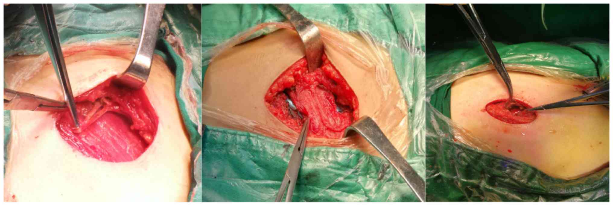
The specific surgical procedures are as follows: The
patient is treated with general anesthesia. The specific position
of the operation and the position of the incision are determined by
the location of the fracture. The skin and subcutaneous tissue are
removed layer by layer, and the tissue is completely freed. The
peripheral blood vessels and nerves are protected. The protection
of the long thoracic nerve, thoracodorsal, intercostobrachial and
intercostal nerve cutaneous branches and thoracic and thoracodorsal
vessels is required (Fig. 1). The
intermuscular space or muscle fibers are followed to the deep part,
exposing the ribs and fracture ends. The exfoliation of the
intercostal muscle is performed on the upper edge of the rib. When
the exfoliation is performed, the periosteum is retained as much as
possible to promote the healing of the fracture. The thoracic
cavity can be entered through the exfoliation point for internal
fixation surgery, thoracic cavity exploration, and hemothorax
removal. If fixed with a rib-embracing device, the intercostal
nerve and vessels are freed, the fracture ends are repositioned,
and the fixation is placed. If the locking plate is used for
fixation, the fracture end can be directly restored, and the
locking plate can be directly placed. Two screws are placed and
locked at the two ends of the fracture, and the titanium alloy rib
locking plate is firmly fixed. Satisfactory surgical results can be
obtained. Exposure limits are often applied to <5 consecutive
rib fractures.
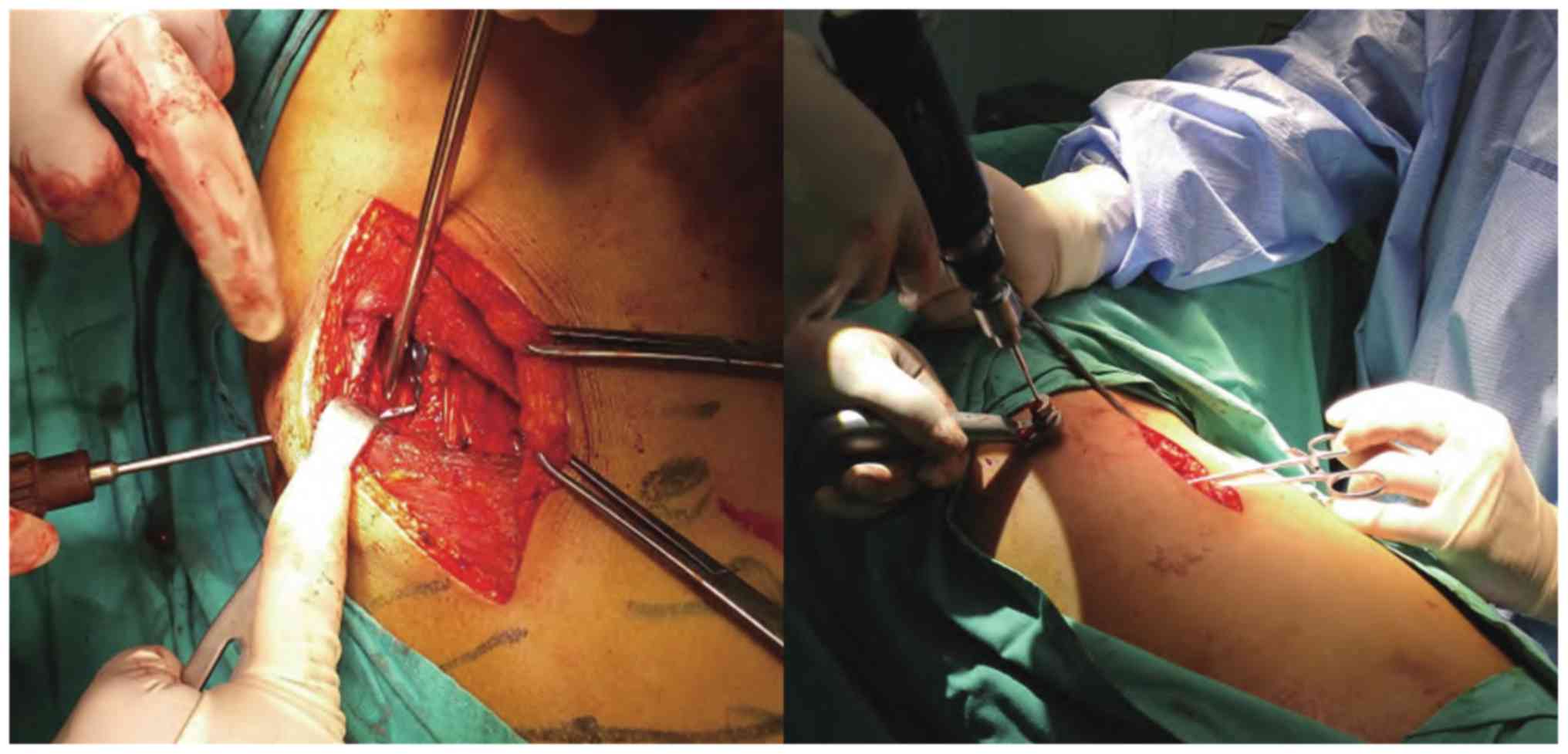
Tunnel-type titanium alloy rib locking plate
rib fracture internal fixation
During the operation, the center position of
multiple rib fractures was selected according to the location and
shape of the fracture (the approximate center point of the maximum
lateral and longitudinal line of the multiple lateral rib fracture
area) (Fig. 2).
For the multiple rib fractures away from the center
of the incision, the tunnel-type rib fracture internal fixation
system is used for fixation. If the fracture is obviously
dislocated, the periosteum is cut along the fracture line and the
reduction clamp is used to pull the reduction. The internal
fixation material is selected from the Synthes MatrixRIB titanium
alloy rib locking plate fixing system (Johnson & Johnson). The
rib cage is selected according to the shape of the rib. The rib
bone plate is drilled and fixed with 5–6 pieces of locking pin with
a length of 8 mm and a diameter of 3 mm, and the fracture end is
carefully avoided. The procedure has the advantages of small
trauma, simple operation, safety, liable fixation, good tissue
compatibility and less complications. It is suitable for rib
fractures in special parts of costal cartilage and paraspinal,
especially ideal for patients with complications, such as floating
thoracic wall, flail chest and blood pneumothorax.
Video-assisted thoracoscopic assisted
minimally invasive rib fracture open reduction and internal
fixation
Video-assisted thoracoscopic techniques have been
applied to thoracic exploration and hemostasis in the treatment of
chest trauma, which have become necessary technical means for the
treatment of chest trauma (11,12).
Video-assisted thoracoscopic surgery can observe the
place where the chest wall floating is more serious, and guide the
reasonable choice of surgical incision. During the operation, the
thoracic cavity, lung and diaphragm muscle injury can be detected
and treated in time. Thoracoscopy is used to remove the
functionalized hemothorax to maximize the space for lung
recruitment. However, there are few reports on the application of
open reduction and internal fixation for rib fractures. The reason
is that the bony thorax is tightly combined with the soft tissue of
the chest wall. There is no conventional internal distraction
surgical instrument to provide operation space for surgery and few
surgical instruments are suitable for minimally invasive rib
fracture internal fixation.
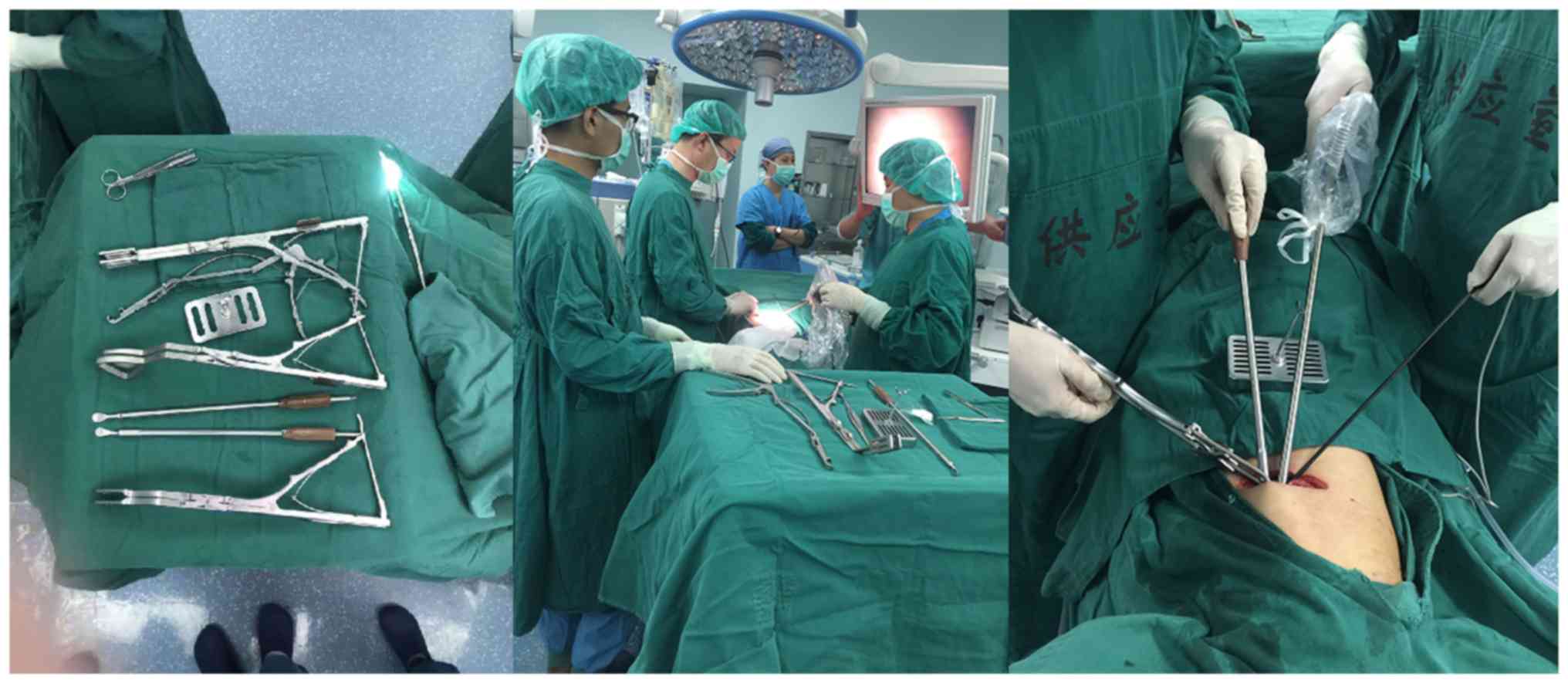
Tunnel-type thoracoscopic rib fracture
fixation
Positive results of the application of endoscopic
techniques have been reported. The author team developed and
designed a surgical device that can be used to open the muscular
chest wall from inside, and the rib plate clamp for the endoscope,
the periosteal free device, and the endoscope bracket (Fig. 3). The chest wall expander can open
the thoracic and muscular thorax, providing an operating space,
making it possible to apply the video-assisted thoracoscopic
technique to the open reduction and internal fixation of the rib
fracture. The endoscopic periosteal free device can absorb a large
amount of smoke during most of the operation to ensure the clarity
of the surgical field of view. The rib plate clamp for the
laparoscopic plate can push the rib fixation plate along the rib
direction and buckle on both ends of the anatomical reduction rib
fracture. At the same time, the special rib clamp is used to fix
the rib fixing plate to the fracture end surface. Due to the small
operation space, the existing clamp can flexibly adjust the angle,
which is suitable for minimally invasive surgery. The skeletal
thoracic tunnel endoscopic rib internal fixation operation is
realized, which can effectively reduce the length of the incision,
reduce the muscle damage of the chest wall, fully reveal the
surgical field, reduce the surgical trauma, and save the operation
time and manpower. Thus, this technique has obvious advantages
compared with conventional incision surgery (13).
The specific surgical procedure is as follows:
General anesthesia by single lumen cannula, the patient's surgical
position is designed according to the rib fracture site and the
number of fractures (supine position, prone position, or left and
right lateral position), and if necessary, the position can be
changed during surgery; the 3D reconstruction data of the ribs, the
surface positioning measuring scale and the CT surface positioning
technique of the CT fractures are combined to mark the surface
location of the fracture end. The surgical incision is located in
combination with the anatomical distribution of the chest wall
muscles of the fracture site, generally taking into consideration
the central position of the multiple rib fracture and the chest
wall muscle space (commonly used approaches: Pectoralis major
approach, breast inferior margin approach, auscultation triangle
approach, erector spine approach and axillary approach).
The incision can be transverse, longitudinal or
oblique, ~3–5 cm long, free subcutaneous and muscular layer,
dissected along the muscle space and muscle texture to the skeletal
thoracic surface, avoiding breaking muscles; to cut part of the
muscle attachment point along the bony thoracic surface; a special
chest wall opener is used to open the surgical instrument to prop
up the muscular thorax in a direction perpendicular to the skeletal
thorax (the chest wall opener has completed the application of the
National utility model patent, application no. 201721524911.X). To
separate the skeletal thoracic and muscular thoracic the
thoracoscope is inserted, the necessary muscular thorax is freed
under the guidance of the endoscope, and form a temporary chest
wall tunnel. After reduction of the fracture of the distal
incision, the rib fixation plate should be buckled at the ends of
the rib fractures on the anatomical reduction along the direction
of the ribs, and the rib fixation plates were circum ferentially
fixed to the fracture end surface using special clamps. The
internal fixation material can be determined according to the
fracture site of the patient and the economic conditions of the
patient. For example, the rib cage locking plate can be combined
with the rib minimally invasive plate system (MatrixRIB-MIPO;
Johnson & Johnson). Compared with conventional surgical
video-assisted thoracoscopic surgery, this procedure can provide
good illumination, visually and comprehensively explore the tissue
structure around the fracture end, determine the specific location,
number and severity of the rib fracture, and effectively avoid
damage to the chest wall muscle and intercostal vessel and nerve,
significantly reducing postoperative complications. Therefore, it
is worthy of clinical promotion.
The Su total thoracoscopic rib fracture bone
plate nail intrathoracic implantation and fixation
Thoracoscopic implanted NiTi memory alloy rib plate
intrathoracic implantation and thoracoscopic absorbable rib nail
fixation were performed.
The indications for laparoscopic surgery were as
follows: i) Patients with fractures in the F-zone, i.e.,
non-occlusion of the axillary, anterior and posterior and lateral
chest walls; ii) single or multiple fractures or simultaneous
laparoscopic repair of vascular injuries. Surgical fractures should
be in the non-occluded area of the axillary, anterior and posterior
and lateral chest walls. The Su total thoracoscopic rib fractures
and bone plate fixation in the thoracic cavity are technically
feasible in selected patients, however, they are not replaceable in
severe cases, such as conventional thoracotomy for open thoracic
surgery, which requires further improvement. Pieracci et al
(14) reported the use of locking
plate fixation in the thoracic cavity to fix the ribs in the
thoracic cavity. The advantage is that the thoracic cavity can be
explored while fixing the ribs, however, the operation in the
thoracic cavity is limited, the fracture reduction is difficult,
and has high requirements.
Minimally invasive Nuss surgery for patients
with severe flail chest with sternal fracture
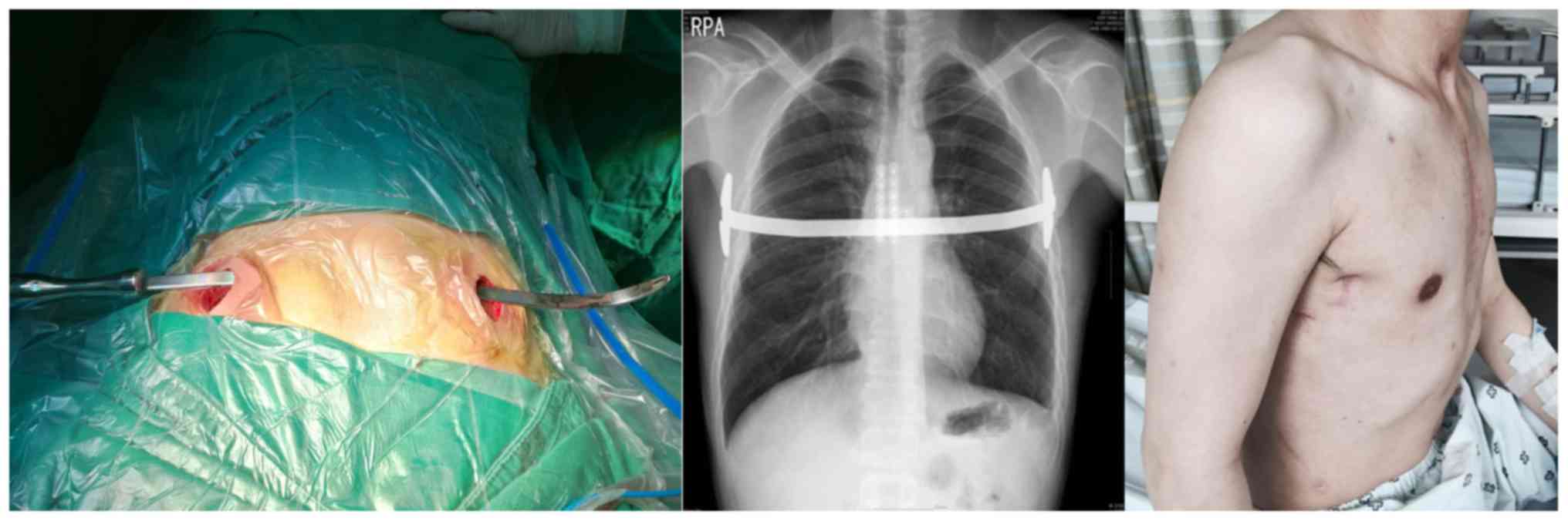
Lee and Kang (15)
reported the use of Nuss method for the treatment of bilateral
vertebral fractures with sternal fractures and sacral thoracotomy
(Fig. 4); combination of sternal
fractures and severe flail chest is a clinically life-threatening
disease, and the conventional sternal approach uses a metal plate
to stabilize the chest wall, which does not achieve the effect of
stabilizing the chest wall. Nuss surgery is used to support the
collapse of the chest wall from the posterior sternum. The chest
wall is stabilized after surgery. The operation uses the midline
incision on both sides of the chest for ~3 cm. A subcutaneous
tunnel is established between the 4th intercostal space. The Kelly
forceps is placed under the sternum and above the mediastinum. The
Kelly forceps is placed from one side of the chest wall incision to
the opposite side, and then 36-cm pectus rod is pre-plasticized to
form a symmetrical arch, pointing to the opposite side along the
subcutaneous tunnel, rotating the Pectus rod, and then the
collapsed thorax is propped up.
This operation is suitable for patients with severe
flail chest combined with thoracic fracture with short operative
time and small surgical injury. It has certain advantages in
thoracic trauma rescue surgery, however, it has poor effect on
local rib fracture reduction. The risk for postoperative delayed
thoracic hemorrhage is high. Unless the patient feels unwell after
surgery, the orthopedic plate left in the body will not be removed.
If the patient has obvious chest discomfort or demands, the
orthopedic plate will be removed 1 year after the thorax is
stabilized.
Introduction of internal fixation materials
suitable for different surgical methods
At present, the commonly used fixing materials in
China include intramedullary fixation, embracing fixation and steel
plate screw fixation, which are respectively represented by
absorbable rib nails, nickel-titanium or memory alloy plates, and
MatrixRIB titanium plates, each with advantages and disadvantages
(16,17). Among them, the nickel-titanium alloy
embracing fixation is a simple operation procedure, which does not
require excessive peeling of the periosteum during surgery, and is
suitable for patients with a large number of fractures.
Because the embracing device easily compresses the
intercostal nerve, there is a possibility that the pain cannot be
relieved after surgery, and fractures near the rib cartilage and
the spine are not suitable for application; pure titanium alloy
embracing device requires fully peeling the periosteum to
accurately reset the fracture end, and the fixation effect is
exact, although the operation is complicated. As a result, it is
not suitable for rib fractures far from the incision, or for rib
fractures near costal cartilage and the spine. Titanium alloy rib
locking plate for internal fixation of rib fractures has a good
therapeutic effect for some cases that are difficult to reset or
with multiple fractures (18). Small
incision or tunnel laparoscopic surgery can be combined with the
rib minimally invasive plate system (MatrixRIB-MIPO), which can
accurately shape and firmly fix rib fractures in multiple
locations, especially for fractures near costal cartilage and the
spine (19). The absorbable rib nail
or plate is made of polylactic acid polymer, which has been
successfully applied to traumatic flail chest and non-traumatic
thoracotomy (20). It has not been
widely clinically popularized because of its limitations in
biomechanics and degradation rate.
Conclusion
At present, surgical treatment of multiple rib
fractures has been accepted by an increasing number of medical
workers (21–23), and surgery tends to be minimally
invasive. Different rib fixing materials have their own advantages
and disadvantages. Nickel-titanium alloy embracing fixator is a new
material with memory function at temperature. It can be stretched
and deformed at low temperature, automatically restored to its
original state under body temperature, without secondary surgery.
It has good histocompatibility, strong corrosion resistance, less
immune rejection, and does not affect magnetic resonance imaging.
The pure titanium claw-shaped bone plate has high strength, good
histocompatibility, and no need for secondary surgery. The
advantages are similar to those of the nickel-titanium alloy
embracing fixator, however, special tools are required for fixing
and removing, and a large space is required for operation. The
absorbable intramedullary nail has a slightly higher flexural
strength than the human cortical bone, and is completely degraded
in 3–5 years. The effective support time is 8–10 months. It does
not require secondary surgery, and the fracture healing can be
stimulated during the decomposition process. However, there are
certain deficiencies in clinical application. The stability of the
internal fixation is slightly worse than that of the
nickel-titanium alloy embracing fixator and the pure titanium
claw-shaped bone plate. For the fractured wedge or comminuted
fracture, it is usually not fixed. The development and clinical
application of absorbable rib fixation materials will be a research
hotspot in the field of chest trauma treatment. The clinical
promotion of absorbable materials in multiple rib fracture surgery
will make the surgery even less invasive, reducing surgical
complications (24,25).
3D printing technology has been widely used in
various medical departments, such as the production of personalized
artificial prostheses and built-in objects (26,27);
especially for some complex rib fractures, 3D printing technology
is adopted. According to the rib fracture reconstruction model one
day before surgery, the corresponding rib internal fixation
materials and models, and individualized plasticity are selected to
complete accurate preoperative planning; thereby reducing or even
eliminating the internal fixation shaping time in tunnel surgery,
further reducing the surgical injury of the patient. The
development of surgical instruments specially used to open the
musculoskeletal thoracic space provides an operating space for
laparoscopic surgery, opening up a new range of clinical use of
thoracoscopic surgery.
In conclusion, with in-depth understanding of the
minimally invasive concept, the accumulation of clinical experience
and the unremitting exploration of technology, the continuous
improvement of fixed materials and surgical instruments for
minimally invasive surgery with less trauma and less operation time
will be used clinically to achieve a satisfactory clinical
outcome.
Acknowledgements
Not applicable.
Funding
No funding was received.
Availability of data and materials
Not applicable.
Authors' contributions
HX, DZ, JL, ZS and DW were involved in the
conception and design of this study. LD, PZ, YZ and XL were
responsible for the literature research and drafted the manuscript.
DZ and DW made revisions from critical perspective for important
intellectual content. The final version was read and approved by
all authors.
Ethics approval and consent to
participate
Not applicable.
Patient consent for publication
Not applicable.
Competing interests
The authors declare that they have no competing
interests.
References
|
1
|
Ho XN, Wee IJ, Syn N, Harrison M, Wilson L
and Choong AM: The endovascular repair of blunt traumatic thoracic
aortic injury in Asia: A systematic review and meta-analysis.
Vascular. 27:213–223. 2019. View Article : Google Scholar : PubMed/NCBI
|
|
2
|
Brasel KJ, Moore EE, Albrecht RA, deMoya
M, Schreiber M, Karmy-Jones R, Rowell S, Namias N, Cohen M, Shatz
DV, et al: Western trauma association critical decisions in trauma:
Management of rib fractures. J Trauma Acute Care Surg. 82:200–203.
2017. View Article : Google Scholar : PubMed/NCBI
|
|
3
|
Pieracci FM, Majercik S, Ali-Osman F, Ang
D, Doben A, Edwards JG, French B, Gasparri M, Marasco S, Minshall
C, et al: Consensus statement: Surgical stabilization of rib
fractures rib fracture colloquium clinical practice guidelines.
Injury. 48:307–321. 2017. View Article : Google Scholar : PubMed/NCBI
|
|
4
|
Bemelman M, de Kruijf MW, van Baal M and
Leenen L: Rib fractures: To fix or not to fix? An evidence-based
algorithm. Korean J Thorac Cardiovasc Surg. 50:229–234. 2017.
View Article : Google Scholar : PubMed/NCBI
|
|
5
|
Kocher GJ, Sharafi S, Azenha LF and Schmid
RA: Chest wall stabilization in ventilator-dependent traumatic
flail chest patients: Who benefits? Eur J Cardiothorac Surg.
51:696–701. 2017.PubMed/NCBI
|
|
6
|
Farquhar J, Almarhabi Y, Slobogean G,
Slobogean B, Garraway N, Simons RK and Hameed SM: No benefit to
surgical fixation of flail chest injuries compared with modern
comprehensive management: Results of a retrospective cohort study.
Can J Surg. 59:299–303. 2016. View Article : Google Scholar : PubMed/NCBI
|
|
7
|
Uchida K, Nishimura T, Takesada H, Morioka
T, Hagawa N, Yamamoto T, Kaga S, Terada T, Shinyama N, Yamamoto H,
et al: Evaluation of efficacy and indications of surgical fixation
for multiple rib fractures: A propensity-score matched analysis.
Eur J Trauma Emerg Surg. 43:541–547. 2017. View Article : Google Scholar : PubMed/NCBI
|
|
8
|
Pieracci FM, Lin Y, Rodil M, Synder M,
Herbert B, Tran DK, Stoval RT, Johnson JL, Biffl WL, Barnett CC, et
al: A prospective, controlled clinical evaluation of surgical
stabilization of severe rib fractures. J Trauma Acute Care Surg.
80:187–194. 2016. View Article : Google Scholar : PubMed/NCBI
|
|
9
|
de Campos JRM and White TW: Chest wall
stabilization in trauma patients: Why, when, and how? J Thorac Dis.
10 (Suppl 8):S951–S962. 2018. View Article : Google Scholar : PubMed/NCBI
|
|
10
|
Taylor BC, French BG and Fowler TT:
Surgical approaches for rib fracture fixation. J Orthop Trauma.
27:e168–e173. 2013. View Article : Google Scholar : PubMed/NCBI
|
|
11
|
Berninger MT, Kellermann F, Woltmann A,
Bühren V and Lang M: Single-port VATS-assisted internal fixation of
serial rib fractures. Unfallchirurg. 121:335–338. 2018.(In German).
View Article : Google Scholar : PubMed/NCBI
|
|
12
|
Fraser SF, Tan C, Kuppusamy MK, Gukop P
and Hunt IJ: The role of a video-assisted thoracic approach for rib
fixation. Eur J Trauma Emerg Surg. 43:185–190. 2017. View Article : Google Scholar : PubMed/NCBI
|
|
13
|
Xia H, Zhu P, Li J, Zhu D, Sun Z, Deng L,
Zhang Y and Wang D: Thoracoscope combined with internal support
system of chest wall in open reduction and internal fixation for
multiple rib fractures. Exp Ther Med. 16:4650–4654. 2018.PubMed/NCBI
|
|
14
|
Pieracci FM, Johnson JL, Stovall RT and
Jurkovich GJ: Completely thoracoscopic, intra-pleural reduction and
fixation of severe rib fractures. Trauma Case Rep. 1:39–43. 2015.
View Article : Google Scholar : PubMed/NCBI
|
|
15
|
Lee SK and Kang K: Nuss procedure for
surgical stabilization of flail chest with horizontal sternal body
fracture and multiple bilateral rib fractures. J Thorac Dis.
8:E390–E392. 2016. View Article : Google Scholar : PubMed/NCBI
|
|
16
|
Klein GT, Lu Y and Wang MY: 3D printing
and neurosurgery - ready for prime time? World Neurosurg.
80:233–235. 2013. View Article : Google Scholar : PubMed/NCBI
|
|
17
|
Nolasco-de lRAL, Mosiñoz-Montes R,
Matehuala-García J, Román-Guzmán E, Quero-Sandoval F and
Reyes-Miranda AL: Unstable thorax fixation with bioabsorbable
plates and screws. Presentation of some cases. Cir Cir. 83:23–28.
2015.PubMed/NCBI
|
|
18
|
Benian AS, Pushkin SIu, Syzrantsev IuV and
Kameev IR: Osteosyntesis of ribs using the technology ‘matrix rib’
in treatment of victims with multiple float rib fractures. Vestn
Khir Im I I Grek. 172:78–79. 2013.(In Russian). PubMed/NCBI
|
|
19
|
Vana PG, Neubauer DC and Luchette FA:
Contemporary management of flail chest. Am Surg. 80:527–535.
2014.PubMed/NCBI
|
|
20
|
Gao E, Li Y, Zhao T, Guo X, He W, Wu W,
Zhao Y and Yang Y: Simultaneous surgical treatment of sternum and
costal cartilage fractures. Ann Thorac Surg. 107:e119–e120. 2019.
View Article : Google Scholar : PubMed/NCBI
|
|
21
|
Galos D, Taylor B and McLaurin T:
Operative fixation of Rib fractures indications, techniques, and
outcomes. Bull Hosp Jt Dis (2013). 75:15–20. 2017.PubMed/NCBI
|
|
22
|
Tarng YW, Liu YY, Huang FD, Lin HL, Wu TC
and Chou YP: The surgical stabilization of multiple rib fractures
using titanium elastic nail in blunt chest trauma with acute
respiratory failure. Surg Endosc. 30:388–395. 2016. View Article : Google Scholar : PubMed/NCBI
|
|
23
|
Dehghan N: Challenges in plate fixation of
chest wall injuries. Injury. 49 (Suppl 1):S39–S43. 2018. View Article : Google Scholar : PubMed/NCBI
|
|
24
|
Morodomi Y, Okamoto T, Tagawa T, Shoji F,
Katsura M, Fujishita T, Fujiyoshi T, Akahoshi T, Yasuda M and
Maehara Y: A novel method of using bioabsorbable materials for the
surgical repair of flail chest. J Trauma Acute Care Surg.
81:984–987. 2016. View Article : Google Scholar : PubMed/NCBI
|
|
25
|
Nolasco-de la Rosa AL, Mosiñoz-Montes R,
Matehuala-García J, Cuautle-Ramírez AA, Román-Guzmán E,
Reyes-Miranda AL and Quero-Sandoval F: Thoracic inestability fixed
with bioabsorbable screws and plates. Acta Ortop Mex. 30:311–315.
2016.(In Spanish). PubMed/NCBI
|
|
26
|
Kim YH, Park Y and Chung KJ:
Considerations for the management of medial orbital wall blowout
fracture. Arch Plast Surg. 43:229–236. 2016. View Article : Google Scholar : PubMed/NCBI
|
|
27
|
Leinicke JA, Elmore L, Freeman BD and
Colditz GA: Operative management of rib fractures in the setting of
flail chest: A systematic review and meta-analysis. Ann Surg.
258:914–921. 2013. View Article : Google Scholar : PubMed/NCBI
|