Introduction
Breast cancer is a common disease that frequently
occurs in females. With the rapid changes in lifestyle, the
incidence of breast cancer has been increasing year by year
(1), particularly in young women
(typically <30 years; ranging between 12-65 years in this
present study) (2). Due to the
atypical manifestations of breast cancer and the fact that breast
cancer with a diameter of <2 cm is relatively difficult to
identify by palpation, the disease is associated with relatively
high mortality (3). Therefore, the
early detection and treatment of breast cancer are of great
importance to reduce the incidence and associated mortality
(4). The imaging performance of
breast cancer, particularly for small tumors, is complex and
diverse, leading to ambiguous diagnostic imaging results (5). Ultrasound detection is characterized by
its fast, convenient, non-invasive, low-cost and repeatable
clinical application; it has been widely accepted as the preferred
screening method for breast cancer and has an irreplaceable role in
its diagnosis (6).
Mammography has become a current research hotspot in
the forefront of the ultrasound field. With the help of the
ultrasonic contrast agent SonoVue tracer and post-processing
technology, real-time dynamic tracking of contrast provides
information on the features of the tumor tissue and blood vessel
perfusion mode, including the time-intensity curve (TIC) (7), perfusion process, washout features and
overall distribution, which reflect differences in the tumor
vessel-associated perfusion time sequence and their spatial
distribution. Through a receiver operating characteristic (ROC)
curve analysis, parameters from the TIC have been indicated to
enhance the diagnostic performance (8).
In recent years, with the rapid development of
contrast-enhanced ultrasound technology, its clinical value in the
differential diagnosis of liver cancer has been widely recognized
(9). However, its application in
diagnosing breast diseases remains under investigation, and only
few studies have assessed ultrasound imaging parameters in the
diagnosis of associated diseases. In order to improve the
differential diagnosis of benign and malignant breast tumors, the
perfusion characteristics of the real-time contrast-enhanced
ultrasound of breast masses were investigated in the present
study.
Materials and methods
Study subjects
A total of 125 female subjects were included in the
present study, with a mean age of 42.6±17.3 years, median age of
34.5 years (range, 12-65 years), who underwent ultrasound
angiography at Ningbo No. 2 Hospital (Ningbo, China) between August
2015 and August 2017. All of these patients had a breast mass
(single lesion) detected on ultrasound, of which 52 cases had
malignant tumors and 73 had benign lesions, as confirmed by
percutaneous biopsy and post-operative pathology. According to the
inclusion criteria, lesion imaging features were selected, and the
differential examination for benign and malignant lesions mainly
focused on the early diagnosis. The mass diameter ranged from 9 to
45 mm. The disease history was carefully checked prior to
examination. Prior written informed consent was obtained from each
patient and the study was approved by the Ethics Review Board of
the Ningbo No. 2 Hospital (Ningbo, Zhejiang, China).
Real-time contrast-enhanced ultrasound
detection
Real-time contrast-enhanced ultrasound was imaging
performed with the DU8 Color Doppler Ultrasonic diagnostic
instrument (EsaoteSpA), equipped with the superficial conventional
linear array probe LA532 with the frequency of 4-13 MHz, as well as
the contrast probe LA532E with the frequency of 4.5-7.5 MHz.
SonoVue microbubble contrast agent (Bracco SpA) was used for
detection. Patients were placed in the supine position. The
morphological boundary of the lesion was detected in the
two-dimensional mode and the blood flow distribution within the
lesion was observed using the color and energy Doppler mode.
Intravenous access was established and 2.4 ml SonoVue was rapidly
injected into each patient via the left elbow vein, followed by
rapid injection of 5 ml saline. The contrast mode was applied and
the dynamic development process of the breast lesion was observed
for 3-5 min. The dynamic imaging acquisition time was >3 min.
The morphology of the lesion, time to initiation of enhancement,
time to peak (TTP) and peak intensity of the TIC were analyzed.
Evaluation criteria
The lesions' morphology on contrast-enhanced US and
perfusion pattern were assessed, with the major features observed
including whether the lesion shape was regular, whether the
boundary was clear and the distorted vascular shape, as well as
contrast agent distribution, filling defects and wash-out mode. The
enhancement morphology of each mass was classified, with the major
categories including scattered punctuate enhancement, circular or
semi-circular enhancement, dendritic enhancement, partial
enhancement and overall enhancement. The surrounding tissue was set
to moderate enhancement and the enhancement pattern at different
levels during different time-periods was recorded and analyzed.
Image analysis
The perfusion processes of breast tissue and lesions
was reviewed and analyzed with the in-built acoustic TIC analysis
software (EsaoteSpA). The region of interest of the breast lesion
and surrounding tissue was manually determined and the TIC was
obtained and analyzed. TICs for malignant and benign breast lesions
were generated and analyzed using the Wash-in/Wash-out software
(Qlab HC795041VS; Philips Medical Systems, Inc.).
Statistical analysis
Values are expressed as the mean ± standard
deviation. SPSS 17.0 software (SPSS, Inc.) was used for statistical
analysis. Quantitative data conformed to the normal distribution.
Comparisons were performed using analysis of variance, the
Student's t-test or the c2 test. The area under the curve (AUC), as
well as the sensitivity and specificity of the ROC were analyzed.
P<0.05 was considered to indicate a statistically significant
difference.
Results
Imaging features of contrast-enhanced
ultrasound
The imaging features of contrast-enhanced ultrasound
of malignant and benign breast lesions were analyzed. For the
malignant breast tumors, typical features included irregular shape
and vascular morphology, uneven contrast agent distribution,
filling defects and contrast agent retention, ‘fast-out’ wash-out
mode, unclear boundaries and uneven internal echo. Conversely,
benign lesions were characterized by ‘slow-out’ or synchronous
wash-out mode. The results indicated obvious differences in the
imaging features of contrast-enhanced ultrasound between the
malignant and benign lesion groups (P<0.05; Table I).
 | Table IContrast-enhanced ultrasound
characteristics of benign and malignant breast lesions. |
Table I
Contrast-enhanced ultrasound
characteristics of benign and malignant breast lesions.
| Feature | Benign lesions
(n=73) | Malignant lesions
(n=52) | χ2 | P-value |
|---|
| Morphology | | | | |
|
Irregular | 24 (32.8) | 38 (73.1) | 19.6 | 0.0002 |
|
Regular | 49 (76.2) | 14 (26.9) | | |
| Boundary | | | | |
|
Unclear | 15 (20.5) | 32 (61.5) | 21.7 | 0.0007 |
|
Clear | 58 (79.4) | 20 (38.4) | | |
| Vascular
morphology | | | | |
|
Irregular | 29 (39.7) | 43 (82.6) | 22.9 | <0.0001 |
|
Regular | 44 (60.2) | 9 (17.3) | | |
| Contrast agent
distribution | | | | |
|
Uneven | 11 (15.0) | 40 (76.9) | 48.1 | 0.0005 |
|
Even | 62 (84.9) | 12 (23.1) | | |
| Filling defects | | | | |
|
Yes | 13 (17.8) | 46 (88.4) | 60.8 | 0.0002 |
|
No | 60 (82.1) | 6 (11.6) | | |
| Wash-out
retention | | | | |
|
Yes | 18 (24.6) | 48 (92.3) | 55.7 | 0.0001 |
|
No | 55 (75.3) | 4 (7.7) | | |
| Wash-out mode | | | | |
|
Fast-out | 8 (10.9) | 35 (67.3) | 42.7 | 0.0005 |
|
Slow-out or
synchronous | 65 (89.1) | 17 (32.7) | | |
TIC analysis of malignant and benign
breast lesions
Parameters of hemodynamics and perfusion for the
malignant and benign breast lesions were next investigated. The
results indicated that, compared with the benign lesions, the
perfusion time and TTP were significantly earlier for the malignant
lesions (P<0.05). However, the wash-out time for the malignant
breast lesions was later than that for the benign breast lesions.
Furthermore, compared with those of the benign breast lesions, a
significantly greater PI, rising slope and AUC were observed for
the malignant breast lesions (P<0.05; Table II).
| Table IIPerfusion parameter analysis of
malignant and benign breast lesions. |
Table II
Perfusion parameter analysis of
malignant and benign breast lesions.
| Parameter | Benign (n=73) | Malignant (n=52) | P-value |
|---|
| Perfusion time
(sec) | 10.34±1.96 | 5.96±1.47 | 0.01 |
| Perfusion intensity
(e-0.02) | 4.15±2.03 | 2.83±1.46 | 0.02 |
| Time to peak
(sec) | 26.73±7.26 | 16.38±3.58 | <0.01 |
| Peak intensity
(e-0.02) | 6.73±4.21 | 10.42±5.47 | 0.03 |
| Wash-out time
(sec) | 103.34±24.67 | 147.72±30.26 | 0.01 |
| Rising slope
(1/sec) | 3.74±2.17 | 9.42±3.74 | 0.01 |
| Declining slope
(1/sec) | 5.04±2.62 | 4.24±1.69 | 0.04 |
Perfusion performance of malignant and
benign breast lesions
A significantly different enhancement performance
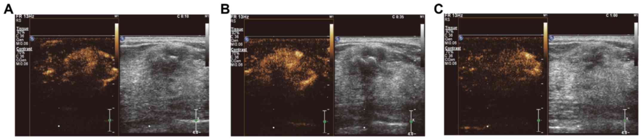
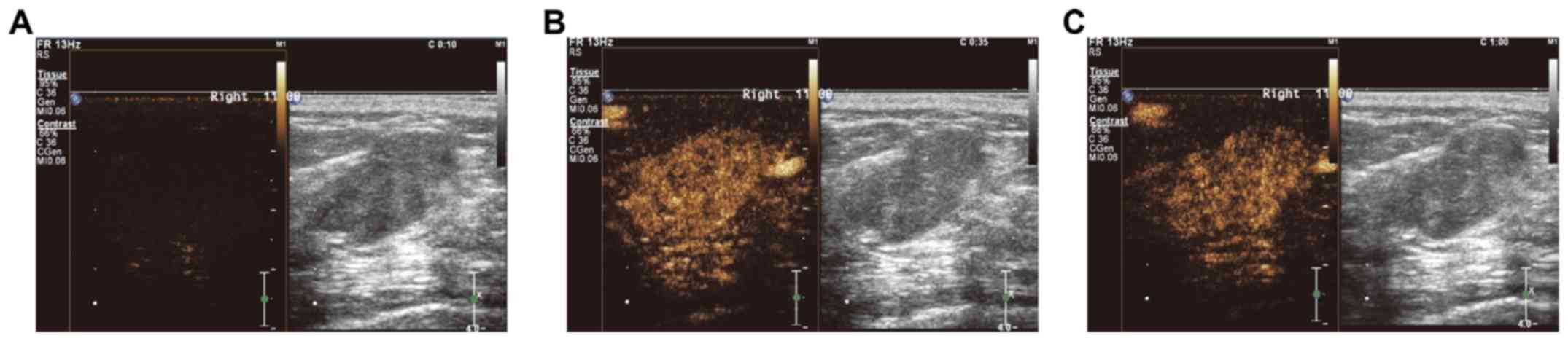
was observed during different time-periods (Fig. 1 and 2). For the malignant breast lesions, uneven
partial enhancement and overall enhancement were mainly observed.
Conversely, the benign breast lesions were mainly characterized by
local punctuate enhancement, circular enhancement and linear
enhancement. Furthermore, in contrast to the findings based on
angiography, all of the malignant breast lesions exhibited an
enlarged focus scope. The breast cancer group comprised 42 cases
with high enhancement, 7 cases with moderate enhancement and 3
cases with low enhancement. However, the focus scope enhancement
was not obvious for the benign breast lesions, comprising 25 cases
of high enhancement, 13 cases of moderate enhancement, 29 cases of
low enhancement and 6 cases of no enhancement.
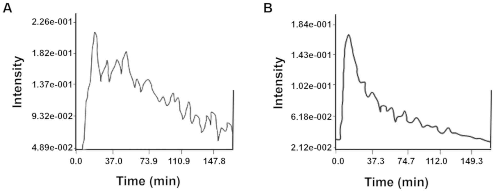
TIC morphology analysis of malignant
and benign breast lesions
TICs for malignant and benign breast lesions
indicated that 88.4% of the TICs for the malignant breast lesions
were of the fast-rising and slow-declining type, while 75.3 and
17.8% of the TICs for the benign breast lesions were of the
slow-rising and fast-declining and of the fast-rising and
fast-declining type, respectively. Significant differences in the
TIC shapes were observed between the malignant and benign breast
lesion groups (P<0.05; Table
III; Fig. 3).
| Table IIITime-intensity curve shape analysis of
malignant and benign breast lesions. |
Table III
Time-intensity curve shape analysis of
malignant and benign breast lesions.
| Parameter | Benign (n=73) | Malignant (n=52) | P-value |
|---|
| Fast-rising and
slow-declining | 5 (6.8) | 46 (88.4) | <0.01 |
| Slow-rising and
fast-declining | 55 (75.3) | 4 (7.6) | <0.01 |
| Fast-rising and
fast-declining | 13 (17.8) | 2 (3.8) | 0.01 |
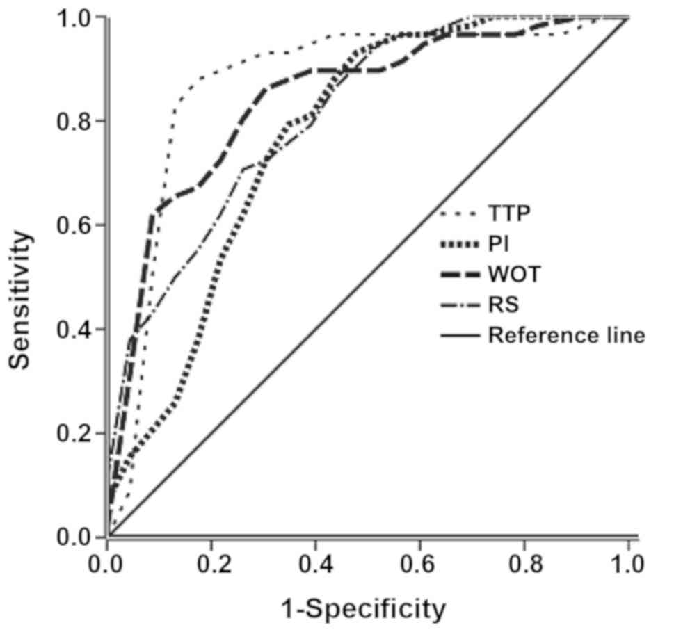
ROC curve analysis of TIC
parameters
ROC curves were constructed based on the TIC
parameters, including peak intensity, TTP, wash-out time and rising
slope. According to the best threshold principle for ROC analysis
(10), the point with the greatest
Youden index (sensitivity + specificity - 1) was considered as the
threshold, i.e., a TTP of ≤14.43 sec, wash-out time of ≥149.76 sec
and rising slope of ≥10.55 1/sec. The sensitivity and specificity
of the various parameters for the diagnosis of malignant breast
lesions are presented in Table IV.
The results suggested that the sensitivity of the wash-out time was
slightly but not significantly lower than that of the TTP and the
rising slope (P>0.05). The AUC values for the peak intensity,
TTP, wash-out time and rising slope for the diagnosis of malignant
breast lesions were 0.696, 0.870, 0.841 and 0.816, respectively.
The results indicated that the TTP, wash-out time and rising slope
have a diagnostic value for malignant breast lesions (Fig. 4).
| Table IVSensitivity and specificity analysis
of TIC perfusion parameters of malignant and benign breast
lesions. |
Table IV
Sensitivity and specificity analysis
of TIC perfusion parameters of malignant and benign breast
lesions.
| TIC parameter | Sensitivity
(%) | 1-specifity
(%) | Youden index | AUC |
|---|
| Time to peak | 89.7 | 17.4 | 14.43 | 0.870 |
| Wash-out time | 85.6 | 14.3 | 149.76 | 0.841 |
| Rising slope | 72.4 | 26.1 | 10.55 | 0.816 |
Discussion
Breast cancers rank first among malignancies in
females and clinical symptoms are mild at the early stage (11). At the time-point of diagnosis, breast
cancer may have progressed to the intermediate or late stage, which
is associated with high mortality. Therefore, early disease
detection and diagnosis may provide a marked benefit for breast
cancer treatment and prognosis, as well as the survival rate and
quality of life of affected patients (12). At present, the diagnosis of breast
cancer mainly depends on imaging methods. Along with the rapid
development of imaging technologies in recent years, the ability of
clinicians to differentiate between malignant and benign breast
lesions has markedly improved (13).
The new-generation contrast agent SonoVue is able to
clearly visualize the microcirculation of the breast lesion
(14), making true microcirculation
imaging possible (15).
Microcirculation imaging, performed as an additional round of
imaging following the conventional ultrasound, is able to reflect
the blood flow perfusion of normally structured and tumor tissue
(16), under color Doppler and
energy Doppler flow imaging. To date, clinical studies have
indicated that application of contrast agents significantly
improves the diagnostic accuracy for breast lesions.
Contrast-enhanced ultrasound is able to display the microvascular
structure in the diameter range of 20-39 μm (17), and is a reliable method for the
evaluation of the tumor microvascular circulation. Kettenbach et
al (18), indicated that
detection of the vascular morphology by the computer-assisted
quantitative assessment of power Doppler US might be one of the
most suitable methods for distinguishing between malignant and
benign breast lesions.
In the present study, the breast lesions whose
diagnosis was not possible by conventional ultrasound were
subjected to contrast-enhanced ultrasound. The enhancement
morphology and perfusion pattern of the breast lesions were
analyzed, and the clinical application value of contrast-enhanced
ultrasound was investigated. All of the included subjects were
finally diagnosed by pathology. The focal lesion morphology, lesion
boundaries, vascular morphology within lesions, contrast agent
distribution, wash-out mode and focal range of these malignant and
benign breast lesions were observed by contrast-enhanced ultrasound
detection. In a previous study, irregular radial boundaries were
observed on 84.6% of the malignant breast lesions on
contrast-enhanced ultrasound, while mass lumps were observed for
benign breast lesions, and only 29.4% of the benign breast lesions
had a regular morphology (19). In a
study by Zhao et al (20),
91.1% of malignant breast lesions exhibited a crab claw-like
enhancement, while 83.9% of the benign breast lesions were
homogeneously enhanced. In the present study, malignant breast
lesions were mainly characterized by an irregular appearance and
vascular morphology on contrast-enhanced ultrasound detection, and
an uneven contrast agent distribution with filling defects and
contrast agent retention was observed. Furthermore, the malignant
breast lesions mainly exhibited uneven partial enhancement and
overall enhancement, with the TICs being of the fast-rising and
slow-declining type, and the fast-out wash-out mode, as well as
unclear boundaries and uneven echo were common. On the contrary,
the benign breast lesions mainly exhibited local punctuate
enhancement, circular enhancement and linear enhancement, and TICs
were of the slow-rising and fast-declining, and fast-rising and
fast-declining types, and the slow-out or synchronous wash-out
modes were mainly observed.
The TIC represents the dynamic process of
contrast-enhanced ultrasound, which accurately reflects the
differential blood perfusion and wash-out processes of malignant
and benign breast lesions. In the present study, TIC analysis
suggested that 88.4% of the TICs of the malignant breast lesions
were of the fast-rising and slow-declining type, which may be
mainly attributed to angiogenesis and abnormal blood supply within
tumors. Abnormal blood vessels are prone to venous thrombus, which
may interfere with the blood supply and lead to contrast agent
retention. Furthermore, the TICs of 75.3% of the benign breast
lesion were of the slow-rising and fast-declining type, while 17.8%
were of the fast-rising and fast-declining type. This phenomenon
may be due to the fact that angiogenesis within benign breast
lesions may result from normal blood vessels, or their hyperplasia
and thickening. There was no abnormal vascular network or
arteriovenous fistula within the benign lesions, and the normal
venous drainage did not result in any contrast agent retention. In
the ROC analysis, the AUC values for the peak intensity, time to
peak, wash-out time and rising slope were 0.696, 0.870, 0.841 and
0.816, respectively, indicating a satisfactory clinical value for
the time to peak, wash-out time and rising slope in diagnosing
malignant breast lesions. These results are in accordance with
those of a previous study (21).
These phenomena may be explained by the notion that, due to the
rich angiogenesis and blood flow perfusion within masses, the
perfusion parameters reflect the top transient amount of
microbubbles and the flow of contrast agent during a certain
period. Regarding the sensitivity and specificity analysis, the
Youden indexes for time to peak, wash-out time and rising slope
were 14.43, 149.76 and 10.55, respectively, while the sensitivities
were 89.7, 85.6 and 72.4, respectively. As a tool for authenticity
assessment, changes in the Youden index may provide imaging
information for distinguishing between malignant and benign breast
lesions in the clinic. Of note, all of the three parameters had
high sensitivities for diagnosing malignant breast lesions,
indicating that these three indicators may reflect the angiogenesis
within the breast masses. However, the present study was only a
preliminary investigation of the diagnostic value of perfusion
parameters from contrast-enhanced ultrasound. Further in-depth
studies with optimized diagnostic indicators and larger sample
sizes are still required in the future.
In conclusion, the present study indicated that the
TIC parameters from contrast-enhanced ultrasound of breast lesions
have promising clinical value in differentiating between malignant
and benign lesions. Time to peak, wash-out time and rising slope
may contribute to the identification of malignant breast lesions,
which may facilitate early treatment and patient prognosis.
Acknowledgements
Not applicable.
Funding
This study was supported by the Science and
Technology Benefit Project (grant no. 2017c50070).
Availability of data and materials
The datasets used and/or analyzed during the present
study are available from the corresponding author on reasonable
request.
Authors' contributions
YZ, MZ, XF and DM contributed to the study design,
experimental performance, data collection/analysis, and manuscript
preparation. All authors read and approved the final
manuscript.
Ethical approval and consent to
participate
Written informed consent was obtained from each
patient and the study was approved by the Ethics Review Board of
the Ningbo No. 2 Hospital (Ningbo, Zhejiang, China).
Patient consent for publication
Not applicable
Competing interests
All authors declare that they have no competing
interests.
References
|
1
|
Grayson M: Breast cancer. Nature.
485(S49)2012.PubMed/NCBI View
Article : Google Scholar
|
|
2
|
Johnson RH, Hu P, Fan C and Anders CK:
Gene expression in ‘young adult’ type breast cancer: A
retrospective analysis. Oncotarget. 6:13688–13702. 2015.PubMed/NCBI View Article : Google Scholar
|
|
3
|
Howell A, Anderson AS, Clarke RB, Duffy
SW, Evans DG, Garcia-Closas M, Gescher AJ, Key TJ, Saxton JM and
Harvie MN: Risk determination and prevention of breast cancer.
Breast Cancer Res. 16(446)2014.PubMed/NCBI View Article : Google Scholar
|
|
4
|
Watanabe T, Kaoku S, Yamaguchi T, Izumori
A, Konno S, Okuno T, Tsunoda H, Ban K, et al: Multicenter
Prospective Study of Color Doppler Ultrasound for Breast Masses:
Utility of Our Color Doppler Method. Ultrasound Med Biol.
45:1367–1379. 2019.PubMed/NCBI View Article : Google Scholar
|
|
5
|
Purushothaman HN, Lekanidi K, Shousha S
and Wilson R: Lesions of uncertain malignant potential in the
breast (B3): What do we know? Clin Radiol. 71:134–140.
2016.PubMed/NCBI View Article : Google Scholar
|
|
6
|
JGuo R and Lu G: Ultrasound Imaging
Technologies for Breast Cancer Detection and Management: A Review.
Ultrasound Med Biol. 44:37–70. 2018.PubMed/NCBI View Article : Google Scholar
|
|
7
|
Wang YM, Fan W, Zhang K, Zhang L, Tan Z
and Ma R: Comparison of transducers with different frequencies in
breast contrast-enhanced ultrasound (CEUS) using SonoVue as
contrast agent. Br J Radiol. 89(20151050)2016.PubMed/NCBI View Article : Google Scholar
|
|
8
|
Sadeghi-Naini A, Sannachi L, Pritchard K,
Trudeau M, Gandhi S, Wright FC, Zubovits J, Yaffe MJ, Kolios MC and
Czarnota GJ: Early prediction of therapy responses and outcomes in
breast cancer patients using quantitative ultrasound spectral
texture. Oncotarget. 5:3497–3511. 2014.PubMed/NCBI View Article : Google Scholar
|
|
9
|
Liu J, Wang D, Li H, Li H, Zhou T, Zhao S
and Ding Z: Clinical value of contrast-enhanced ultrasound in
diagnosis of hyperechoic liver lesions. Med Sci Monit.
21:2845–2850. 2015.PubMed/NCBI View Article : Google Scholar
|
|
10
|
Obuchowski NA and Bullen JA: Receiver
operating characteristic (ROC) curves: review of methods with
applications in diagnostic medicine. Phys Med Biol.
63(07TR01)2018.PubMed/NCBI View Article : Google Scholar
|
|
11
|
Johnson CJ, Graff R, Moran P, Cariou C and
Bordeaux S: Breast cancer stage, surgery, and survival statistics
for Idaho's National Breast and Cervical Cancer Early Detection
Program population, 2004-2012. Prev Chronic Dis.
12(E36)2015.PubMed/NCBI View Article : Google Scholar
|
|
12
|
Cance WG, Carey LA, Calvo BF, Sartor C,
Sawyer L, Moore DT, Rosenman J, Ollila DW and Graham M II:
Long-term outcome of neoadjuvant therapy for locally advanced
breast carcinoma: Effective clinical downstaging allows breast
preservation and predicts outstanding local control and survival.
Ann Surg. 236:295–302; discussion 302-303. 2002.PubMed/NCBI View Article : Google Scholar
|
|
13
|
Alunni-Fabbroni M, Müller V, Fehm T, Janni
W and Rack B: Monitoring in metastatic breast cancer: Is imaging
outdated in the era of circulating tumor cells? Breast Care
(Basel). 9:16–21. 2014.PubMed/NCBI View Article : Google Scholar
|
|
14
|
Xiao X, Ou B, Yang H, Wu H and Luo B:
Breast contrast-enhanced ultrasound: Is a scoring system feasible?
A preliminary study in China. PLoS One. 9(e105517)2014.PubMed/NCBI View Article : Google Scholar
|
|
15
|
Madjar H, Prömpeler HJ, Del Favero C,
Hackelöer BJ and Llull JB: A new Doppler signal enhancing agent for
flow assessment in breast lesions. Eur J Ultrasound. 12:123–130.
2000.PubMed/NCBI View Article : Google Scholar
|
|
16
|
Wang J, Qin B, Chen X, Wagner WR and
Villanueva FS: Ultrasound mol imaging angiogenesis using vasc
endothelial growth factor-conjugated microbubbles. Mol
Pharmaceutics. 14:781–790. 2017.PubMed/NCBI View Article : Google Scholar
|
|
17
|
Forsberg F, Kuruvilla B, Pascua MB,
Chaudhari MH, Merton DA, Palazzo JP and Goldberg BB: Comparing
contrast-enhanced color flow imaging and pathological measures of
breast lesion vascularity. Ultrasound Med Biol. 34:1365–1372.
2008.PubMed/NCBI View Article : Google Scholar
|
|
18
|
Kettenbach J, Helbich TH, Huber S, Zuna I
and Dock W: Computer-assisted quantitative assessment of power
Doppler US: Effects of microbubble contrast agent in the
differentiation of breast tumors. Eur J Radiol. 53:238–244.
2005.PubMed/NCBI View Article : Google Scholar
|
|
19
|
Gity M, Arabkheradmand A, Taheri E and
Shakiba M: Diagnostic investigation of breast magnetic resonance
imaging in malignant and benign mass lesions. Arch Med Sci.
14:1061–1069. 2018.PubMed/NCBI View Article : Google Scholar
|
|
20
|
Zhao H, Xu R, Ouyang Q, Chen L, Dong B and
Huihua Y: Contrast-enhanced ultrasound is helpful in the
differentiation of malignant and benign breast lesions. Eur J
Radiol. 73:288–293. 2010.PubMed/NCBI View Article : Google Scholar
|
|
21
|
Amioka A, Masumoto N, Gouda N, Kajitani K,
Shigematsu H, Emi A, Kadoya T and Okada M: Ability of
contrast-enhanced ultrasonography to determine clinical responses
of breast cancer to neoadjuvant chemotherapy. Jpn J Clin Oncol.
46:303–309. 2016.PubMed/NCBI View Article : Google Scholar
|