Introduction
NNEDV are a group of chronic diseases characterized
by degeneration and pigmentation of the skin and mucosal tissues of
the vulva, including squamous hyperplasia (SH), lichen sclerosus
(LS) and mixed lesions of SH and LS, which are common refractory
gynecological diseases that often cause severe and torturous vulvar
pruritus. At present, the pathogenesis of these disorders is
unclear, and NNEDV may undergo malignant transformation. As early
as 2008, NNEDV were identified as precancerous lesions (1), and it has been reported that vulvar
squamous cell carcinomas develop via two different pathways. One is
closely associated with human papillomavirus (HPV) infection, which
is known as the classical pathway, and the other is associated with
inflammatory dermatitis, which is known as the differentiated or
simple pathway, in which HPV infection is not involved. The latter
accounts for <5% of identifiable vulvar squamous cell carcinomas
(1). Inflammatory dermatosis
occurs in the epithelium adjacent to the cancerous tissue, which is
often continuous with LS or chronic simple lichen disease. NNEDV
can become malignant through the differentiated pathway; however,
the mechanism by which it becomes malignant remains unknown.
Previous studies have explored infection, genetics, hormone levels,
autoimmune and metabolic factors, as well as local stimulation, but
the mechanisms of carcinogenesis have yet to be clearly identified
(2-4).
Molecular biology provides a novel approach to the investigation of
the pathogenesis of NNEDV with regard to changes in the mechanism
of cell cycle regulation.
The abnormal proliferation of cells caused by
dysregulation of the cell cycle is the basis of tumorigenesis and
development. The most important cell cycle regulation system of
eukaryotic cells is that comprising cyclins, cyclin-dependent
kinases (CDKs) and CDK inhibitors (CKIs). In the normal cell cycle,
a cyclin combines with a CDK to form a complex that regulates cell
proliferation and differentiation from the G1 phase to
the S phase. However, CKIs inhibit the activity of the cyclin-CDK
complexes and block the cell cycle at the G1 phase,
which keeps the system in dynamic equilibrium and maintains the
normal operation of the cell cycle. Disruption of the balance of
this system can lead to a shortened cell cycle, with uncontrolled
cell growth and proliferation, and may also result in tumorigenesis
(5).
Cyclin D1, CDK4 and CKI P27 (P27) have been found to
be important for the regulation of the cell cycle (5). Therefore, the purpose of the present
study was to investigate the expression and significance of cyclin
D1, CDK4 and P27 in NNEDV and serve as a reference for the clinical
diagnosis and treatment of these disorders.
Materials and methods
NNEDV and control groups
Patients admitted to the Department of Gynecology
and Obstetrics of The Affiliated Hospital of Southwest Medical
University (Luzhou, China) from August 2020 to July 2021 were
enrolled in the present study. A diagnosis of NNEDV was confirmed
based on pathology results by two independent pathologists. The
inclusion criteria were patients who fulfilled the pathological
diagnostic criteria set by the International Society for the Study
of Vulvar Diseases (ISSVD) and the International Society of
Gynecological Pathologists (ISGYP) in 1987(6), with clinical manifestations including
repeated vulvar pruritus and burning sensation, and who volunteered
to participate in the study. The exclusion criteria were patients
who were pregnant or lactating, as well as those with a history of
hypertension, coronary heart disease, diabetes, hyperlipidemia,
thyroid disease, recurrent abortion, adverse pregnancy, immune
diseases and malignant tumors, all types of acute vaginitis and
acute vulvar infection. In addition, those patients who had
received hormone or drug therapy within 1 month, had a mental
illness, neurological dysfunction or a long history of smoking were
also excluded.
In total, 36 patients [age, 22-75 years; mean age,
39±11.53 years; body mass index (BMI) 18-24 kg/m2; mean
BMI, 21.04±1.06 kg/m2] were included in the NNEDV group,
including 20 cases of SH, 10 cases of LS and 6 cases of mixed
lesions. The course of the disease ranged from 6 months to 20
years, with an average of 4.67 years. In the control group, normal
vulva skin samples from 20 patients undergoing perineal repair were
collected (mean age, 41.10±13.17 years; BMI, 18-24
kg/m2; mean BMI, 21.01±1.14 kg/m2). No
significant differences in general data between the NNEDV and
control groups were identified (P>0.05).
Skin tissues (diameter, 0.5 cm; depth, 0.5 cm) were
excised under local anesthesia from patients in the control and
NNEDV groups. The samples were immediately fixed in 10% neutral
formalin and then embedded in paraffin.
Reagents
The immunohistochemical streptavidin peroxidase
(S-P) method was used to detect the expression of cyclin D1, CDK4,
and P27 in skin lesions. The primary antibodies used include: i)
Rabbit anti-human cyclin D1 monoclonal antibody (cat. no. ZA-0101;
0.1 ml; dilution, 1:50); ii) rabbit anti-human CDK4 monoclonal
antibody (cat. no. ZA-0614; 0.1 ml; dilution, 1:150); iii) rabbit
anti-human P27 monoclonal antibody (cat. no. ZA-0557; 0.1 ml;
dilution, 1:20). The S-P immunohistochemical series kit (cat. no.
SP-9000) includes: i) Normal goat serum blocking solution; ii)
secondary antibody: biotin labeled sheep anti-rabbit IgG working
solution (dilution, 1:1); iii) horseradish enzyme labeled chain
enzyme ovalbumin working solution (dilution, 1:1). All the
aforementioned reagents were purchased from Beijing Zhongshan
Jinqiao Biotechnology Co., Ltd.
Main instruments
Leica pathological tissue slicer (Leica Microsystems
GmbH), Heating and baking machine (Tk-218; Hubei Xiaogan Taiwei
Electronic Equipment Co., Ltd.), refrigerator (Bco-220; Changhong
Meiling Co., Ltd.), constant temperature incubator (wk891, 37˚C;
Ningbo David Medical Device Co., Ltd.), super high-temperature
pressure cooker (diameter, 38 cm; Wuhan Suber Pressure Pot Co.,
Ltd.), Confocal microscope (Ax70; Olympus Corporation) and image
processing system (KS400; Zeiss AG) were used for processing the
samples as well as visualizing and analyzing the results.
Detection methods All tissues were
fixed in 10% neutral formalin at 36-38˚C for 24 h
After dehydration and being rendered translucent,
the tissues were waxed, embedded in paraffin and routinely
sectioned to a thickness of 4 µm. Four sections from each tissue
were collected, one of which was stained with hematoxylin and eosin
at 36-38˚C, the entire dyeing process takes 1 h and 30 sec, while
the remaining sections were prepared for immunohistochemistry using
the S-P method, which process are as follows: i) Continuously slice
paraffin specimens at 4 µm and place in oven at 60˚C for 3-5 h; ii)
leave paraffin sections at 22˚C for 5 min, then dipped into xylene
I and II for 10 min, respectively; iii) immerse in 100, 95, 90, 80
and 70% alcohol solution for 10 min, respectively; iv) rinse three
times with double distilled water for 1 min each time; v) antigen
repair: Put the tissue section into antigen repair solution (PH
6.0), steam under high pressure for 3 min and cool to room
temperature; vi) rinse with 0.01 M PBS solution (pH 7.4) three
times for 10 min each time; vii) endogenous peroxidase was blocked
by hydrogen peroxide (3%) and incubated at 37˚C for 10 min; viii)
rinse with 0.01 M PBS solution (pH 7.4) three times for 5 min each
time; ix) add 5% normal goat serum blocking solution (purchased
from Beijing Zhongshan Jinqiao Biotechnology Co., Ltd.) and place
in a wet box in the incubator at 37˚C for 20 min to eliminate
non-specific staining; x) pour out the serum and add the primary
antibody (rabbit anti-human cyclin D1, CDK4 or P27 monoclonal
antibody). Place in incubator at 37˚C for 30 min and transfer to
refrigerator at 4˚C overnight; xi) rewarm the following day, rinse
with 0.01 M PBS solution three times for 5 min each time; xii) add
biotin-labeled secondary antibody (sheep anti-rabbit IgG) working
solution dropwise and place in a wet box in the incubator at 37˚C
for 25 min; xiii) rinse with 0.01 M PBS solution (pH 7.4) three
times for 5 min each time; xiv) add 100 µl of horseradish enzyme
labeled chain enzyme ovalbumin working solution and put it in wet
box in incubator at 37˚C for 15 min; xv) rinse with 0.01 M PBS
solution (pH 7.4) three times for 5 min each time; xvi) DAB color
development: Color development at room temperature and control the
reaction time under the microscope; xvii) rinse thoroughly with tap
water for 10 min; xviii) hematoxylin was counterstained at 25˚C for
5 min, rinse thoroughly with tap water for 5 min, then add
hydrochloric acid (0.5%) and alcohol solution to differentiate
saturated lithium carbonate returns to blue; xix) use conventional
gradient alcohol dehydration, immerse in xylene and seal the sheet
with neutral gum. Observe and photograph under the microscope. As
the quality control standard, 0.01 M PBS solution (pH 7.4) was used
as the negative control instead of the primary antibody.
Pathological diagnostic criteria of
NNEDV
Diagnostic criteria jointly created by the ISSVD and
ISGYP in 1987 were applied to diagnose NNEDV. The 20 cases of SH
exhibited cell proliferation of the squamous epithelium, obvious
keratosis and incomplete keratosis of the epidermis, hypertrophy of
the prickle cell layer, extended epithelial feet, evident dermal
papilla between the epithelial feet, as well as a small amount of
lymphocyte and plasma cell infiltration around the blood vessels in
the superficial dermis. The 10 cases of LS presented with epidermal
hyperkeratosis and keratin embolism of the hair follicles, thinning
of the prickle cell layer with liquefaction degeneration of the
basal cells, reduced melanocyte counts, the thickening or
disappearance of epithelial feet, and homogenization of the dermis,
as well as the infiltration of lymphocytes and plasma cells in
homogeneous bands. The 6 cases of mixed type presented with the
pathological features of both types of lesions (Fig. 1).
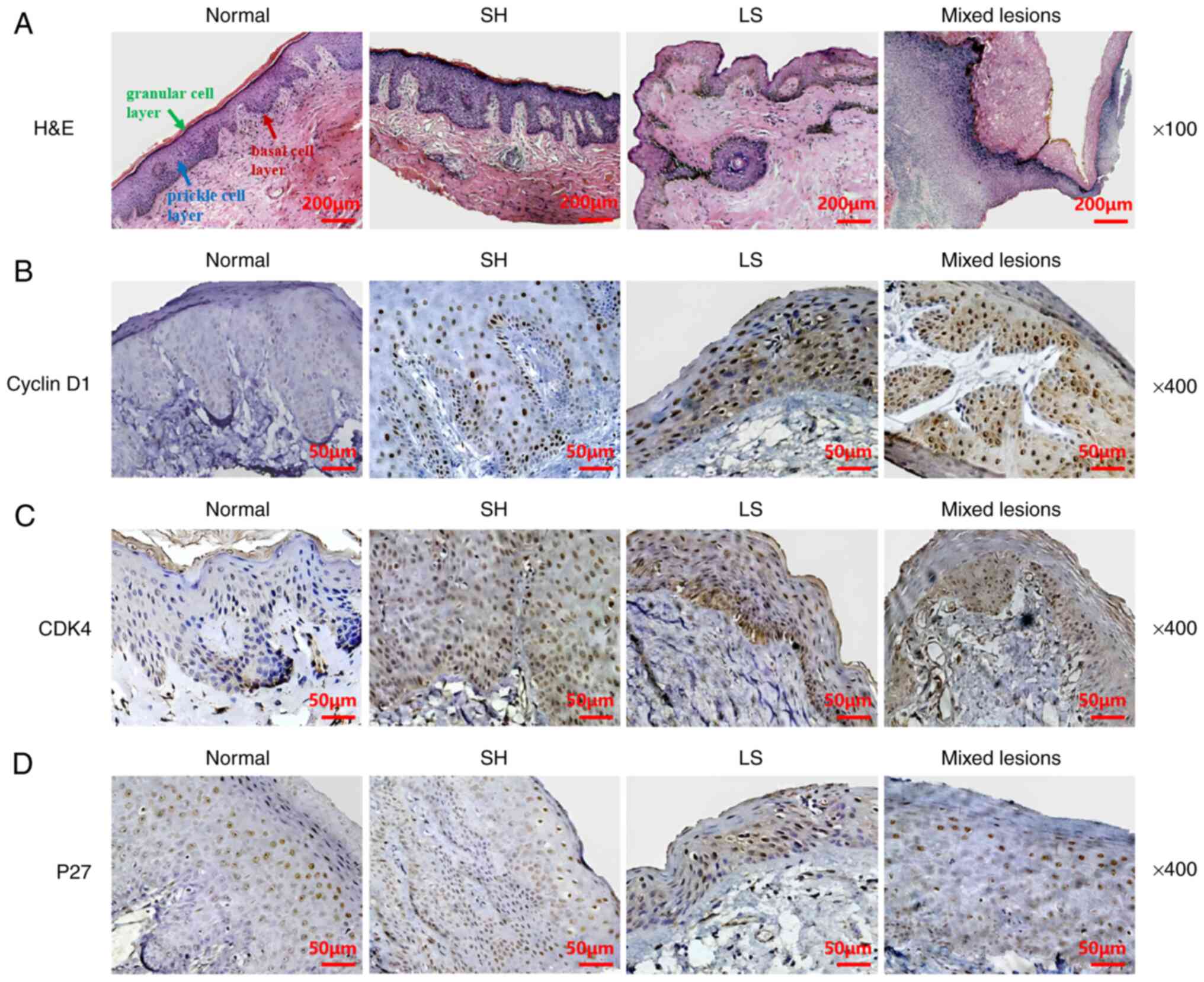 | Figure 1Expression of cyclin D1, CDK4 and P27
in normal vulvar tissue and vulvar tissues from patients with
non-neoplastic epithelial disorders of the vulva. (A) H&E
staining of normal, SH, LS and mixed lesion tissues, respectively.
Scale bars, 200 µm. Immunohistochemical staining of (B) cyclin D1,
(C) CDK4 and (D) P27 in normal, SH, LS and mixed lesion tissues,
respectively. Scale bars, 50 µm. CDK4, cyclin-dependent kinase 4;
P27, cyclin-dependent kinase inhibitor P27; SH, squamous
hyperplasia; LS, lichen sclerosus; H&E, hematoxylin and
eosin. |
Determination of immunohistochemical
results
Positive staining was indicated by tan or yellow
particles. The nucleus and/or cytoplasm were sites of positive
staining for cyclin D1 and CDK4, while the nucleus was the site of
positive staining for P27. For each section, five typical fields
were observed using a confocal microscope at a magnification of
x100, and photographic images were captured at a magnification of
x400. The positive expression of protein was evaluated based on the
mean optical density (MOD). Image Pro 6.0 Professional Image
Analysis Software (Media Cybernetics, Inc.) was used to assess the
MOD. MOD analysis was performed blindly by three different people
and the mean value was taken.
Statistical methods
The SPSS Statistics 22.0 software package (IBM
Corp.) was used to perform the statistical analysis. Data are
expressed as the mean ± standard deviation. Multiple sample means
were compared using one-way analysis of variance followed by
Tukey's post hoc test for pairwise comparisons. Two sample means
were compared using the unpaired t-test, with a=0.05 (bilateral)
being set as the test level. P<0.05 was considered to indicate a
statistically significant result.
Results
Sites of cyclin D1, CDK4 and P27
expression in normal and NNEDV tissues
Staining for cyclin D1 and CDK4 was observed in the
nucleus and/or cytoplasm. In normal tissues, the positive cells
were mainly distributed in the epithelial basal cell layer, and
also present in the prickle cell layer. However, in NNEDV tissues,
the positive cells were mainly distributed in the prickle cell
layer. Positive cells were also observed in the granular cell layer
in some cases, with cyclin D1 positive cells in 7 cases of SH and 4
cases of LS, as well as CDK4 positive cells in 8 cases of SH, 3
cases of LS and 2 cases of mixed lesions. P27 staining was visible
in the nucleus and was mainly distributed in the basal cell and
prickle cell layers of the normal and NNEDV tissues (Fig. 1).
Expression intensity of cyclin D1,
CDK4 and P27 in normal and NNEDV tissues
The MOD of cyclin D1 in the control, SH, LS and
mixed groups was 0.53±0.76, 2.36±1.62, 2.25±1.71 and 2.38±0.92,
respectively. The MOD of CDK4 in the control, SH, LS and mixed
groups was 1.71±0.52, 4.03±1.81, 3.69±2.21 and 4.17±1.12,
respectively. The MOD of P27 in the control, SH, LS and mixed
groups was 4.15±2.33, 2.31±1.35, 2.48±1.11 and 2.35±1.35,
respectively. The MOD values of cyclin D1 and CDK4 in the three
pathological types of NNEDV (SH, LS and mixed lesions) were
significantly higher compared with those of the control group (all
P<0.05). The MOD values of P27 in the three types of NNEDV were
lower than that of the control group, but no significant difference
was detected (all P>0.05). No significant difference in the MOD
values of cyclin D1, CDK4 or P27 were detected among the three
pathological types of NNEDV (all P>0.05; Table I).
 | Table IMean optical density of cyclin D1,
CDK4 and P27. |
Table I
Mean optical density of cyclin D1,
CDK4 and P27.
| Group | Number | Cyclin D1 | CDK4 | P27 |
|---|
| SH | 20 |
2.36±1.62a |
4.03±1.81a | 2.31±1.35 |
| LS | 10 |
2.25±1.71a |
3.69±2.21a | 2.48±1.11 |
| Mixed lesions | 6 |
2.38±0.92a |
4.17±1.12b | 2.35±1.35 |
| Control | 20 | 0.53±0.76 | 1.71±0.52 | 4.15±2.33 |
| F-value | | 3.60 | 3.67 | 1.83 |
| P-value | | 0.03 | 0.02 | 0.18 |
Changes in the ratios of the MOD of
cyclin D1, CDK and P27 in the prickle cell layer to those in the
basal cell layer
The distribution of cyclin D1 and CDK4 positive
cells in the epithelium of the control group was different from
that of the NNEDV group. The ratio of the MOD of cyclin D1 in the
prickle cell layer to that in the basal cell layer was 0.43±0.30 in
the control group and 2.29±0.77 in the NNEDV group. The ratio of
the MOD of CDK4 in the prickle cell layer to that in the basal cell
layer was 0.87±0.50 in the control group and 2.41±0.86 in the NNEDV
group. For both cyclin D1 and CDK4, the ratio of the MOD in the
prickle cell layer to that in the basal cell layer was
significantly higher in the NNEDV group compared with the control
group (both P<0.01). The ratio of the MOD of P27 in the prickle
cell layer to that in the basal cell layer was 1.56±1.17 in the
control group and 1.39±0.77 in the NNEDV group; no significant
difference in this ratio was found between the two groups
(P>0.05; Table II).
| Table IIRatios of the mean optical density of
cyclin D1, CDK4 and P27 in the prickle cell layer to those in the
basal cell layer. |
Table II
Ratios of the mean optical density of
cyclin D1, CDK4 and P27 in the prickle cell layer to those in the
basal cell layer.
| Group | Number | Cyclin D1 | CDK4 | P27 |
|---|
| Control | 20 | 0.43±0.30 | 0.87±0.50 | 1.56±1.17 |
| NNEDV | 36 | 2.29±0.77 | 2.41±0.86 | 1.39±0.77 |
| t-value | | 9.55 | 4.08 | 0.44 |
| P-value | | <0.00 | 0.00 | 0.67 |
Discussion
Each stage of the cell cycle is precisely regulated.
Hartwell et al (7) proposed
the concept of the cell cycle regulatory checkpoint in the 1970s.
There are two key regulatory points in the cell cycle:
G1/S and G2/M (8). Cell cycle progression is primarily
driven by CDKs, which bind to cyclins to form a complex and are
activated by phosphorylation or dephosphorylation, thereby
promoting the expression of genes associated with the cell cycle
(9-12).
To date, a number of CDKs, including CDK1, 2, 4, 6,
7 and 9 and cyclins, including cyclins A, B and D-H, have been
identified and are known to play an important role in cell cycle
regulation: Cyclin D-CDK4/6 complex initiates the cell cycle
process, cyclin E-CDK2 complex regulates entry to the S phase,
cyclin A-CDK2 complex regulates S-phase DNA replication and cyclin
A/B-CDK1 complex triggers mitosis. Each cyclin-CDK complex can
trigger the expression of the next cyclin-CDK complex to regulate
the cell cycle at all stages (13,14).
The reason for selecting cyclin D1, CDK4 and P27 for
examination in the present study was that cyclin D (cyclin
D1/D2/D3) and CDK4/6 are the core molecules that drive the
initiation of the cell cycle. Once cyclin D1-CDK4 complexation
occurs, the cell cycle is triggered. The abnormal expression of
cyclin D1 leads to DNA damage and activation of checkpoint kinase
1, resulting in abnormal cell growth (13,14).
P27 is a broad-spectrum CKI that inhibits most CDKs (15,16).
When P27 binds to a cyclin D-CDK complex, the activity of the CDK
is inhibited and retinoblastoma-associated protein phosphorylation
is inhibited, leading to cell-cycle arrest in the G phase and the
cessation of cell growth (15,16).
In normal tissues, the levels of CDK4, cyclin D1 and P27 are in
balance. However, the upregulation of CDK4 and cyclin D, as well as
the downregulation of P27, can promote cell division and
proliferation disorders, and thereby induce numerous types of human
tumors (for example, esophageal squamous cell carcinoma, lung
cancer, oral squamous cell carcinoma, renal pelvis and ureter
cancer, vulvar cancer, etc.) (17-25).
Therefore, investigation of the expression of cyclin D1, CDK4 and
P27 in patients with NNEDV is potentially of great importance.
The characteristics and significance of cyclin D1,
CDK4 and P27 expression in NNEDV tissues were investigated in the
present study. It was observed that the MODs of cyclin D1 and CDK4
in the three pathological types of NNEDV were significantly higher
than those in the control group. No statistically significant
differences in the MODs of cyclin D1 and CDK4 were detected among
the three pathological types. The MOD of P27 in the control group
was higher than that in the three pathological groups, but the
differences were not found to be statistically significant.
Furthermore, no statistically significant difference in the MOD of
P27 was detected among the three pathological groups. These results
indicate that the proliferation of epithelial cells in NNEDV
tissues is likely to be more active than that in normal tissues and
suggest that high expression of cyclin D1 and CDK4 plays a key role
in the transformation of normal tissue to NNEDV.
Previous studies have confirmed that cyclin D1 and
CDK4 are protooncogenes and are upregulated in numerous types of
tumors (26-29).
One study (20) revealed that the
expression levels of cyclin D1 tended to increase as vulvar tissue
progressed from normal to NNEDV, vulvar intraepithelial neoplasia
(VIN) and vulvar squamous cell carcinoma (SCCV), and the expression
of cyclin D1 was significantly increased in vulvar carcinoma. These
findings indicate that the high expression of cyclin D1 is
associated with the occurrence and development of vulvar carcinoma.
In the present study, the expression levels of cyclin D1 and CDK4
were significantly increased in NNEDV tissues compared with normal
tissues. This suggests that although NNEDV is a benign lesion, the
high expression of cyclin D1 and CDK4 in the lesion may cause the
normal cell cycle to become dysregulated and cell proliferation to
be activated, ultimately resulting in carcinogenesis. Therefore,
the high expression levels of cyclin D1 and CDK4 may be important
in the formation of NNEDV, and the possibility of malignant
transformation of NNEDV should be monitored clinically. In
addition, the expression levels of these proteins could be used as
molecular markers to distinguish pathological tissues from normal
tissues.
As an important tumor suppressor gene, P27 is a
widely studied prognostic factor, and the deletion of P27 is an
important index for judging the nature and prognosis of numerous
diseases. Kagawa et al (30) observed that decreased expression of
P27 was associated with the progression and poor prognosis of
breast, colon and gastric carcinomas. In addition, Yanagi et
al (24) observed that P27 was
expressed in the normal epidermis, and the expression level of P27
in squamous cell carcinoma (SCC) was significantly lower than that
in normal epidermis. Yanagi et al also reported that the
absence of P27 expression was a frequent event in SCC. In the
present study, the expression of P27 in NNEDV was not significantly
decreased compared with that in the control tissue and did not
differ according to the pathological type of NNEDV. This result is
consistent with the findings of Zamparelli et al (31), which showed that there was no
significant difference in P27 protein expression between normal
vulva skin, NNEDV and VIN, although a decreasing trend was
observed. However, in another study, Zannoni et al (32) reported a significant difference in
P27 expression between SCCV and precancerous lesions, and a
significant reduction of P27 expression in NNEDV tissues compared
with the normal control group, with a decreasing trend in P27
expression in the following sequence: Normal skin
tissue-NNEDV-VIN-SCCV.
Based on these findings, it is hypothesized that the
high expression of cyclin D1 and CDK4 disrupted the balance between
cyclin D1-CDK4 complex and P27 levels, resulting in the epithelial
cell proliferation in NNEDV being more active than that in normal
tissues, indicating that NNEDV has the potential to undergo
malignant transformation. The occurrence and development of NNEDV
may be associated with acceleration of the cell cycle, and cyclin
D1, CDK4 and P27 are involved in regulation of the cell cycle in
NNEDV.
It is possible that preventing the proliferation of
abnormal skin cells via control of the cell cycle, achieved by
concurrently inhibiting cyclin D1 and CDK4 as well as promoting the
activity of P27, may become a useful molecular strategy for the
therapy of NNEDV. As P27 is a broad spectrum CKI with an important
role in the G1 and S phases of the cell cycle, the
promotion of P27 activity may be beneficial.
The characteristics and significance of the
distribution of cyclin D1, CDK4 and P27 in normal and NNEDV tissues
were also investigated in the present study. The results
demonstrated that for cyclin D1 and CDK4, the ratio of the MOD in
the prickle cell layer to that in the basal cell layer was higher
in the NNEDV group compared with the control group (P<0.001 and
P=0.001, respectively). However, for P27, the ratio of the MOD in
the prickle cell layer to that in the basal cell layer showed no
significant difference between the two groups (P>0.05). These
results suggest that the distributions of cyclin D1- and
CDK4-positive cells in the epithelial tissues of the control and
NNEDV groups differed. The cyclin D1- and CDK4-positive cells were
mainly distributed in the basal cell layer in normal tissues, which
was a normal distribution. In the basal layer, which is also known
as the germinal layer, cells divide actively to constantly produce
new cells that move up to the stratum corneum where they replenish
the aged and shed keratinocytes. Thus, basal cells play a role in
epidermal repair and germination. However, in NNEDV tissues, cyclin
D1 and CDK4 positive cells were mainly distributed in the prickle
cell layer, otherwise known as the stratum spinosum, which suggests
that cell division and proliferation in the prickle cell layer were
more active than those in the basal cell layer. P27 positive cells
were mainly distributed in the basal cell and prickle cell layers
of the normal and NNEDV tissues, which is consistent with the
findings of some previous studies. For example, Pu et al
(33) found that the expression of
proliferating cell nuclear antigen in LS tissues was mainly
distributed in the prickle cell and granular cell layers, with
lower levels of expression in the basal cell layer. The results
indicated that the thinned prickle layer in LS had stronger
proliferative capacity than normal skin. In addition, Liu et
al (34) found that the
prickle cell layer of NNEDV tissues had strong growth and
differentiation potential. Therefore, cell proliferation appears to
be abnormal in NNEDV tissues, with an imbalance of cell
proliferation activity between different layers of the epithelium.
The results suggest that the cells in the basal cell layer retained
normal proliferation activity whereas those in the prickle cell
layer were likely to proliferate more actively. Even in the thinned
prickle layer in LS, the proliferative ability of the cells
remained strong. Therefore, the change in the balance of epithelial
cell proliferation between different layers of NNEDV tissue may
indicate the possibility of malignant transformation.
The clinical significance of cyclin D1, CDK and P27
expression in NNEDV merits consideration. Although there are
differences in the clinical manifestations and pathological changes
of SH, LS and mixed lesions, no significant differences in the MOD
of cyclin D1, CDK4 and P27 were detected among these three
pathological types and changes in cyclin D1, CDK4 and P27 were not
significantly associated with the pathology of NNEDV. Therefore,
the three pathological types may have a common or similar
pathogenesis. As proto-oncogenes, cyclin D1 and CDK4 play an
important role in the occurrence, development, biological behavior
and prognosis of some malignant tumors. The results of the present
study suggest the possibility of malignant transformation occurring
in cases of NNEDV. Therefore, it is recommended that patients with
NNEDV are subjected to long-term follow-ups. In addition, cyclin D1
and CDK4 could potentially be used as indicators for the follow-up
of lesion progression.
The ultimate purpose of the present study on NNEDV
was to provide information useful in the clinic and serve as a
basis for clinical drug development, diagnosis and treatment, or
further experimental research. The study provides a theoretical
basis for the development of more effective treatments for
retarding or reversing the progress of NNEDV and preventing
malignant transformation. We hypothesize that cyclin D1, CDK4 and
P27 are potential targets for the development of new clinical
therapeutic drugs for the treatment of patients with NNEDV.
However, the study has certain limitations. Although
cyclin D1 and CDK4 are protooncogenes associated with NNEDV, the
clinical malignant transformation rate of NNEDV is not high, which
indicates that other regulatory may influence the cell cycle. The
relationships between other regulatory factors in the cell cycle
and the exact mechanism of NNEDV require further evaluation.
Acknowledgements
Not applicable.
Funding
Funding: This study was supported by the Young Program of
National Natural Science Foundation of China (grant no.
71501035).
Availability of data and materials
The datasets used and/or analyzed during the current
study are available from the corresponding author on reasonable
request.
Authors' contributions
HL was responsible for experimental design,
conceptualization, research implementation, data
analysis/interpretation and administrative, technical and material
support. FZ contributed to research implementation, data collection
and critical review of the knowledge content of the article. ZL
performed data collection and statistical analysis. HL and ZL
confirm the authenticity of all the raw data. All authors read and
approved the final manuscript.
Ethics approval and consent to
participate
The study protocol was approved by the Ethics
Committee of Southwest Medical University (ref. no. 2020-61). This
study was conducted in accordance with The Declaration of Helsinki.
Written informed consent was obtained from all participants.
Patient consent for publication
Informed consent was obtained from all patients
regarding the publication of the data and associated images.
Competing interests
The authors declare that they have no competing
interests.
References
|
1
|
Nieuwenhof H, Avoort I and Hullu J: Review
of squamous premalignant vulvar lesions. Crit Rev Oncol Hematol.
68:131–156. 2008.PubMed/NCBI View Article : Google Scholar
|
|
2
|
Ruan L, Xie Z, Wang H, Jiang J, Shi H and
Xu J: High-intensity focused ultrasound treatment for
non-neoplastic epithelial disorders of the vulva. Int J Gynecol
Obstet. 109:167–170. 2010.PubMed/NCBI View Article : Google Scholar
|
|
3
|
Pu D and Hou M: Etiology and pathogenesis
of white lesions of the vulva. J Practical Obstetrics Gynecol.
19:2–3. 2003.(In Chinese).
|
|
4
|
Li GT, Cao JH and Fu YJ: Expression of
cyclin D1 and p16 protein in vulvar white lesion. Zhonghua Fu Chan
Ke Za Zhi. 41:322–325. 2006.PubMed/NCBI(In Chinese).
|
|
5
|
Bantie L, Tadesse S, Likisa J, Yu M, Noll
B, Heinemann G, Lokman NA, Ricciardelli C, Oehler MK, Beck A, et
al: A first-in-class CDK4 inhibitor demonstrates in vitro, ex-vivo
and in vivo efficacy against ovarian cancer. Gynecol Oncol.
159:827–838. 2020.PubMed/NCBI View Article : Google Scholar
|
|
6
|
Le J: Gynecology and Obstetrics (7th
edition). People's Health Publishing House. 231-235:2008.(In
Chinese).
|
|
7
|
Hartwell L H, Culotti J, Pringle JR and
Reid BJ: Genetic control of the cell division cycle in yeast: A
model to account for the order of cell cycle events is deduced from
the phenotypes of yeast mutants. Science. 183:46–51. 1974.
|
|
8
|
Frederick TJ and Wood TL: IGF-I and FGF-2
coordinately enhance cyclin D1 and cyclin E-cdk2 association and
activity to promote G1 progression in oligodendrocyte progenitor
cells. Mol Cell Neurosci. 25:480–492. 2004.PubMed/NCBI View Article : Google Scholar
|
|
9
|
Du Q, Guo X, Wang M, Li Y, Sun X and Li Q:
The application and prospect of CDK4/6 inhibitors in malignant
solid tumors. J Hematol Oncol. 13(41)2020.PubMed/NCBI View Article : Google Scholar
|
|
10
|
Qie S and Diehl JA: Cyclin D degradation
by E3 ligases in cancer progression and treatment. Semin Cancer
Biol. 67:159–170. 2020.PubMed/NCBI View Article : Google Scholar
|
|
11
|
Billard-Sandu C, Tao YG, Sablin MP,
Dumitrescu G, Billard D and Deutsch E: CDK4/6 inhibitors in
P16/HPV16-negative squamous cell carcinoma of the head and neck.
Eur Arch Otorhinolaryngol. 277:1273–1280. 2020.PubMed/NCBI View Article : Google Scholar
|
|
12
|
Deng L, Xu M, Fu L, Du G and Wang L:
Effects of CDK4/6 inhibitor palbociclib on proliferation, cell
cycle and apoptosis of human melanoma cells. Tumor. 40:521–530.
2020.(In Chinese).
|
|
13
|
Liu J, Peng Y and Wei W: Cell cycle on the
crossroad of tumorigenesis and cancer therapy. Trends Cell Biol.
32:30–44. 2022.PubMed/NCBI View Article : Google Scholar
|
|
14
|
Bury M, Le Calvé B, Ferbeyre G, Blank V
and Lessard F: New insights into CDK regulators: Novel
opportunities for cancer therapy. Trends Cell Biol. 31:331–344.
2021.PubMed/NCBI View Article : Google Scholar
|
|
15
|
Janumyan Y, Cui Q, Yan L, Sansam CG,
Valentin M and Yang E: G0 function of BCL2 and BCL-xL requires BAX,
BAK, and p27 phosphorylation by mirk, revealing a novel role of BAX
and BAK in quiescence regulation. J Biol Chem. 283:34108–34120.
2008.PubMed/NCBI View Article : Google Scholar
|
|
16
|
Lee S, Kim S, Chung H, Moon JH, Kang SJ
and Park CG: Mesenchymal stem cell-derived exosomes suppress
proliferation of T cells by inducing cell cycle arrest through
p27kip1/Cdk2 signaling. Immunol Lett. 225:16–22. 2020.PubMed/NCBI View Article : Google Scholar
|
|
17
|
Kalu NN and Johnson FM: Do CDK4/6
inhibitors have potential as targeted therapeutics for squamous
cell cancers? Expert Opin Investig Drugs. 26:207–217.
2017.PubMed/NCBI View Article : Google Scholar
|
|
18
|
Zhou J, Wu Z, Wong G, Pectasides E,
Nagaraja A, Stachler M, Zhang H, Chen T, Zhang H, Liu JB, et al:
CDK4/6 or MAPK blockade enhances efficacy of EGFR inhibition in
oesophageal squamous cell carcinoma. Nat Commun.
8(13897)2017.PubMed/NCBI View Article : Google Scholar
|
|
19
|
Fang L, Xu X, Zheng W, Wu L and Wan H: The
expression of microRNA-340 and cyclin D1 and its relationship with
the clinicopathological characteristics and prognosis of lung
cancer. Asian J Surg. 44:1363–1369. 2021.PubMed/NCBI View Article : Google Scholar
|
|
20
|
Laisom A, Pukhrambam G, Shameen Y, Akoijam
N and Sen R: Immunohistochemical study on pten and cyclin D1 in
non-neoplastic and neoplastic endometrial lesions. Int J Med Biomed
Studies. 5:2021.
|
|
21
|
Thambiah LJ, Bindushree RV, Anjum A,
Pugazhendi SK, Babu L and Nair RP: Evaluating the expression of p16
and p27 in oral epithelial dysplasias and oral squamous cell
carcinoma: A diagnostic marker for carcinogenesis. J Oral
Maxillofac Pathol. 22:59–64. 2018.PubMed/NCBI View Article : Google Scholar
|
|
22
|
Kamai T, Takagi K, Asami H, Ito Y, Arai K
and Yoshidal KI: Prognostic significance of p27Kip1 and Ki-67
expression in carcinoma of the renal pelvis and ureter. BJU Int.
86:14–19. 2000.PubMed/NCBI View Article : Google Scholar
|
|
23
|
Hao S, Wu X and Li H: Expression and
significance of cyclin D1 in vulvar carcinogenesis. China Modern
Doctor. 47:15–16. 2009.(In Chinese).
|
|
24
|
Yanagi T, Hata H, Mizuno E, Kitamura S,
Imafuku K, Nakazato S, Wang L, Nishihara H, Tanaka S and Shimizu H:
PCTAIRE1/CDK16/PCTK1 is overexpressed in cutaneous squamous cell
carcinoma and regulates p27 stability and cell cycle. J Dermatol
Sci. 86:149–157. 2017.PubMed/NCBI View Article : Google Scholar
|
|
25
|
Shi X, Zhu K, Ye Z and Yue J: VCP/p97
targets the nuclear export and degradation of p27Kip1 during G1 to
S phase transition. FASEB J. 34:5193–5207. 2020.PubMed/NCBI View Article : Google Scholar
|
|
26
|
Li Y, Xiao X, Chen H, Chen Z, Hu K and Yin
D: Transcription factor NFYA promotes G1/S cell cycle transition
and cell proliferation by transactivating cyclin D1 and CDK4 in
clear cell renal cell carcinoma. Am J Cancer Res. 10:2446–2463.
2020.PubMed/NCBI
|
|
27
|
Zhou L, Liu R, Liang X, Zhang S, Bi W,
Yang M, He Y, Jin J, Li S, Yang X, et al: lncRNA RP11-624L4.1 is
associated with unfavorable prognosis and promotes proliferation
via the CDK4/6-cyclin D1-Rb-E2F1 pathway in NPC. Mol Ther Nucleic
Acids. 22:1025–1039. 2020.PubMed/NCBI View Article : Google Scholar
|
|
28
|
Nawroth R, Koch J, Mantwill K, Hindupuhr S
and Holm PS: CDK4/6 inhibitors improve therapy of oncolytic
adenovirus by manipulation of RB-E2F regulated transcription.
Urologic Oncol. 38:908–909. 2020.
|
|
29
|
Li W, Li Y, Wang Z, Ren H, Zhang Y, Yang
P, Pan S and Wang Y: Genistein promotes the proliferation of
ovarian cancer OVCAR-5 cells by upregulating cyclin D1 and CDK4
expressions. J Xian Jiaotong University (Medical Sciences).
42:59–64. 2021.PubMed/NCBI View Article : Google Scholar : (In Chinese).
|
|
30
|
Kagawa Y, Yoshida K, Hirai T and Toge T:
Significance of the expression of p27Kip1 in esophageal squamous
cell carcinomas. Dis Esophagus. 13:179–184. 2000.PubMed/NCBI View Article : Google Scholar
|
|
31
|
Zamparelli A, Masciullo V, Bovicelli A,
Santini D, Ferrandina G, Minimo C, Terzano P, Costa S, Cinti C,
Ceccarelli C, et al: Expression of cell-cycle -associated proteins
pRB2/p130 and p27kip in vulvar squamous cell carcinomas. Hum
Pathol. 32:4–9. 2001.PubMed/NCBI View Article : Google Scholar
|
|
32
|
Zannoni GF, Faraglia B, Tarquini E,
Camerini A, Vrijens K, Migaldi M, Cittadini A and Sgambato A:
Expression of the CDK inhibitor p27kip1 and oxidative DNA damage in
non-neoplastic and neoplastic vulvar epithelial lesions. Mod
Pathol. 19:504–513. 2006.PubMed/NCBI View Article : Google Scholar
|
|
33
|
Pu D, Yin L, Hu F and Li N: DNA content,
PCNA and P53 expression vulve lichen sclerosus and their relation
with cell proliferation. Chinese J Histochem Cytochem. 2:169–173.
2001.(In Chinese).
|
|
34
|
Liu H, Xue X, Su B and Zhang X: Expression
and significance of EGFR a and anti-oncogene protein p53 in vulva
dystrophy and carcinoma of old women. China J Modern Med. 7:4–7.
2004.(In Chinese).
|


