Spandidos Publications Logo
  • About
    • About Spandidos
    • Aims and Scopes
    • Abstracting and Indexing
    • Editorial Policies
    • Reprints and Permissions
    • Job Opportunities
    • Terms and Conditions
    • Contact
  • Journals
    • All Journals
    • Oncology Letters
      • Oncology Letters
      • Information for Authors
      • Editorial Policies
      • Editorial Board
      • Aims and Scope
      • Abstracting and Indexing
      • Bibliographic Information
      • Archive
    • International Journal of Oncology
      • International Journal of Oncology
      • Information for Authors
      • Editorial Policies
      • Editorial Board
      • Aims and Scope
      • Abstracting and Indexing
      • Bibliographic Information
      • Archive
    • Molecular and Clinical Oncology
      • Molecular and Clinical Oncology
      • Information for Authors
      • Editorial Policies
      • Editorial Board
      • Aims and Scope
      • Abstracting and Indexing
      • Bibliographic Information
      • Archive
    • Experimental and Therapeutic Medicine
      • Experimental and Therapeutic Medicine
      • Information for Authors
      • Editorial Policies
      • Editorial Board
      • Aims and Scope
      • Abstracting and Indexing
      • Bibliographic Information
      • Archive
    • International Journal of Molecular Medicine
      • International Journal of Molecular Medicine
      • Information for Authors
      • Editorial Policies
      • Editorial Board
      • Aims and Scope
      • Abstracting and Indexing
      • Bibliographic Information
      • Archive
    • Biomedical Reports
      • Biomedical Reports
      • Information for Authors
      • Editorial Policies
      • Editorial Board
      • Aims and Scope
      • Abstracting and Indexing
      • Bibliographic Information
      • Archive
    • Oncology Reports
      • Oncology Reports
      • Information for Authors
      • Editorial Policies
      • Editorial Board
      • Aims and Scope
      • Abstracting and Indexing
      • Bibliographic Information
      • Archive
    • Molecular Medicine Reports
      • Molecular Medicine Reports
      • Information for Authors
      • Editorial Policies
      • Editorial Board
      • Aims and Scope
      • Abstracting and Indexing
      • Bibliographic Information
      • Archive
    • World Academy of Sciences Journal
      • World Academy of Sciences Journal
      • Information for Authors
      • Editorial Policies
      • Editorial Board
      • Aims and Scope
      • Abstracting and Indexing
      • Bibliographic Information
      • Archive
    • International Journal of Functional Nutrition
      • International Journal of Functional Nutrition
      • Information for Authors
      • Editorial Policies
      • Editorial Board
      • Aims and Scope
      • Abstracting and Indexing
      • Bibliographic Information
      • Archive
    • International Journal of Epigenetics
      • International Journal of Epigenetics
      • Information for Authors
      • Editorial Policies
      • Editorial Board
      • Aims and Scope
      • Abstracting and Indexing
      • Bibliographic Information
      • Archive
    • Medicine International
      • Medicine International
      • Information for Authors
      • Editorial Policies
      • Editorial Board
      • Aims and Scope
      • Abstracting and Indexing
      • Bibliographic Information
      • Archive
  • Articles
  • Information
    • Information for Authors
    • Information for Reviewers
    • Information for Librarians
    • Information for Advertisers
    • Conferences
  • Language Editing
Spandidos Publications Logo
  • About
    • About Spandidos
    • Aims and Scopes
    • Abstracting and Indexing
    • Editorial Policies
    • Reprints and Permissions
    • Job Opportunities
    • Terms and Conditions
    • Contact
  • Journals
    • All Journals
    • Biomedical Reports
      • Information for Authors
      • Editorial Policies
      • Editorial Board
      • Aims and Scope
      • Abstracting and Indexing
      • Bibliographic Information
      • Archive
    • Experimental and Therapeutic Medicine
      • Information for Authors
      • Editorial Policies
      • Editorial Board
      • Aims and Scope
      • Abstracting and Indexing
      • Bibliographic Information
      • Archive
    • International Journal of Epigenetics
      • Information for Authors
      • Editorial Policies
      • Editorial Board
      • Aims and Scope
      • Abstracting and Indexing
      • Bibliographic Information
      • Archive
    • International Journal of Functional Nutrition
      • Information for Authors
      • Editorial Policies
      • Editorial Board
      • Aims and Scope
      • Abstracting and Indexing
      • Bibliographic Information
      • Archive
    • International Journal of Molecular Medicine
      • Information for Authors
      • Editorial Policies
      • Editorial Board
      • Aims and Scope
      • Abstracting and Indexing
      • Bibliographic Information
      • Archive
    • International Journal of Oncology
      • Information for Authors
      • Editorial Policies
      • Editorial Board
      • Aims and Scope
      • Abstracting and Indexing
      • Bibliographic Information
      • Archive
    • Medicine International
      • Information for Authors
      • Editorial Policies
      • Editorial Board
      • Aims and Scope
      • Abstracting and Indexing
      • Bibliographic Information
      • Archive
    • Molecular and Clinical Oncology
      • Information for Authors
      • Editorial Policies
      • Editorial Board
      • Aims and Scope
      • Abstracting and Indexing
      • Bibliographic Information
      • Archive
    • Molecular Medicine Reports
      • Information for Authors
      • Editorial Policies
      • Editorial Board
      • Aims and Scope
      • Abstracting and Indexing
      • Bibliographic Information
      • Archive
    • Oncology Letters
      • Information for Authors
      • Editorial Policies
      • Editorial Board
      • Aims and Scope
      • Abstracting and Indexing
      • Bibliographic Information
      • Archive
    • Oncology Reports
      • Information for Authors
      • Editorial Policies
      • Editorial Board
      • Aims and Scope
      • Abstracting and Indexing
      • Bibliographic Information
      • Archive
    • World Academy of Sciences Journal
      • Information for Authors
      • Editorial Policies
      • Editorial Board
      • Aims and Scope
      • Abstracting and Indexing
      • Bibliographic Information
      • Archive
  • Articles
  • Information
    • For Authors
    • For Reviewers
    • For Librarians
    • For Advertisers
    • Conferences
  • Language Editing
Login Register Submit
  • This site uses cookies
  • You can change your cookie settings at any time by following the instructions in our Cookie Policy. To find out more, you may read our Privacy Policy.

    I agree
Search articles by DOI, keyword, author or affiliation
Search
Advanced Search
presentation
Experimental and Therapeutic Medicine
Join Editorial Board Propose a Special Issue
Print ISSN: 1792-0981 Online ISSN: 1792-1015
Journal Cover
September-2023 Volume 26 Issue 3

Full Size Image

Sign up for eToc alerts
Recommend to Library

Journals

International Journal of Molecular Medicine

International Journal of Molecular Medicine

International Journal of Molecular Medicine is an international journal devoted to molecular mechanisms of human disease.

International Journal of Oncology

International Journal of Oncology

International Journal of Oncology is an international journal devoted to oncology research and cancer treatment.

Molecular Medicine Reports

Molecular Medicine Reports

Covers molecular medicine topics such as pharmacology, pathology, genetics, neuroscience, infectious diseases, molecular cardiology, and molecular surgery.

Oncology Reports

Oncology Reports

Oncology Reports is an international journal devoted to fundamental and applied research in Oncology.

Experimental and Therapeutic Medicine

Experimental and Therapeutic Medicine

Experimental and Therapeutic Medicine is an international journal devoted to laboratory and clinical medicine.

Oncology Letters

Oncology Letters

Oncology Letters is an international journal devoted to Experimental and Clinical Oncology.

Biomedical Reports

Biomedical Reports

Explores a wide range of biological and medical fields, including pharmacology, genetics, microbiology, neuroscience, and molecular cardiology.

Molecular and Clinical Oncology

Molecular and Clinical Oncology

International journal addressing all aspects of oncology research, from tumorigenesis and oncogenes to chemotherapy and metastasis.

World Academy of Sciences Journal

World Academy of Sciences Journal

Multidisciplinary open-access journal spanning biochemistry, genetics, neuroscience, environmental health, and synthetic biology.

International Journal of Functional Nutrition

International Journal of Functional Nutrition

Open-access journal combining biochemistry, pharmacology, immunology, and genetics to advance health through functional nutrition.

International Journal of Epigenetics

International Journal of Epigenetics

Publishes open-access research on using epigenetics to advance understanding and treatment of human disease.

Medicine International

Medicine International

An International Open Access Journal Devoted to General Medicine.

Journal Cover
September-2023 Volume 26 Issue 3

Full Size Image

Sign up for eToc alerts
Recommend to Library

  • Article
  • Citations
    • Cite This Article
    • Download Citation
    • Create Citation Alert
    • Remove Citation Alert
    • Cited By
  • Similar Articles
    • Related Articles (in Spandidos Publications)
    • Similar Articles (Google Scholar)
    • Similar Articles (PubMed)
  • Download PDF
  • Download XML
  • View XML
Case Report Open Access

Intraoral approach for the treatment of non‑infiltrating angiolipoma of the floor of the mouth in an elderly patient: A case report with review of the literature

  • Authors:
    • Yei-Jin Kang
    • Sang-Shin Lee
    • Seong-Gon Kim
  • View Affiliations / Copyright

    Affiliations: Department of Oral and Maxillofacial Surgery, Gangneung‑Wonju National University Dental Hospital, Gangneung, Gangwon 25457, Republic of Korea, Department of Oral and Maxillofacial Pathology, College of Dentistry, Gangneung‑Wonju National University, Gangneung, Gangwon 25457, Republic of Korea, Department of Oral and Maxillofacial Surgery, College of Dentistry, Gangneung‑Wonju National University, Gangneung, Gangwon 25457, Republic of Korea
    Copyright: © Kang et al. This is an open access article distributed under the terms of Creative Commons Attribution License.
  • Article Number: 458
    |
    Published online on: August 4, 2023
       https://doi.org/10.3892/etm.2023.12157
  • Expand metrics +
Metrics: Total Views: 0 (Spandidos Publications: | PMC Statistics: )
Metrics: Total PDF Downloads: 0 (Spandidos Publications: | PMC Statistics: )
Cited By (CrossRef): 0 citations Loading Articles...

This article is mentioned in:


Abstract

A 75‑year‑old male patient visited Gangneung‑Wonju National University Dental Hospital (Gangneung, South Korea) with a 35‑mm fluctuant lesion on the floor of the mouth. It was a dome‑shaped exophytic lesion with its top on Wharton's duct orifice area. The encapsulated lesion was excised cautiously and the final diagnosis was non‑infiltrating angiolipoma. To the best of our knowledge, this is the first report of an intraoral approach for the treatment of a non‑infiltrating angiolipoma on the floor of the mouth in an elderly patient. Differentiating it from a ranula is essential for the surgical approach and, as there is a higher transition to infiltrating angiolipoma, definite treatment should be considered in elderly patients.

Introduction

Angiolipomas are benign mesenchymal tumours composed of mature lipocytes and vessels and are a subtype of lipomas (1,2). They represent 5-17% of lipomas (2) and are infrequent in the oral area (1). According to previous literature, the mean age of affected patients is 37 years (other oral lipomas: Fifties and sixties; cutaneous lipoma: Younger patients) (3,4). Angiolipomas are classified as non-infiltrating and less frequently infiltrating (5-7). The non-infiltrating lesions are encapsulated and lack evidence of adjacent tissue invasion. The diameter of these lesions is generally #x003C;4 cm and they are more common in adolescents and young adults (5). Infiltrating angiolipomas lack a circumferential capsule and are characterised by adjacent structure invasion and difficulty separating the masses from the surrounding tissues (8). Although benign, inadequate excision may lead to recurrence (9). Infiltrating angiolipomas appear more frequent in elderly patients (9). The present study reported a rare case of a non-infiltrating angiolipoma on the floor of the mouth in an elderly patient. To the best of our knowledge, this is the first report of an intraoral approach for the treatment of non-infiltrating angiolipoma of the floor of the mouth in an elderly patient. This study was approved by the Institutional Review Board (IRB) of Gangneung-Wonju National University Dental Hospital (Gangneung, Korea; no. GWNUDH-IRB2022-A013).

Case report

A 75-year-old male patient with normal body weight visited Gangneung-Wonju National University Dental Hospital 
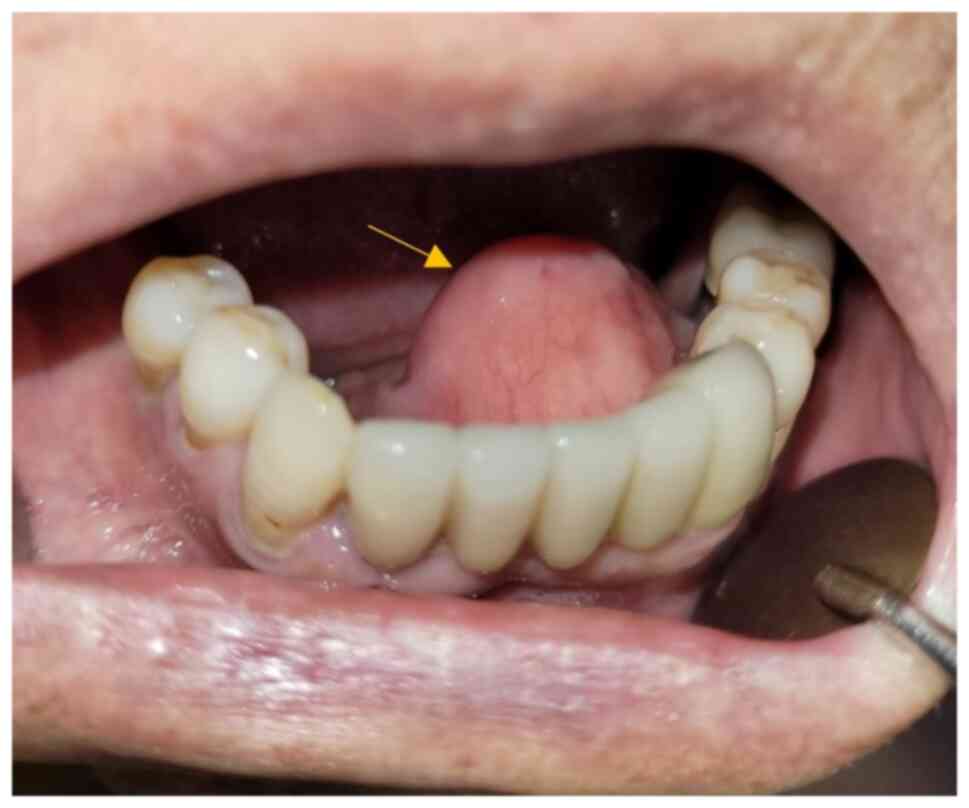
(Gangneung, South Korea) in April 2022 with a 35 mm-sized fluctuant pink lesion on the floor of the mouth. The lesion was dome-shaped and exophytic, with its top on the orifice area of Wharton's duct (Fig. 1). The patient was asymptomatic and had noticed the tumour 30 years before the visit, and it had been gradually increasing in size. When the first interviewer asked about pain before meals to rule out salivary gland-related lesions, the patient reported no food intake-related pain. However, the patient had dry mouth symptoms and the lesion elevated the tongue and interfered with speech and swallowing. Physical examination of the lesion revealed soft, mobile, tender and slow-growing masses, and the following differential diagnoses were considered: Ranula, haemangioma, lipoma, leiomyoma and neurilemmoma (9).

Figure 1

Pre-operative intra-oral view of the patient's condition. The lesion in question, identified as an angiolipoma, is prominently displayed (arrow). It exhibits a distinctive dome-like shape, indicative of its exophytic nature, which refers to its growth outward from the tissue surface. The apex of this lesion is specifically located at the orifice area of Wharton's duct, a crucial anatomical structure in the mouth floor responsible for the drainage of saliva from the submandibular glands. The visual representation aids in understanding the initial state of the lesion before any surgical intervention was performed.

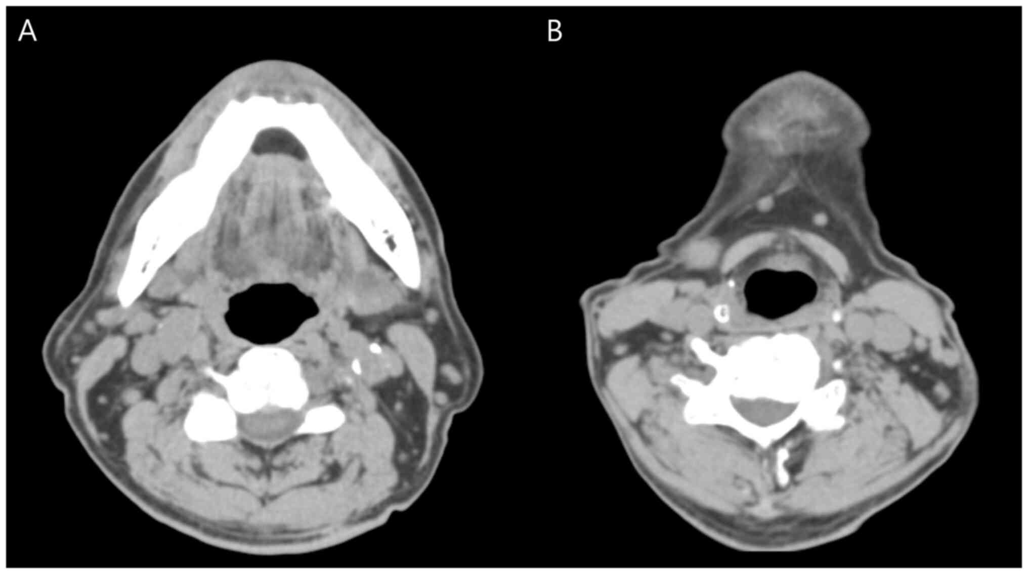
Enhanced computed tomography was performed for further evaluation. However, it is impossible to differentiate between ranula and lipoma based on Hounsfield units (ranula, 100; lipoma, 65-120), and due to artefacts from the dental crown, evaluation of soft tissue swelling in the mouth floor was impossible. The peripheral bone tissue exhibited no abnormal signs. Nodal enlargement in the submandibular and submental areas was detected and considered as reactive lymph nodes (Fig. 2). Given the features of the tumor, including its slow growth rate and clear margin, our initial assessment indicated a high likelihood of it being a benign mass. Consequently, no pre-operative plan for conducting frozen pathology was established. However, it is important to note that, while the tumor exhibited characteristics of a benign lesion, it is still possible for long-standing benign masses to undergo malignant transformation over time. Recently, the patient had experienced xerostomia and it was not possible to see whether the lesion had a yellowish color due to its deep location. The floor of the mouth contains numerous salivary glands, including the sublingual gland and the Warton's duct is also present. In addition, there was a possibility for dehiscence in the mylohyoid muscle, allowing mucus to drain into the submandibular space, resulting in a condition known as a ‘plunging ranula’. The clinical diagnosis before surgery was ranula.

Figure 2

Pre-operative enhanced CT scan. (A) The scan reveals the complexity of distinguishing between a ranula and a lipoma based solely on Hounsfield units, a measure of radiodensity. The Hounsfield units for a ranula typically register around 100, while a lipoma ranges between 65 and 120. This overlap in values makes differentiation challenging. Further complicating the evaluation is the presence of artefacts resulting from the dental crown, which obstructs the clear assessment of soft tissue swelling in the mouth floor. Despite these limitations, the CT scan does not indicate any specific abnormalities in the peripheral bone tissue. (B) Of note, the scan does reveal nodal enlargement in the submandibular and submental regions. This enlargement is interpreted as a reactive lymph node, a common immune response to infection or inflammation. CT, computed tomography.

Despite the patient's age, ranula, which is prevalent in children, could not be ruled out, and for this reason, marsupialisation and fibrin glue injection would have been required 
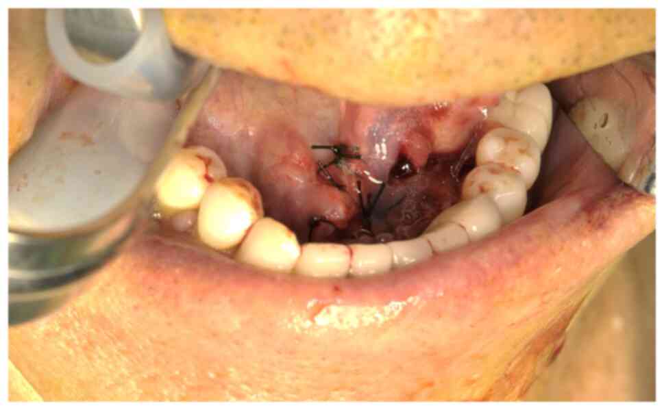
(10). Under general anaesthesia, an intraoral incision was made in the lesion's periphery as a trapdoor, preserving the Wharton's duct (Fig. 3). When a small incision was made at the margin of the exophytic lesion on the floor of the mouth, the soft yellowish tissue extruded slightly. As this yellowish mass resembled adipose tissue, the treatment plan was changed from 
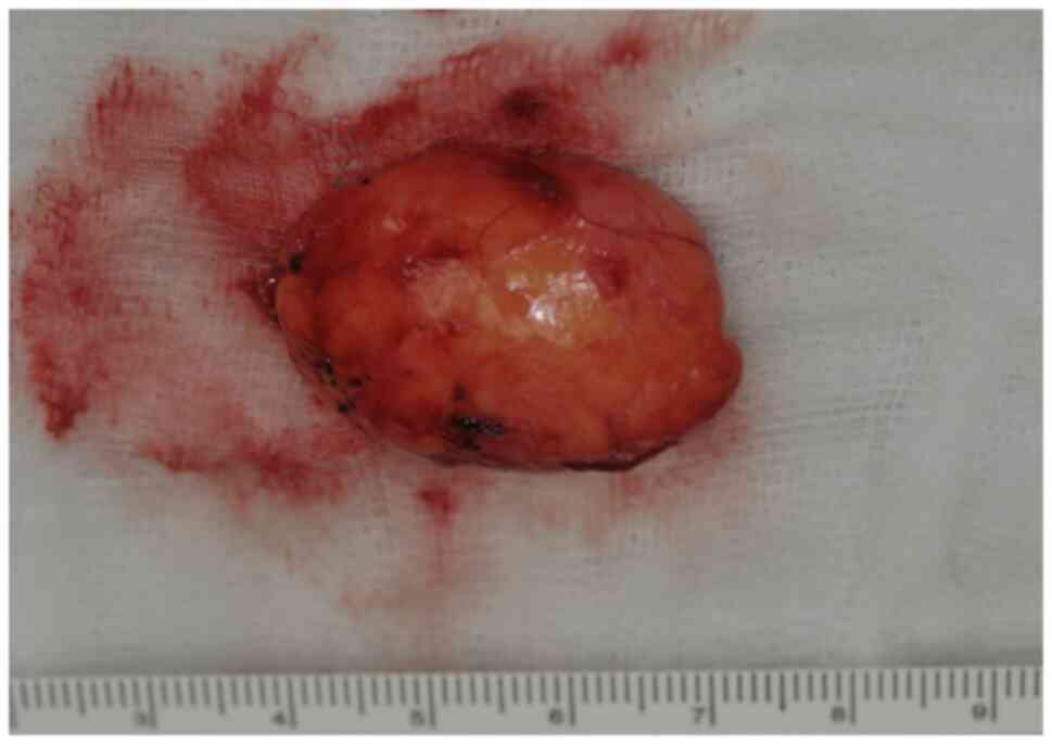
marsupialisation to surgical excision. The mass was well encapsulated. The encapsulated lesion was excised (Fig. 4) and referred for pathological examination with an adjacent tissue (trapdoor) sample.

Figure 3

Peri-operative intra-oral view following the surgical intervention. The surgical site in the floor of the mouth was reconstructed using circumferential tissue, a technique that involves the strategic placement of tissue to mimic the original anatomical structure. This method was employed to restore the mouth floor to its normal function and appearance. Following the procedure, the wound underwent a healing process known as secondary intention. This process allows the wound to heal naturally from the inside out, without the need for sutures or other closure methods. The image provides a visual representation of circumferential tissue used to approximate for healing by secondary intention and the successful reconstruction of the mouth floor.

Figure 4

Gross appearance of the excised mass following the surgical procedure. The mass (~3.5 cm) is characterized by a well-defined encapsulation, a protective layer that often surrounds such growths. This encapsulated lesion was carefully excised, ensuring minimal disturbance to the surrounding tissues. Along with the lesion, an adjacent tissue sample, often referred to as a ‘trapdoor’ sample due to its shape and method of extraction, was also removed. Both the excised lesion and the trapdoor sample were subsequently sent for pathological examination. This step is crucial for confirming the diagnosis and understanding the nature of the lesion in greater detail.

For pathological evaluation, Harris hematoxylin & eosin (regressive) stain was performed according to standard procedures. Prior to staining, samples were fixed using 10% neutral buffered formalin for 24 h at room temperature (20˚C). The thickness of the sections was 4 µm and staining was performed at room temperature (20˚C; 1 and 5 min). An Olympus BX50 muti microscope (Olympus Corp.) was used for analysis. The specimens revealed large fibrous stroma-encapsulated fatty tissue without atypia. Numerous capillaries proliferated into the fatty tissue. The capillaries contained fibrin thrombi (11) and fibrofatty changes (12). The adjacent tissue exhibited loose collagenised fibrous epithelial tissue with capillary proliferation. No infiltration of adipose cells was observed in this specimen and no atypical changes were observed (Fig. 5).
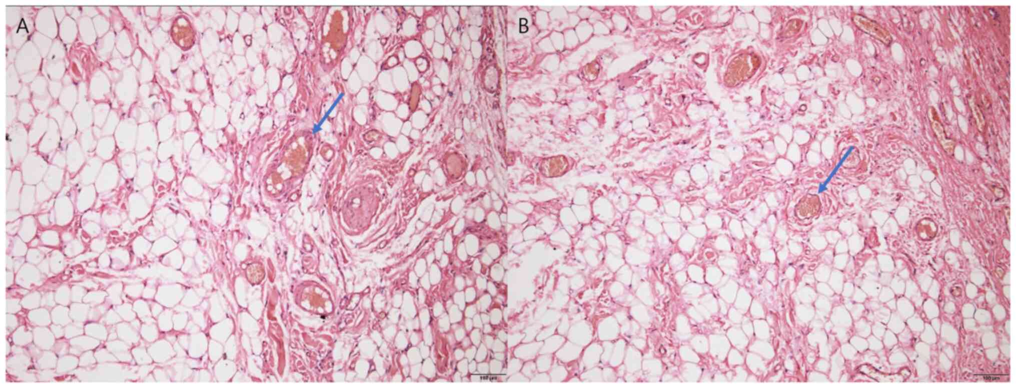
Figure 5

Pathological evaluation of the excised mass. (A) The evaluation revealed a large fibrous stroma, a connective tissue framework, encapsulating fatty tissue. Of note, this fatty tissue did not exhibit any signs of atypia, suggesting the absence of abnormal or dysplastic cells. Within this fatty tissue, a significant proliferation of capillaries was observed, indicating an active blood supply to the area (arrow). (B) These capillaries were found to contain fibrin thrombi (arrow), clots formed by fibrin proteins, and exhibited fibrofatty changes. These changes typically involve the replacement or transformation of normal tissue structures with fibrous and fatty tissues. The image provides a detailed visual representation of these pathological findings (haematoxylin and eosin stain; magnification, x100; scale bars, 100 µm).

The final diagnosis was non-infiltrating angiolipoma. The symptoms of dry mouth may have been a consequence of mass compression. The patient recovered without any complications and no evidence of recurrence or discomfort was observed 15 months postoperatively.

Discussion

The present case study reported on a rare case of non-infiltrating angiolipoma that was confused with a ranula, as the patient had a dry mouth. Recurrence has been frequently reported in cases of the infiltrating form of the lesion (4,13). Histopathologic evaluation revealed fibrous encapsulated tumour formation composed of >50% mature lipocytes with angiomatous proliferation consisting of several small-to-medium-sized congested capillaries containing fibrin thrombi (3,14).

Infiltrating angiolipoma is more common in elderly patients; however, the patient of the present study was a rare case of non-infiltrating angiolipoma on the floor of the mouth in an elderly patient. There are various hypotheses regarding the aetiology of angiolipoma: i) History of trauma, ii) lipomatous differentiation by hormones at puberty, iii) fatty degeneration of central haemangioma, and iv) vascular proliferation of congenital lipoma (15). Considering age, one possible aetiology is vascular proliferation on the pre-existing oral lipoma.

It may be enlarged, such as a ‘plunging ranula’, potentially causing airway problems if it was a ranula. Magnetic resonance imaging (MRI) is the gold standard for further differential diagnosis of the ranula. The T2-weighted images demonstrate a characteristic heterogeneously increased signal within the lesion (16). However, if the ranula has a high protein concentration, the signal intensities may be high for all of the imaging sequences (16). In such cases, a differential diagnosis of lipomas is difficult (17). The relatively high cost of MRI must also be considered. In addition, if the lesion shows a prominent arterial supply on angiography, it is a candidate for preoperative embolization (18).

Marsupialisation has been reported for the management of ranula, odontogenic cysts (odontogenic keratocyst) and benign odontogenic tumours (ameloblastoma followed by enucleation). However, the recurrence rate continues to be of concern (14-67, 12 and 29.3%, respectively) (19,20). In marsupialisation, the cyst lining is sutured to the oral mucosa and the mouth floor is reconstructed. However, in the present study, the mass was resected, including the capsule and the overlying mucosa. This enables pathological diagnosis and may reduce the possibility of recurrence without any additional surgery. Circumferential tissue was used to approximate and reconstruct the floor of the mouth and the wound was healed by secondary intention.

In the literature, the overall prognosis for these lesions is good, as no malignant transformation has been reported (4,21). However, these benign tumours do not spontaneously regress and may become larger, more tender and more disfiguring (9,21). In addition, as there is a higher incidence of infiltrating angiolipoma, definite treatment should be considered in elderly patients.

In conclusion, as non-infiltrating angiolipoma of the floor of the mouth in an elderly patient is a rare pathology, it is important to diagnose the lesion exactly and treat it by intraoral approach without the possibility of complications.

Acknowledgements

Not applicable.

Funding

Funding: This study was supported financially by Gangneung-Wonju National University Dental Hospital (Gangneung, Gangwon, Korea).

Availability of data and materials

All data generated or analyzed during this study are included in this published article.

Authors' contributions

YJK and SGK performed the surgical treatment. Pathological examination and diagnosis was performed by SSL. Additional pathological analysis and literature review was completed by YJK and SGK. YJK wrote the manuscript. SSL and SGK confirm the authenticity of all the raw data. SGK supervised the study. All authors have read and approved the final version of the manuscript.

Ethics approval and consent to participate

This study was approved by the IRB of Gangneung-Wonju National University Dental Hospital (Gangneung, Korea; no. GWNUDH-IRB2022-A013).

Patient consent for publication

Not applicable.

Competing interests

The authors declare that they have no competing interests.

References

1 

Chandrasekaran D, Chinnaswami R, Narasimhan M, Kumar AEN and Natarajan P: Non infiltrating angiolipoma of the palate in geriatric patient: A case report with review of literature. J Clin Diagn Res. 10:ZD01–ZD02. 2016.PubMed/NCBI View Article : Google Scholar

2 

Hoeft S, Luettges J and Werner JA: Infiltrating angiolipoma of the M. temporalis. Auris Nasus Larynx. 27:265–269. 2000.PubMed/NCBI View Article : Google Scholar

3 

Silva-Junior GO, Picciani BL, Costa RC, Barbosa SM, Silvares MG, Souza RB, Cantisano MH and Pires FR: Oral soft-tissue angiolipoma: Report of two cases of rare oral lipomatous lesion with emphasis on morphological and immunohistochemical features. J Oral Sci. 55:85–88. 2013.PubMed/NCBI View Article : Google Scholar

4 

Yanase S, Nomura J, Matsumura Y, Kato H, Takeoka T, Imura H, Matsuura R, Nakanishi K and Tagawa T: Angiolipoma of the cheek: A case report with a literature review. Asian J Oral Maxillofac Surg. 23:35–37. 2011.

5 

Saydam L, Bozkurt MK, Ugur MB, Ozcelik T and Kutluay L: Angiolipoma of the neck: A case report. Ear Nose Throat J. 84:375–377. 2005.PubMed/NCBI

6 

Auo HJ and Kang JM: Infiltrating angiolipoma of the nasopharynx: Adjacent to an aberrant internal carotid artery. Auris Nasus Larynx. 36:247–250. 2009.PubMed/NCBI View Article : Google Scholar

7 

Lin JJ and Lin F: Two entities in angiolipoma. A study of 459 cases of lipoma with review of literature on infiltrating angiolipoma. Cancer. 34:720–727. 1974.PubMed/NCBI View Article : Google Scholar

8 

Gerard N and Schultz DA: Angiolipoma of the upper lip: Report of a case. J Oral Maxillofac Surg. 67:1340–1341. 2009.PubMed/NCBI View Article : Google Scholar

9 

Reiser V, Haj Yahya B, Chaushu G, Kaplan I and Hamzani Y: Angiolipoma in the head and neck: Imaging, diagnosis and management. Medicina (Kaunas). 56(283)2020.PubMed/NCBI View Article : Google Scholar

10 

Kokong D, Iduh A, Chukwu I, Mugu J, Nuhu S and Augustine S: Ranula: Current concept of pathophysiologic basis and surgical management options. World J Surg. 41:1476–1481. 2017.PubMed/NCBI View Article : Google Scholar

11 

Ohnishi Y, Watanabe M, Fujii T, Yasui H, Kubo H and Kakudo K: Infiltrating angiolipoma of the lower lip: A case report and literature review. Oncol Lett. 9:833–836. 2015.PubMed/NCBI View Article : Google Scholar

12 

Hemavathy S, Roy S and Kiresur A: Intraosseous angiolipoma of the mandible. J Oral Maxillofac Pathol. 16:283–287. 2012.PubMed/NCBI View Article : Google Scholar

13 

Flaggert JJ III, Heldt LV and Keaton WM: Angiolipoma of the palate: Report of a case. Oral Surg Oral Med Oral Pathol. 61:333–336. 1986.PubMed/NCBI View Article : Google Scholar

14 

Shah VS, Harish M, Patel JR and Shah N: Infiltrating angiolipoma of the cheek. BMJ Case Rep. 2013(bcr2013200041)2013.PubMed/NCBI View Article : Google Scholar

15 

Palaia G, Gaimari G, Giudice RL, Galanakis A, Tenore G and Romeo U: Excision of an oral angiolipoma by KTP laser: A case report. Ann Stomatol (Roma). 2:28–31. 2011.PubMed/NCBI

16 

Gupta A and Karjodkar F: Plunging ranula: A case report. ISRN Dent. 2011(806928)2011.PubMed/NCBI View Article : Google Scholar

17 

Brandwein M and Som P: Salivary glands: Anatomy and pathology. In: Som RM, Curtin HD (eds). Head and Neck Imaging, 4th edition. St. Louis, MO: Mosby, pp2039-2050, 2003.

18 

Rabin D, Hon BA, Pelz DM, Ang LC, Lee DH and Duggal N: Infiltrating spinal angiolipoma: A case report and review of the literature. J Spinal Disord Tech. 17:456–461. 2004.PubMed/NCBI View Article : Google Scholar

19 

Hong J, Yun PY, Chung IH, Myoung H, Suh JD, Seo BM, Lee JH and Choung PH: Long-term follow up on recurrence of 305 ameloblastoma cases. Int J Oral Maxillofac Surg. 36:283–288. 2007.PubMed/NCBI View Article : Google Scholar

20 

Kademani D and Tiwana P: Atlas of oral and maxillofacial surgery. 1st edition. Elsevier Health Sciences, 2015.

21 

Alvi A, Garner C and Thomas W: Angiolipoma of the head and neck. J Otolaryngol. 27:100–103. 1998.PubMed/NCBI

Related Articles

  • Abstract
  • View
  • Download
  • Twitter
Copy and paste a formatted citation
Spandidos Publications style
Kang Y, Lee S and Kim S: Intraoral approach for the treatment of non‑infiltrating angiolipoma of the floor of the mouth in an elderly patient: A case report with review of the literature. Exp Ther Med 26: 458, 2023.
APA
Kang, Y., Lee, S., & Kim, S. (2023). Intraoral approach for the treatment of non‑infiltrating angiolipoma of the floor of the mouth in an elderly patient: A case report with review of the literature. Experimental and Therapeutic Medicine, 26, 458. https://doi.org/10.3892/etm.2023.12157
MLA
Kang, Y., Lee, S., Kim, S."Intraoral approach for the treatment of non‑infiltrating angiolipoma of the floor of the mouth in an elderly patient: A case report with review of the literature". Experimental and Therapeutic Medicine 26.3 (2023): 458.
Chicago
Kang, Y., Lee, S., Kim, S."Intraoral approach for the treatment of non‑infiltrating angiolipoma of the floor of the mouth in an elderly patient: A case report with review of the literature". Experimental and Therapeutic Medicine 26, no. 3 (2023): 458. https://doi.org/10.3892/etm.2023.12157
Copy and paste a formatted citation
x
Spandidos Publications style
Kang Y, Lee S and Kim S: Intraoral approach for the treatment of non‑infiltrating angiolipoma of the floor of the mouth in an elderly patient: A case report with review of the literature. Exp Ther Med 26: 458, 2023.
APA
Kang, Y., Lee, S., & Kim, S. (2023). Intraoral approach for the treatment of non‑infiltrating angiolipoma of the floor of the mouth in an elderly patient: A case report with review of the literature. Experimental and Therapeutic Medicine, 26, 458. https://doi.org/10.3892/etm.2023.12157
MLA
Kang, Y., Lee, S., Kim, S."Intraoral approach for the treatment of non‑infiltrating angiolipoma of the floor of the mouth in an elderly patient: A case report with review of the literature". Experimental and Therapeutic Medicine 26.3 (2023): 458.
Chicago
Kang, Y., Lee, S., Kim, S."Intraoral approach for the treatment of non‑infiltrating angiolipoma of the floor of the mouth in an elderly patient: A case report with review of the literature". Experimental and Therapeutic Medicine 26, no. 3 (2023): 458. https://doi.org/10.3892/etm.2023.12157
Follow us
  • Twitter
  • LinkedIn
  • Facebook
About
  • Spandidos Publications
  • Careers
  • Cookie Policy
  • Privacy Policy
How can we help?
  • Help
  • Live Chat
  • Contact
  • Email to our Support Team