Introduction
The global prevalence of dementia has exponentially
increased due to prolonged life expectancy and an aging population.
Currently, >55 million people are suffering from dementia
globally (1). The syndrome of
dementia is caused by a various diseases that eventually destroy
nerve cells, thereby damaging the brain, and causing cognitive
ability deterioration compared with that expected from the normal
biological aging course (1,2).
Mild cognitive impairment (MCI), can be considered the
‘transitional zone’ between normal cognition and dementia and has
become a novel topic in clinical research (3).
MCI is an early stage that can result in dementia.
MCI can be reversed and diagnosis at an early stage is crucial to
control the progression to dementia. Currently, neuropsychological
testing (4), transcranial
ultrasonography (5), near-infrared
spectroscopy that enables measurement of hemoglobin concentration
changes in the brain (6), magnetic
resonance imaging (7), detection
of cerebrospinal fluid biomarkers, detection of such as amyloid
beta and phosphorylated tau protein in spinal fluid (8,9),
electroencephalography (10), and
olfaction tests (11) are the
diagnostic modalities used to diagnose dementia. However, all of
these testing methods are useful in diagnosing dementia, but they
cannot detect MCI with sensitivity.
MicroRNAs, which are small RNA molecules that
regulate gene expression in cells, are found in blood and are
mainly encapsulated in extracellular vesicles such as exosomes
(12,13). In recent years, many researchers
have revealed serum microRNAs as biomarkers for early disease
detection (14,15). This study aimed to clarify changes
in serum microRNAs in cognitively impaired mice.
Materials and methods
Mice
We purchased male C57BL/6NJcl mice from CLEA Japan.
All mice were provided solid diet CE-2 (CLEA Japan) and water ad
libitum and were housed in a conventional animal room with 12-h
light/dark cycles. Mice were housed up to 5 mice per gauge, and
bedding, feed, and water were changed weekly. In this study, 10
4-week-old (4W), 9 8-week-old (8W), 5 36-week-old (36W) and 6
58-week-old (58W) mice were used. We consulted the report of
Ackert-Bicknell et al in determining the age groups
(16). The weight of each mouse
was measured using an electronic balance and is shown in the
results. Mice were observed 2-3 times per day for monitoring, and
no abnormalities in mouse health or behavior were observed during
the rearing period. Blood samples from all mice were collected by
cardiac blood sampling was collected under anesthesia with the
inhalation anesthetic solution isoflurane (Pfizer) at the end of
the 4W, 8W, 36W and 58W time points. Small animal anesthesia
machines (Muromachi Kikai) were used to anesthetize the mice, and
isoflurane vaporized to a concentration of 4-5% was inhaled into
the mice and maintained at 2-3% throughout the experiment, and
blood was drawn from the mouse hearts. After anesthesia,
approximately 0.5-1.0 ml of blood was drawn from the heart, and
mice were promptly cervically dislocated to minimize distress as a
humane endpoint. From the start of anesthesia to the end of blood
collection took less than 10 min per animal. Death was confirmed by
respiratory arrest and cardiac arrest. All mice used were
euthanized immediately after the experiment. Blood samples were
then placed in a Microtainer (Becton Dickinson), and the coagulated
blood was centrifuged at 6000 G for 3 min for serum separation. The
Hirosaki University Ethics Committee for Animal Experiments
approved this experiment which was conducted under the Hirosaki
University Guidelines for Animal Experiments (Approval No.
AE01-2023-004).
Biochemical examination
Serum alkaline phosphatase (ALP) and total
cholesterol levels were measured to evaluate age-related
biochemical changes. LabAssay™ ALP (Fujifilm Wako Shibayagi Co.,
Ltd.) was used to measure ALP, following the kit protocol.
LabAssay™ Cholesterol (Fujifilm Wako Shibayagi Co., Ltd.) was used
to measure total cholesterol following the kit protocol.
Morris water maze test
The Morris water maze test, which was conducted in a
circular pool of 120 cm in diameter and 80 cm high, was used to
assess the cognitive ability of mice (17,18).
Markers were placed around the pool, and the mice were allowed to
swim in the pool filled with water with a platform for evacuation
under the water surface at one location only. The maximum time to
reach the platform was 60 sec. Mice that reached the platform were
returned to the gauge after letting them stay on the platform for
20 sec. Mice which failed to reach the platform within 60 sec were
led to the platform and allowed to stay for 20 sec to memorize the
platform location. This was repeated for five days, starting with
three locations per day, and the reduced time taken to reach the
platform was used to evaluate cognitive ability.
Total RNA extraction
Isogen II reagent and ethachinmate (Nippongene) were
used to extract total RNAs from 200-µl of sera following the
manufacturer's instructions. A Qubit 4 Fluorometer (ThermoFisher
Scientific) and Qubit™ microRNA Assay kits (ThermoFisher
Scientific) were used to measure the total RNA concentration
extracted from sera. An Agilent 2100 Bioanalyzer and an Agilent RNA
6000 Pico Kit (Agilent Technologies) were used to determine and
confirm the peaks characteristic of the total RNAs, following the
manufacturer's instructions.
Microarray analysis
SurePrint G3 Mouse miRNA 8 x 60-K Microarray
(Agilent Technologies) was used to analyze serum microRNA
expression. Cyanine 3 labeling was conducted with 1 ng of serum
microRNA using the miRNA Complete Labeling and Hyb kit (Agilent
Technologies), following the manufacturer's instructions.
Hybridization, microarray slide washing, and fluorescence image
scanning were performed using the procedure described in our
previous report (19). MicroRNAs
whose expression varied >1.5-fold (P<0.05) were
screened using fold change analysis and Student's t-test (unpaired)
at 58W compared with 8W using GeneSpring 14.9 software (Agilent
Technologies). The obtained microarray data were registered with
Gene Expression Omnibus (GSE249248).
Statistical analysis
Statcel 3 software (OMS Publishing Inc.) was used
for statistical analyses. One-way analysis of variance was
performed to compare the results of the three groups and the
Tukey-Kramer test was used for multiple comparisons. P<0.05 was
considered to indicate a statistically significant difference.
Results
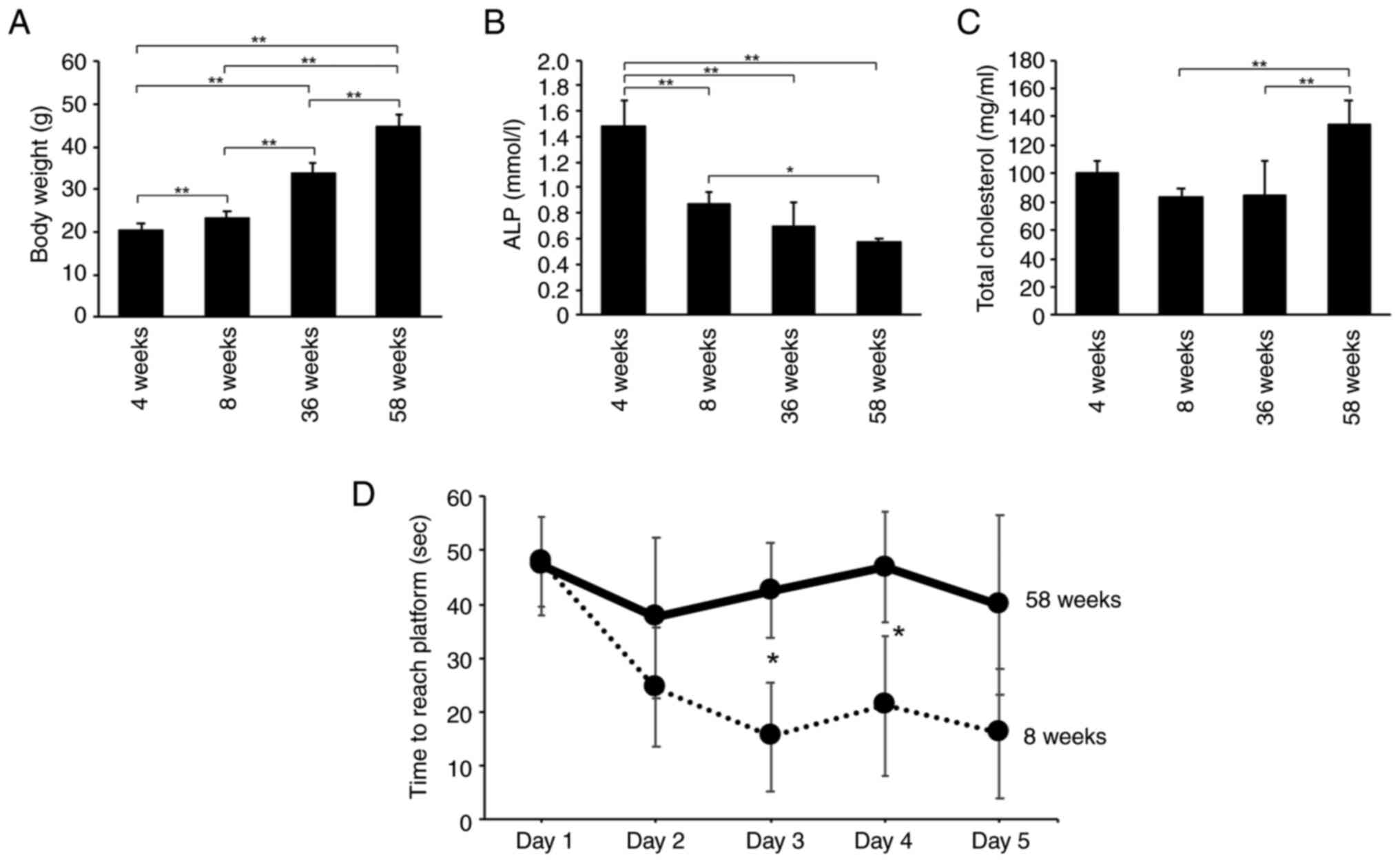
Changes in body weight, serum ALP,
serum total cholesterol and cognitive ability during aging
An electronic balance was used to weigh 4, 8, 36 and
58W mice. The weight of the mice increased with aging, with a
statistically significant difference in body weight between 8W and
58W (each n=4) (P<0.01) (Fig.
1A). The mice underwent serum biochemical tests serum was
conducted on the mice, which demonstrated highest serum ALP in 4W
mice and decreased with aging, with a statistically significant
difference in ALP between 4W or 8W and 58W mice (each n=3-4)
(P<0.05) (Fig. 1B). Serum total
cholesterol was highest in 58W mice and was observed to increase
with aging, with a statistically significant difference in total
cholesterol between 8W and 58W mice (each n=3-4) (P<0.01)
(Fig. 1C). The other parameters,
including red blood cell count, white blood cell count, hemoglobin
concentration, hematocrit, mean corpuscular volume, mean
corpuscular hemoglobin, mean corpuscular hemoglobin concentration,
neutrophil count, lymphocyte count, and monocyte count, between the
groups did not demonstrate statistically significant differences
(Fig. S1).
The Morris water maze test revealed a significantly
delayed case reaching the platform in 58W compared to 8W on the
third and fourth day (each n=5-6) (P<0.05) (Fig. 1D). These results indicated that the
58W mice demonstrated signs of aging and were beginning to decline
in cognitive ability.
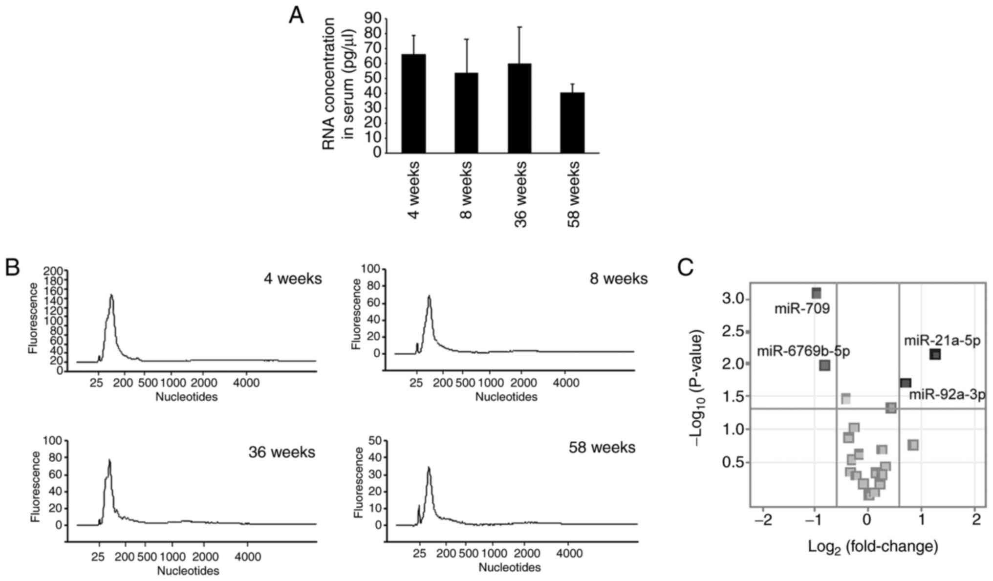
Changes in microRNA expression in
mouse serum with aging
The total RNA was extracted from the serum and
analyzed to identify the aging-associated changes occurring in
serum RNA. Serum RNA concentrations in 4W, 8W, 36W and 58W mice
were evaluated, and statistically significant differences were not
noted in serum RNA concentrations respectively (each n=4) (Fig. 2A). Next, RNA electrophoresis was
conducted to determine the size of RNA in the serum of 4W, 8W, 36W
and 58W mice using an Agilent bioanalyzer. A single peak of small
RNA ranging from 25 to 200 nucleotides in size was detected in the
serum RNA of all 4W, 8W, 36W and 58W mice (Fig. 2B).
We then performed miRNA microarrays using serum RNA
from 8W and 58W mice (each n=4). The results indicated that the
serum of the 58W mice demonstrated 1.5 times greater expression of
two microRNAs (miR-21a-5p and miR-92a-3p) and 1.5 times lowered
expression of two microRNAs (miR-6769b-5p and miR-709) compared
with the serum of 8W mice (Fig.
2C). These results indicate the presence of microRNAs in serum
with altered expression corresponding to a decline in cognitive
ability.
Discussion
This study revealed microRNAs whose expression was
altered in the serum of middle-aged 58W mice, which demonstrated
reduced cognitive ability compared with young 8W mice with normal
cognitive ability.
In general, women are at higher risk of developing
dementia than men, but we used male mice in this study to examine
changes in blood composition during aging because of the article by
Hahm et al (20). Although
not included in the present data, we conducted experiments using
mice older than 58W, but because we could not obtain sufficient
samples of older mice due to spontaneous mouse death and other
factors, we used mice up to 58W in this study for this study.
Changes in body weight, serum biochemistry, and
cognitive performance with aging were examined in 4W, 8W, 36W and
58W mice. A statistically significant increase in body weight was
found among each mouse as they aged (Fig. 1A). Previous reports revealed that
the body weight of mice increased with age (21). Further, serum ALP is higher in
young age, and serum total cholesterol is higher in old age
(21,22). Hence, serum ALP and serum total
cholesterol were measured to infer the age of the mice used. Serum
ALP was statistically significantly lower in the 58W group than in
the 4W or 8W group (Fig. 1B).
Furthermore, serum total cholesterol was significantly higher in
the 58W group than in the 8W group (Fig. 1C). These results are consistent
with those of previous studies. A decrease in cognitive ability was
observed in 58W mice compared with 8W mice when the cognitive
abilities of 8W and 58W mice with statistically significant
differences in serum ALP and serum total cholesterol were confirmed
(Fig. 1D). We then examined the
difference in serum microRNA expression between 8W and 58W mice
with significant differences in cognitive ability (Fig. 2C). We identified miR-21a-5p and
miR-92a-3p as microRNAs that were upregulated and miR-6769b-5p and
miR-709 as microRNAs that were downregulated in the serum of 58W
mice compared with 8W mice. In this study, swimming time was
assessed in the Morris water maze test. In order to fully evaluate
cognitive function, it is necessary to measure and evaluate swim
path, swim speed, and crossing number. In the future, we would like
to evaluate cognitive function using mice aged more than 58W as
well as various measurement methods in the Morris water maze
test.
This study revealed elevated serum miR-21a-5p
expression in mice with impaired cognitive ability (Fig. 2C). Very few studies have revealed
the association between cognitive ability and serum miR-21a-5p
levels. Yuan et al revealed an increased expression of serum
miR-21 in patients with cognitive impairment following the
incidence of stroke compared with patients with normal cognition
following stroke (23). Other
previously conducted studies have revealed that miR-21 plays a
crucial role in cognitive ability regulation because an increase in
miR-21 in the brains of Alzheimer's model mice was observed to
restore cognitive ability (24,25).
This result indicates that the loss of miR-21 in the brain and its
leakage into the serum is a factor associated with cognitive
decline. Further studies that explore the relationship between
cognitive ability and miR-21 expression should be conducted in the
future.
Several studies reported on the association between
serum miR-92a-3p levels and dementia, and several investigators
have reported an association between miR-92a-3p levels and
cognitive ability. Siedlecki-Wullich et al demonstrated a
statistically significant increase in serum miR-92a-3p in the
patient group with Alzheimer's disease compared with the group with
normal cognitive abilities and indicated an increasing trend in the
incidence of this serum in the MCI group (26). However, Peña-Bautista et al
contradicted this claim by revealing a decrease in plasma
miR-92a-3p levels in patients with early Alzheimer's disease
(27).
This study revealed that a decrease in serum miR-709
levels reduces in mice with cognitive decline. This is a novel
result because previous reports of an association between serum
miR-709 levels and cognitive ability are limited. Further studies
are warranted to confirm and elaborate the association between
serum miR-709 levels and cognitive ability. In the future, we would
like to further analyze aged mice to reveal novel MCI
biomarkers.
Supplementary Material
Changes in blood cell count and
fractionation. (A) Red blood cell count (each n=4); (B) hemoglobin
concentration (each n=4); (C) hematocrit (each, n=5-10); (D) MCV
(each n=4); (E) MCH (each n=4); (F) MCHC (each n=4); (G) white
blood cell count (each n=3-4); (H) neutrophil count (each n=3-8);
(I) lymphocyte count (each n=3-7); (J) monocyte count (each n=3-5).
MCH, mean corpuscular hemoglobin; MCHC, mean corpuscular hemoglobin
concentration; MCV, mean corpuscular volume; W, weeks-old.
Acknowledgements
Not applicable.
Funding
Funding: This work was supported by JST SPRING (grant no.
JPMJSP2152).
Availability of data and materials
The microarray data generated in the present study
may be found in the Gene Expression Omnibus under accession number
GSE249248 or at the following URL: https://www.ncbi.nlm.nih.gov/geo/query/acc.cgi?acc=GSE249248.
The other data generated in the present study may be requested from
the corresponding author.
Authors' contributions
KY and MC confirm the authenticity of all the raw
data. KY, KoM, MF and MC were major contributors in performing the
experiments and writing the manuscript. WK, HO and KaM helped
conduct the experiments. All authors read and approved the final
manuscript.
Ethics approval and consent to
participate
All experiments were performed in accordance with
The Guideline for Animal Experimentation of Hirosaki University.
The Animal Research Committee of Hirosaki University (approval no.
AE01-2023-097-1) approved and monitored the procedures.
Patient consent for publication
Not applicable.
Competing interests
The authors declare that they have no competing
interests.
References
|
1
|
World Health Organization (WHO): Dementia.
https://www.who.int/en/news-room/fact-sheets/detail/dementia.
Accessed August 19, 2023.
|
|
2
|
Błaszczyk JW: Pathogenesis of dementia.
Int J Mol Sci. 24(543)2022.PubMed/NCBI View Article : Google Scholar
|
|
3
|
Wolf A, Tripanpitak K, Umeda S and
Otake-Matsuura M: Eye-tracking paradigms for the assessment of mild
cognitive impairment: A systematic review. Front Psychol.
14(1197567)2023.PubMed/NCBI View Article : Google Scholar
|
|
4
|
Del Bene VA, Gerstenecker A and Lazar RM:
Formal neuropsychological testing: Test batteries, interpretation,
and added value in practice. Clin Geriatr Med. 39:27–43.
2023.PubMed/NCBI View Article : Google Scholar
|
|
5
|
Di Lazzaro V, Bella R, Benussi A, Bologna
M, Borroni B, Capone F, Chen KS, Chen R, Chistyakov AV, Classen J,
et al: Diagnostic contribution and therapeutic perspectives of
transcranial magnetic stimulation in dementia. Clin Neurophysiol.
132:2568–2607. 2021.PubMed/NCBI View Article : Google Scholar
|
|
6
|
Devezas MÂM: Shedding light on
neuroscience: Two decades of functional near-infrared spectroscopy
applications and advances from a bibliometric perspective. J
Neuroimaging. 31:641–655. 2021.PubMed/NCBI View Article : Google Scholar
|
|
7
|
Botz J, Lohner V and Schirmer MD: Spatial
patterns of white matter hyperintensities: A systematic review.
Front Aging Neurosci. 15(1165324)2023.PubMed/NCBI View Article : Google Scholar
|
|
8
|
Papaliagkas V, Kalinderi K, Vareltzis P,
Moraitou D, Papamitsou T and Chatzidimitriou M: CSF biomarkers in
the early diagnosis of mild cognitive impairment and Alzheimer's
disease. Int J Mol Sci. 24(8976)2023.PubMed/NCBI View Article : Google Scholar
|
|
9
|
Hansson O, Blennow K, Zetterberg H and
Dage J: Blood biomarkers for Alzheimer's disease in clinical
practice and trials. Nat Aging. 3:506–519. 2023.PubMed/NCBI View Article : Google Scholar
|
|
10
|
Ulbl J and Rakusa M: The importance of
subjective cognitive decline recognition and the potential of
molecular and neurophysiological biomarkers-A systematic review.
Int J Mol Sci. 24(10158)2023.PubMed/NCBI View Article : Google Scholar
|
|
11
|
Fatuzzo I, Niccolini GF, Zoccali F,
Cavalcanti L, Bellizzi MG, Riccardi G, de Vincentiis M, Fiore M,
Petrella C, Minni A, et al: Neurons, nose, and neurodegenerative
diseases: Olfactory function and cognitive impairment. Int J Mol
Sci. 24(2117)2023.PubMed/NCBI View Article : Google Scholar
|
|
12
|
Jahromi FNA, Dowran R and Jafari R: Recent
advances in the roles of exosomal microRNAs (exomiRs) in
hematologic neoplasms: Pathogenesis, diagnosis, and treatment. Cell
Commun Signal. 21(88)2023.PubMed/NCBI View Article : Google Scholar
|
|
13
|
Jain G, Das P, Ranjan P, Neha Valderrama F
and Cieza-Borrella C: Urinary extracellular vesicles miRNA-A new
era of prostate cancer biomarkers. Front Genet.
14(1065757)2023.PubMed/NCBI View Article : Google Scholar
|
|
14
|
Bielak C, Arya A and Savill S: Circulating
microRNA as potential diagnostic and prognostic biomarkers of
well-differentiated thyroid cancer: A review article. Cancer
Biomark. 36:193–205. 2023.PubMed/NCBI View Article : Google Scholar
|
|
15
|
Nestler T, Schoch J, Belge G and Dieckmann
KP: MicroRNA-371a-3p-The novel serum biomarker in testicular germ
cell tumors. Cancers (Basel). 15(3944)2023.PubMed/NCBI View Article : Google Scholar
|
|
16
|
Ackert-Bicknell CL, Anderson LC, Sheehan
S, Hill WG, Chang B, Churchill GA, Chesler EJ, Korstanje R and
Peters LL: Aging research using mouse models. Curr Protoc Mouse
Biol. 5:95–133. 2015.PubMed/NCBI View Article : Google Scholar
|
|
17
|
Othman MZ, Hassan Z and Che Has AT: Morris
water maze: A versatile and pertinent tool for assessing spatial
learning and memory. Exp Anim. 71:264–280. 2022.PubMed/NCBI View Article : Google Scholar
|
|
18
|
Vorhees CV and Williams MT: Morris water
maze: Procedures for assessing spatial and related forms of
learning and memory. Nat Protoc. 1:848–858. 2006.PubMed/NCBI View Article : Google Scholar
|
|
19
|
Chiba M, Monzen S, Iwaya C, Kashiwagi Y,
Yamada S, Hosokawa Y, Mariya Y, Nakamura T and Wojcik A: Serum
miR-375-3p increase in mice exposed to a high dose of ionizing
radiation. Sci Rep. 8(1302)2018.PubMed/NCBI View Article : Google Scholar
|
|
20
|
Hahm JR, Jo MH, Ullah R, Kim MW and Kim
MO: Metabolic stress alters antioxidant systems, suppresses the
adiponectin receptor 1 and induces Alzheimer's like pathology in
mice brain. Cells. 9(249)2020.PubMed/NCBI View Article : Google Scholar
|
|
21
|
Mazzaccara C, Labruna G, Cito G, Scarfò M,
De Felice M, Pastore L and Sacchetti L: Age-related reference
intervals of the main biochemical and hematological parameters in
C57BL/6J, 129SV/EV and C3H/HeJ mouse strains. PLoS One.
3(e3772)2008.PubMed/NCBI View Article : Google Scholar
|
|
22
|
Hu Y, Wang R, Wang S, Yuan C, Yuan Q,
Zhang Y, Ren J, He Y, Wu X, Dai W, et al: Selenium-enriched
polysaccharides from Pyracantha fortuneana (SePFP) ameliorated
hepatic lipid accumulation through enhancing mitochondrial fatty
acid β-oxidation in aging mice. J Oleo Sci. 72:929–938.
2023.PubMed/NCBI View Article : Google Scholar
|
|
23
|
Yuan M, Guo YS, Zhang XX, Gao ZK, Shen XY,
Han Y and Bi X: Diagnostic performance of miR-21, miR-124, miR-132,
and miR-200b serums in post-stroke cognitive impairment patients.
Folia Neuropathol. 60:228–236. 2022.PubMed/NCBI View Article : Google Scholar
|
|
24
|
Cui GH, Wu J, Mou FF, Xie WH, Wang FB,
Wang QL, Fang J, Xu YW, Dong YR, Liu JR and Guo HD: Exosomes
derived from hypoxia-preconditioned mesenchymal stromal cells
ameliorate cognitive decline by rescuing synaptic dysfunction and
regulating inflammatory responses in APP/PS1 mice. FASEB J.
32:654–668. 2018.PubMed/NCBI View Article : Google Scholar
|
|
25
|
Gao X, Xiong Y, Li Q, Han M, Shan D, Yang
G, Zhang S, Xin D, Zhao R, Wang Z, et al: Extracellular
vesicle-mediated transfer of miR-21-5p from mesenchymal stromal
cells to neurons alleviates early brain injury to improve cognitive
function via the PTEN/Akt pathway after subarachnoid hemorrhage.
Cell Death Dis. 11(363)2020.PubMed/NCBI View Article : Google Scholar
|
|
26
|
Siedlecki-Wullich D, Català-Solsona J,
Fábregas C, Hernández I, Clarimon J, Lleó A, Boada M, Saura CA,
Rodríguez-Álvarez J and Miñano-Molina AJ: Altered microRNAs related
to synaptic function as potential plasma biomarkers for Alzheimer's
disease. Alzheimers Res Ther. 11(46)2019.PubMed/NCBI View Article : Google Scholar
|
|
27
|
Peña-Bautista C, Tarazona-Sánchez A,
Braza-Boils A, Balaguer A, Ferré-González L, Cañada-Martínez AJ,
Baquero M and Cháfer-Pericás C: Plasma microRNAs as potential
biomarkers in early Alzheimer disease expression. Sci Rep.
12(15589)2022.PubMed/NCBI View Article : Google Scholar
|