Introduction
Esophageal squamous cell carcinoma (ESCC) is a
predominant histological subtype of esophageal cancer. ESCC ranks
as the sixth most common cancer among males and the ninth most
common cancer among females (1).
Worldwide, ESCC is also one of the most fatal malignancies, as
progression occurs without symptoms and many tumors are
significantly advanced by the time of diagnosis (2,3).
Despite recent advances in surgical techniques and therapy combined
with chemotherapy or chemo-radiotherapy (4–6),
many ESCC patients develop metastatic disease. In particular,
distant metastasis hinders a favorable outcome for patients with
ESCC by limiting the surgical cure (7,8). As
a result, ESCC patient prognosis remains poor, with a 5-year
survival rate of less than 20% worldwide (9,10).
In order to improve the poor prognosis of ESCC, it is essential to
identify an easy-to-use marker for predicting the spread of ESCC
cells into distant organs such as the lung and the abdominal lymph
node.
Cell-free DNA (cfDNA) circulating in the blood has
recently received much attention as an easy-to-use tool for the
evaluation of the malignant potential of various cancers (11–13).
Tomita et al showed for the first time, the diagnostic
efficacy of cfDNA circulating in the blood of 24 patients with ESCC
(14). However, they used only a
very small cohort and failed to investigate the relationship
between cfDNA levels and distant metastasis of ESCC. This prompted
us to examine whether circulating cfDNA levels in the blood may
serve as a prognostic indicator in a larger cohort of ESCC
patients.
Materials and methods
Patients and samples
Between April 1998 and October 2006, 101 patients
underwent surgical treatment for ESCC at Yamaguchi University
Hospital. The clinical characteristics of these patients were based
on the TNM classification (15) of
the Union Internationale Contra le Cancer (UICC) and are presented
in Table I. To explore the
relationship between serum cfDNA levels and patient outcome, we
excluded 10 patients who exhibited residual tumors. The remaining
91 patients were entered into our study and followed up as
described previously (4). Briefly,
all 91 patients were appraised at least once every three months
postoperatively by routine X-ray, ultrasonography (US), computed
tomography (CT) or magnetic resonance imaging, and the levels of
tumor markers such as squamous cell carcinoma (SCC) antigens and
cytokeratin 19 fragments (CYFRA 21-1) were examined.
 | Table I.Patient characteristics and serum
cell-free DNA level. |
Table I.
Patient characteristics and serum
cell-free DNA level.
| Cell-free DNA amount
| |
|---|
| Low (≤135 ng/ml) | High (>135
ng/ml) | P-value |
|---|
| Gendera | | | 0.034 |
| Male (n=85) | 56 | 29 | |
| Female (n=16) | 15 | 1 | |
| Age (year)b | 62.9±8.9 | 60.3±10.4 | 0.202 |
| Tumor
differentiationa | | | 0.565 |
| Well (G1)
(n=12) | 10 | 2 | |
| Moderately (G2)
(n=74) | 50 | 24 | |
| Poorly (G3)
(n=15) | 11 | 4 | |
| UICC TNM
stagea | | | 0.057 |
| I (n=27) | 22 | 5 | |
| IIA/IIB (n=26) | 21 | 5 | |
| III (n=24) | 16 | 8 | |
| IVA/IVB (n=24) | 12 | 12 | |
As controls, we used 46 serum samples taken from 46
age- and gender-matched patients with benign disease who were
recruited from outpatient clinics at the Yamaguchi University
School of Medicine between April 1998 and October 2006. Laboratory
tests and imaging studies including US and CT did not reveal any
ESCC in the 46 control patients.
The study protocol was approved by the Institutional
Review Board for the Use of Human Subjects at the Yamaguchi
University School of Medicine, and written informed consent was
obtained from each patient prior to their entry into the study.
Extraction and quantification of DNA in
sera
Blood samples were collected as described previously
(16). After clotting, which
occurred within 1 h of collection, samples were centrifuged at 3000
rpm (1600 × g) for 10 min at room temperature. Sera was then stored
at −80°C until required. DNA was extracted from 1 ml of serum using
the DNA Extractor SP Kit for Serum and Plasma (Wako Pure Chemical
Industries, Ltd., Osaka, Japan) according to the manufacturer’s
instructions. DNA was quantified using the Quant-iT™
PicoGreen® dsDNA Reagent and Kit (Invitrogen, Paisley,
UK).
Statistical analysis
The Student’s t-test, the Mann-Whitney U test and
the Fisher’s exact test were used to analyze differences between
the two groups. To identify independent factors for distant
metastasis, five factors including gender, age, cfDNA levels, CYFRA
21-1 and SCC antigen levels were analyzed using logistic regression
analysis. The values for cfDNA were transformed logarithmically
prior to the statistical analysis. All analyses were performed
using SPSS 11.0J software (SPSS, Inc., Chicago, IL, USA). P-values
of <0.05 were considered statistically significant.
Results
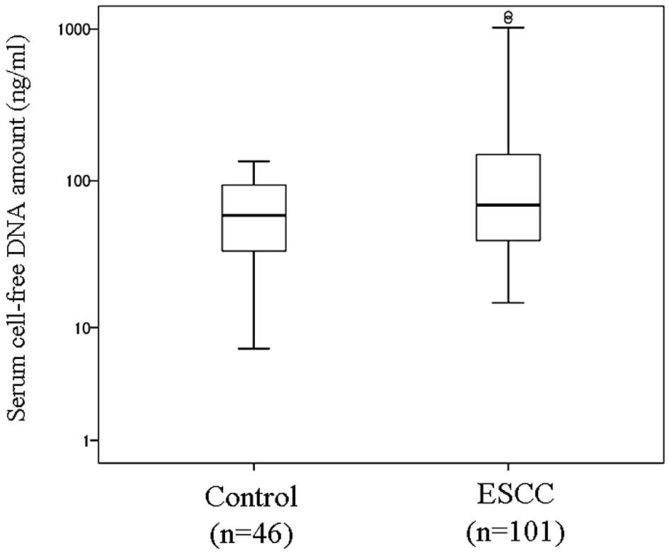
Serum cfDNA levels were significantly higher in the
ESCC patients (median, 67.5 ng/ml; range, 15–1236 ng/ml) than in
control patients without diagnosed ESCC (median, 59 ng/ml; range,
7–135 ng/ml; P=0.034, by the Mann-Whitney U test; Fig. 1).
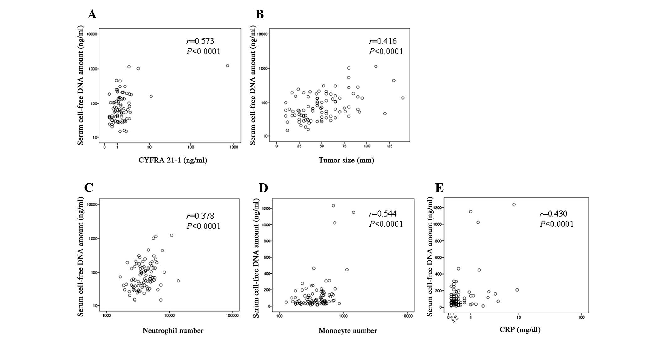
In the ESCC patients, serum cfDNA levels were
positively associated with tumor size and CYFRA 21-1 levels
(r=0.416 and r=0.573, respectively; P<0.0001 for both; Fig. 2), but not with SCC antigen levels
(r=0.050, P=0.642). In addition, serum cfDNA levels were associated
with the host inflammation status such as C-reactive protein (CRP)
levels (r=0.430, P<0.0001) and the number of neutrophils and
monocytes present in the peripheral blood (r=0.378 and r=0.544,
respectively; P<0.0001 for both; Fig. 2). Serum cfDNA levels tended to be
higher in advanced stage tumors than in the early stage tumors
(P=0.057 by the Fisher’s exact test; Table I). Serum cfDNA levels were also
significantly higher in male than female ESCC patients (P=0.034 by
the Fisher’s exact test; Table I).
Serum cfDNA levels were not associated with gender in the control
patients without ESCC (data not shown).
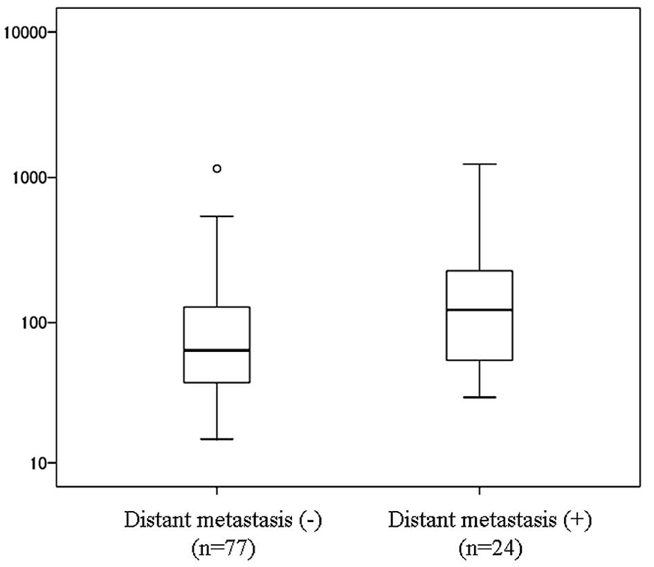
We also identified that serum cfDNA levels were
significantly higher in ESCC patients with distant metastasis
(median, 83 ng/ml; range, 30–1236 ng/ml) than in those without
distant metastasis (median, 74 ng/ml; range, 15–1152 ng/ml;
P=0.011, by the Mann-Whitney U test; Fig. 3). Serum cfDNA levels were not
associated with additional clinicopathologic factors including
disease-free periods and overall survival (data not shown).
Logistic regression analysis revealed that serum
cfDNA levels represented only one independent risk factor for
distant metastasis from the five factors tested, which included
gender, age, cfDNA levels, CYFRA-21-1 and SCC antigen levels
(P=0.0414, relative risk of 1.700; 95% CI, 1.021–2.831).
Discussion
Distant metastasis hinders a favorable outcome in
ESCC patients by limiting the surgical cure (7,8), and
accounts for the poor 5-year survival rate of less than 20%
worldwide (9,10). In order to improve the poor
prognosis for ESCC, it is essential to identify an easy-to-use
marker for predicting metastasis and recurrence of ESCC in distant
organs including the abdominal lymph node. In a large cohort of
ESCC patients, we showed for the first time that serum cfDNA levels
were significantly higher in patients with distant metastasis than
in those without. Using logistic regression analysis, we also
revealed that serum cfDNA levels represented only one independent
risk factor for distant metastasis among the five factors
tested.
In 1863, Rudolf Virchow proposed that inflammation
may be a hallmark of cancer after he identified leukocytes within
cancer tissues. Since then, it has been generally accepted that if
a genetic abnormality is the ‘match that lights the fire’ of
cancer, inflammation corresponds to the ‘fuel that feeds the
flames’ (17). Thus, host
inflammation status contributes greatly to cancer progression. As
shown in our previous study (3),
increased serum levels of inflammatory cytokines such as
interleukin-6 in ESCC patients support this hypothesis. In
addition, our present data demonstrated positive associations
between increased levels of serum cfDNA and the total number of
neutrophils and monocytes present in the peripheral blood of ESCC
patients. This result is consistent with the results of our
previous work, where we found that increased serum cfDNA levels
were related closely to cancer-specific inflammation in patients
with hepatocellular carcinoma (HCC) associated with hepatitis C
virus (HCV) infection (18).
Interestingly, that study revealed no association between the
levels of serum cfDNA and CRP in HCC patients. In contrast, our
current findings show a positive association between the levels of
serum cfDNA and CRP in ESCC patients. This discrepancy is
reasonable, given that CRP values in HCC patients may be largely
affected by liver dysfunction rather than inflammation, as all of
the HCC patients also exhibited HCV-related chronic liver
disease.
CYFRA 21-1 belongs to the intermediate filament
protein family and serves as a particularly useful diagnostic tool
for many cancers including ESCC (19). It has been shown that increased
levels of CYFRA 21-1 are linked to cancer progression and poor
outcome of ESCC (20,21). Therefore, our present finding that
serum levels of cfDNA were positively associated with CYFRA 21-1
levels and tumor size gives strength to the clinical efficacy of
cfDNA as an easy-to-use assay for monitoring ESCC. This hypothesis
is further supported by our finding that serum cfDNA levels
represent only one independent risk factor for distant metastasis
in ESCC patients.
Collectively, the findings arising from the present
study suggest that enhanced inflammation may easily destroy cancer
cells, thus releasing higher levels of cellular DNA and CYFRA 21-1
into the blood stream. Therefore, cfDNA levels may serve as a
predictive marker for distant metastasis in ESCC. Further
investigation is required to precisely define the molecular basis
underlying cfDNA release into the blood and to evaluate its
efficacy as a biomarker for ESCC in a routine clinical setting.
Acknowledgements
This study was sponsored by grants
from the Ministry of Education, Culture, Sports, Science and
Technology of Japan (nos. 18390366 and 17591406).
References
|
1.
|
Parkin DM, Bray F, Ferlay J and Pisani P:
Global cancer statistics. CA Cancer J Clin. 55:74–108. 2005.
View Article : Google Scholar
|
|
2.
|
Enzinger PC and Mayer RJ: Esophageal
cancer. N Engl J Med. 349:2241–2252. 2003. View Article : Google Scholar : PubMed/NCBI
|
|
3.
|
Oka M, Yamamoto K, Takahashi M, Hakozaki
M, Abe T, Iizuka N, Hazama S, Hirazawa K, Hayashi H, Tangoku A,
Hirose K, Ishihara T and Suzuki T: Relationship between serum
levels of interleukin 6, various disease parameters and
malnutrition in patients with esophageal squamous cell carcinoma.
Cancer Res. 56:2776–2780. 1996.PubMed/NCBI
|
|
4.
|
Iizuka N, Hirose K, Noma T, Hazama S,
Tangoku A, Hayashi H, Abe T, Yamamoto K and Oka M: The nm23-H1 gene
as a predictor of sensitivity to chemotherapeutic agents in
oesophageal squamous cell carcinoma. Br J Cancer. 81:469–475. 1999.
View Article : Google Scholar : PubMed/NCBI
|
|
5.
|
Ishikura S, Nihei K, Ohtsu A, Boku N,
Hironaka S, Mera K, Muto M, Ogino T and Yoshida S: Long-term
toxicity after definitive chemoradiotherapy for squamous cell
carcinoma of the thoracic esophagus. J Clin Oncol. 21:2697–2702.
2003. View Article : Google Scholar : PubMed/NCBI
|
|
6.
|
Kleinberg L and Forastiere AA:
Chemoradiation in the management of esophageal cancer. J Clin
Oncol. 25:4110–4117. 2007. View Article : Google Scholar : PubMed/NCBI
|
|
7.
|
Chen G, Wang Z, Liu XY, Zhang MY and Liu
FY: Abdominal lymph node metastasis in patients with mid thoracic
esophageal squamous cell carcinoma. World J Surg. 33:278–283. 2009.
View Article : Google Scholar : PubMed/NCBI
|
|
8.
|
Shimada H, Okazumi S, Matsubara H, Nabeya
Y, Shiratori T, Shimizu T, Shuto K, Hayashi H and Ochiai T: Impact
of the number and extent of positive lymph nodes in 200 patients
with thoracic esophageal squamous cell carcinoma after three-field
lymph node dissection. World J Surg. 30:1441–1449. 2006. View Article : Google Scholar : PubMed/NCBI
|
|
9.
|
Montesano R, Hollstein M and Hainaut P:
Genetic alterations in esophageal cancer and their relevance to
etiology and pathogenesis: a review. Int J Cancer. 3:225–235. 1996.
View Article : Google Scholar : PubMed/NCBI
|
|
10.
|
Parkin DM, Bray F, Ferlay J and Pisani P:
Estimating the world cancer burden: Globocan 2000. Int J Cancer.
94:153–156. 2001. View
Article : Google Scholar : PubMed/NCBI
|
|
11.
|
Gautschi O, Bigosch C, Huegli B, Jermann
M, Marx A, Chasse E, Ratschiller D, Weder W, Joerger M, Betticher
DC, Stahel RA and Ziegler A: Circulating deoxyribonucleic acid as
prognostic marker in non-small cell lung cancer patients undergoing
chemotherapy. J Clin Oncol. 22:4157–4164. 2004. View Article : Google Scholar : PubMed/NCBI
|
|
12.
|
Tokuhisa Y, Iizuka N, Sakaida I, Moribe T,
Fujita N, Miura T, Tamatsukuri S, Ishitsuka H, Uchida K, Terai S,
Sakamoto K, Tamesa T and Oka M: Circulating cell-free DNA as a
predictive marker for distant metastasis of hepatitis C
virus-related hepatocellular carcinoma. Br J Cancer. 97:1399–1403.
2007. View Article : Google Scholar : PubMed/NCBI
|
|
13.
|
Umetani N, Giuliano AE, Hiramatsu SH,
Amersi F, Nakagawa T, Martino S and Hoon DS: Prediction of breast
tumor progression by integrity of free circulating DNA in serum. J
Clin Oncol. 24:4270–4276. 2006. View Article : Google Scholar : PubMed/NCBI
|
|
14.
|
Tomita H, Ichikawa D, Ikoma D, Sai S, Tani
N, Ikoma H, Fujiwara H, Kikuchi S, Okamoto K, Ochiai T and Otsuji
E: Quantification of circulating plasma DNA fragments as tumor
markers in patients with esophageal cancer. Anticancer Res.
27:2737–2741. 2007.
|
|
15.
|
Sobin LH and Wittekind C: TNM
Classification of Malignant Tumours. 6th edition. UICC, Wiley-Liss;
pp. 81–83. 2002
|
|
16.
|
Iizuka N, Sakaida I, Moribe T, Fujita N,
Miura T, Stark M, Tamatsukuri S, Ishitsuka H, Uchida K, Terai S,
Sakamoto K, Tamesa T and Oka M: Elevated levels of circulating
cell-free DNA in the blood of patients with hepatitis C
virus-associated hepatocellular carcinoma. Anticancer Res.
26:4713–4719. 2006.PubMed/NCBI
|
|
17.
|
Balkwill F and Mantovani A: Inflammation
and cancer: back to Virchow? Lancet. 357:539–545. 2001. View Article : Google Scholar : PubMed/NCBI
|
|
18.
|
Iida M, Iizuka N, Sakaida I, Moribe T,
Fujita N, Miura T, Tamatsukuri S, Ishitsuka H, Uchida K, Terai S,
Tokuhisa Y, Sakamoto K, Tamesa T, Miyamoto T, Hamamoto Y and Oka M:
Relation between serum levels of cell-free DNA and inflammation
status in hepatitis C virus-related hepatocellular carcinoma. Oncol
Rep. 20:761–765. 2008.PubMed/NCBI
|
|
19.
|
Barak V, Goike H, Panaretakis KW and
Einarsson R: Clinical utility of cytokeratins as tumor markers.
Clin Biochem. 37:529–540. 2004. View Article : Google Scholar : PubMed/NCBI
|
|
20.
|
Yamamoto K, Oka M, Hayashi H, Tangoku A,
Gondo T and Suzuki T: CYFRA 21-1 is a useful marker for esophageal
squamous cell carcinoma. Cancer. 79:1647–1655. 1997. View Article : Google Scholar : PubMed/NCBI
|
|
21.
|
Brockmann JG, St Nottberg H, Glodny B,
Heinecke A and Senninger NJ: CYFRA 21-1 serum analysis in patients
with esophageal cancer. Clin Cancer Res. 6:4249–4252.
2000.PubMed/NCBI
|