Introduction
Rheumatoid arthritis (RA), an autoimmune disorder,
evolves over time causing progressive damage of synovial-lined
joints and variable extra-articular manifestations (1). RA is the most common autoimmune
disease, with an incidence higher than psoriasis, Crohn's disease,
type I (insulin-dependent) diabetes, lupus and multiple sclerosis
(2). The progression of RA is
characterized by symmetric polyarticular inflammation of the
synovium, resulting in pain and stiffness and over a period of time
leads to joint damage resulting in loss of function (3). The etiology of the autoimmune
disorder leading to RA remains unknown, yet the mechanisms of how
it evolves have been decoded to a great extent (4–7).
RA affects 1% of the world population; in 2010 the global
prevalence of this disease was 0.24% and was found to be more
common in females (8–10). In Australian, Western European and
North American regions the modeled age-standardized prevalence was
found to be highest and the lowest was found to be in Asia and
North African regions (10). In
the USA, RA affects 1.3 million people, making it the third common
type of arthritis among Americans (11).
The pathophysiology of this disease has been linked
to genetic and environmental factors. These factors lead to a
clinical syndrome spanning several disease subsets that require
inflammatory cascades (12–14). The overexpression of tumor
necrosis factor-α (TNF-α) is a prime inflammatory cascade involved
in RA. This cascade leads to the overexpression of cytokines such
as interleukin-1 (IL-1), IL-6 and IL-10, which drives persistent
inflammation and joint destruction (15–18). These components of the chronic
inflammatory response have been used for designing RA management
therapies. To date, the management of RA has been achieved using
disease-modifying antirheumatic drugs (DMARDs). These drugs are
either of biological origin or are synthetic chemical compounds and
based on this classification are termed as biological (bDMARDs) and
synthetic (sDMARDs), respectively. Both bDMARDs and sDMARDs are
characterized by their capacity to reduce or reverse signs and
symptoms of RA. They interfere with the pathophysiology of this
disease, which leads to improvements in disability, quality of
life, ability to work, and suppression of joint damage (19–21).
DMARDs include etanercept, infliximab, methotrexate
(MTX), sulfasalazine, leflunomide, rituximab, abatacept and others.
However, these DMARDs are associated with numerous side effects
(20). In order to overcome this
factor more and more research is being conducted on natural comp
ounds (21). Curcumin
(diferuloylmethane) extracted from Curcuma longa is one of
the many natural products that have been reported to be potential
bDMARDs (22–25). Here in this study, we employed
in silico techniques to investigate the natural compound
curcumin and its derivatives as possible bDMARDs. Molecular docking
simulations of curcumin and its derivatives with p38α, one of the
prominent p38 mitogen-activated protein (MAP) kinase that regulates
the expression of TNF-α and thus plays a role in RA was performed
(26). p38α is involved in the
p38 MAP kinase pathway that regulates a number of cellular
processes critical to the etiology of RA. The activation and
infiltration of leukocytes as well as the production of
inflammatory cytokines are p38-dependent processes. Targeting of
p38α has demonstrated efficacy in animal models of RA and various
p38α inhibitors are currently in phase II and I clinical trials for
RA. Thus using p38α as a therapeutic target for the development of
a novel compound is a viable option. The 4 top complexes generated
by targeting p38α with curcumin and its derivatives, were subjected
to molecular mechanics Poisson-Boltzmann surface area (MM-PBSA)
calculations. The top compound, cyclocurcumin, was further studied,
and inhibition of the release of TNF-α from LPS-stimulated
macrophages by cyclocurcumin treatment confirmed its role as a
potent p38α inhibitor.
Materials and methods
Data set
The X-ray crystallographic structure of p38α MAP
kinase was downloaded from the RCSB Protein Data Bank (http://www.rcsb.org/pdb/home/home.do).
The structure was cocrystallized with the pyrimidine-urea analogue
15 (PDB ID 2GHL, 2.10 Å X-ray resolution). The structure was
stripped of the pyrimidine-urea analogue, where after removing
water molecules, the charge and polar hydrogen atoms were added.
The incomplete residues were corrected and the energy minimization
of the modified structure was set to the default cut-off root mean
square deviation (RMSD) value of 0.3 Å using OPLS 2001 force field
by SPDB viewer (27). The
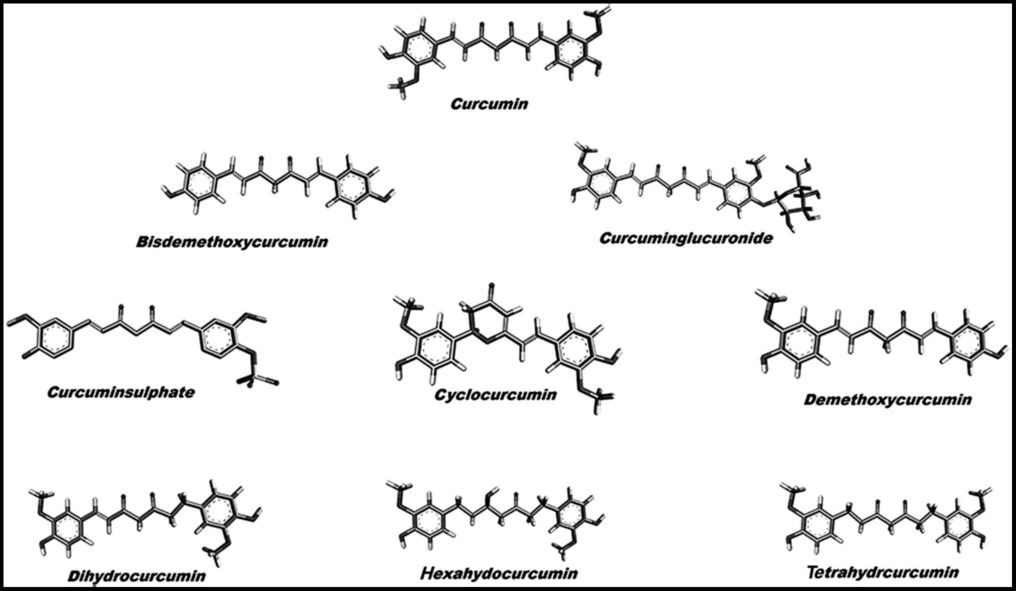
structure of curcumin and its derivatives were retrieved from the
PubChem database (Fig. 1).
Molecular docking simulations
Molecular docking simulation was carried out using
AutoDock 4.2.6 tool (28). This
tool uses binding free energy evaluation for mining the top
compound-protein pose. The tool uses binding site definition to
create a rectangular grid. The execution of the docking runs are
performed within the grid. The energy values calculated by this
suite are the culmination of the intermolecular energy of the
protein, the internal energy of the ligand and the torsional free
energy. The default parameters were set, and the maximum
evaluations were performed to elucidate the top 10 binding modes
for each run. The best compound among curcumin and its derivatives
was selected for further evaluation at the atomic level using
molecular dynamics simulation. The results were analyzed using
Discovery Studio Visualizer (29).
Molecular dynamics simulation
GROningen MAchine for Chemical Simulations GROMACS
4.5.6 atomic simulation tool was used to evaluate the ligand-p38α
complexes over 100 nanoseconds (nsec) (30). GROMOS 43a1 force field was used
for p38α and the force field for the top ligand was calculated
using PRODRG webserver (31,32). The complexes were kept in a water
bath, where water molecules were represented by simple point charge
(SPC216) model. The system was ensured to be overall charge neutral
before being subjected to energy minimization process, where
position restrain procedure was performed with NVT and NPT
ensembles, both of which follow Berendsen coupling scheme. Time
duration of 1 nsec with coupling constant of 0.1 psec at a constant
temperature of 300 K was used for NVT ensemble. This was followed
by NPT ensemble with a time duration of 1 nsec; here a constant
pressure of 1 bar was subjected to a coupling constant of 5 psec.
The complexes were then subjected to simulation for 100 nsec at 300
K. For each run, the SHAKE algorithm for bond lengths and particle
mesh Ewald method for long-range electrostatic interactions was
employed, where a 14 Å cut-off for van der Waals interactions, a 12
Å cut-off for Coulomb interaction with updates every 2 steps
(30,33). The trajectories generated were
subjected to GROMACS inbuild tools for evaluation. The results
generated were visualized using Pymol, Ligplus and VMD (34-36) and the graphs were plotted by using
Grace program (37).
MM-PBSA calculations
The trajectories generated were used for MM-PBSA
calculations (38,39). For each trajectory 1001 complexes
were retrieved, each after 100 psec. Binding modes were analyzed
using Grace program (37). The
tool gave us the binding free energy of the four complexes. The
binding free energy (ΔGbind) calculated by the tool
includes the following species:
ΔGbind=Gcomplex−Gprotein−Gligand=ΔEMM+ΔGsol−TΔS
ΔEMM=ΔEval+ΔEele+ΔEvdw
ΔGsol=ΔGp+ΔGnp
ΔGnp=γSASA+β
In the equations Gcomplex,
Gprotein and Gligand represent the free
energy of the respective species, ΔEMM, ΔGsol
and TΔS represent gas phase energy, solvation energy and an entropy
term. The gas phase energy is the summation of the internal energy
of the bonds, angle and torsion (ΔEval), electrostatic
interaction energy (ΔEele), and van der Waals
interaction energy (ΔEvdw). The salvation free energy
comprises the polar salvation free energy (ΔGp), and the
nonpolar salvation free energy (ΔGnp).
Principal component analysis
g_covar, g_anaeig in-build tools of GROMACS suite
were used for indentifying the dominant motions of the selected
complexes. PCA technique was used to trace these high-amplitude
concerted motions in the p38α-curcumin-based complexes (40). Eigenvectors were generated from
the covariance matrix generated by simple linear transformation.
The first two eigenvectors (EV1 and EV2) having cosine content
<0.2 were used to study free energy landscape (FEL). g_sham
in-build tool was used to calculate FEL.
Cell culture
THP-1 monocytes were procured from American Type
Culture Collection (ATCC; Manassas, VA, USA) and cultured in
RPMI-medium (Sigma-Aldrich, St. Louis, MO, USA) supplemented with
10% FBS and 100 U/ml PenStrep (Gibco, Grand Island, NY, USA). The
cells were grown under standard culture conditions at 37°C under 5%
CO2 in a humidified incubator. For macrophage
differentiation, monocytes were cultured with macrophage CSF
(M-CSF) at 100 ng/ml. Stock solution of 100 mM cyclocurcumin
(Sigma-Aldrich) was prepared in RPMI-medium and diluted to
different concentrations. Cells were stimulated with 10 ng/ml
lipopoly-saccharide (LPS) (Sigma-Aldrich) for TNF-α production.
Cell proliferation assay
3-(4,5-Dimethylthiazol-2-yl)-2,5-diphenyltetrazolium
bromide (MTT) assay is a standard test to measure cell viability
and is based on the conversion of MTT to formazan crystals by
active mitochondrial dehydrogenases. Briefly, a cell suspension
containing ~3×10⁴ cells was plated into each well of a
96-well plate and allowed to attach for 24 h. Cyclocurcumin
prepared in DMEM was added to the wells at a concentration of 10,
20, 40, and 60, 80 and 100 µM for 24 h. After treatment, 100
µl of MTT (Sigma-Aldrich) solution was added to each well at
a concentration of 0.1 mg/ml (dissolved in PBS). After 4 h of
incubation at 37°C in dark, the solution was removed and 100
µl of dimethyl sulfoxide (DMSO) was added to solubilize the
produced formazan. Absorbance was measured at 570 nm using an
automated microplate reader (BioTek Instruments, Winooski, VT,
USA). The results are expressed as percentages relative to the
controls. The percentage of proliferation inhibition was calculated
as = (1 - ODsample/ODcontrol) ×100%.
TNF-α, enzyme-linked immunosorbent assay
(ELISA)
Following treatment with cyclocurcumin and
stimulation of human macrophages with LPS, the supernatants were
harvested. Concentrations of TNF-α (BD Pharmingen, San Diego, CA,
USA) were determined by ELISA, following the manufacturer's
instructions. Absorbance was read at 450 nm on an ELISA plate
reader (BioTek Instruments) using an in-built software program.
Statistical analysis
Statistical analysis was carried out by one-way
ANOVA and the level of significance was tested at a P-value ranging
from 0.01 to 0.001. Data are expressed as the mean ± SD of three
independent experiments.
Results and Discussion
Molecular docking analysis
Molecular docking analysis was carried out using
AutoDock 4.2 tool; 100 conformations for each compound were
generated and the binding energy (kcal/mol) was set as the criteria
for the cut-off for the molecular dynamics simulation analysis.
Table I shows the results
generated by docking curcumin and its selected derivatives against
the active site of p38α. The top 4 compounds with the least binding
energy were curcumin, bisdemethoxycurcumin, cyclocurcumin and
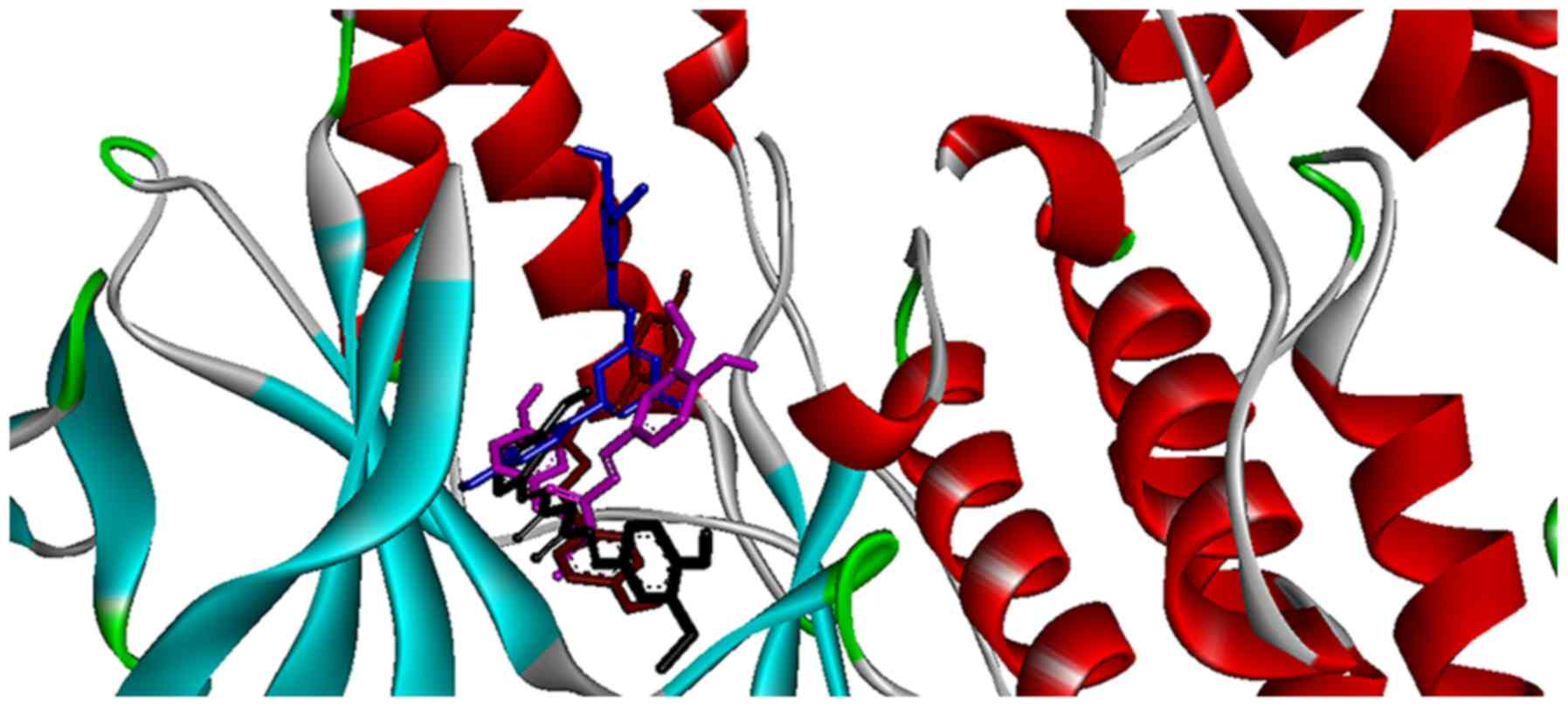
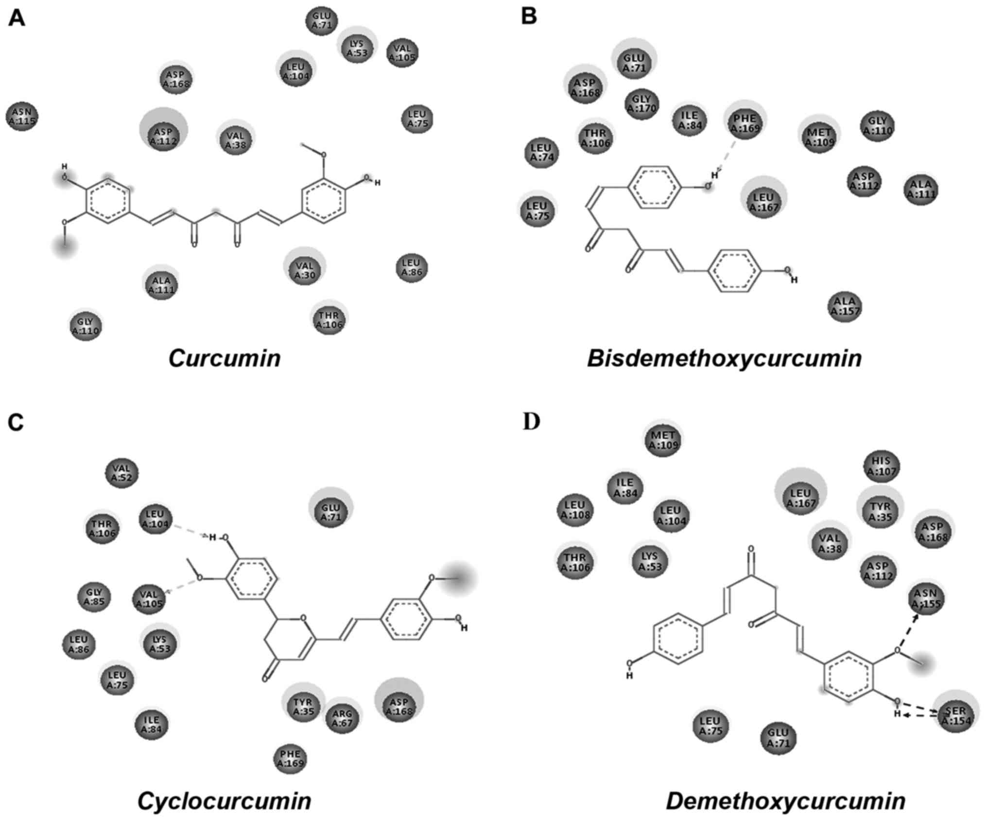
demethoxycurcumin. Protein/ligand complexes from the molecular
docking were subjected to post docking analysis, where weak
interactions involved in the complex were studied (Fig. 2). Four complexes shortlisted for
this analysis showed the favorable binding energy with a range -5
to -7 kcal/mol (Table II). The
effective interactions studied here were supported by energies
including final intermolecular energy, electrostatic energy and van
der Waals energy. The effective interactions of the curcumin-p38α
complex were evaluated using Discovery Studio Visualizer 4.5. The
binding pocket of the complex involved GLY110, ASN115, ASP112,
ASP168, VAL38, LEU104, GLU71, LYS53, VAL105, LEU75, LEU86, VAL30,
THR106 and ALA111. The binding energy of curcumin-p38α was
calculated to be the highest among the studied complexes, with no
non-covalent interaction observed (Fig. 3A). The binding energy of the
bisdemethoxycurcumin-p38α complex at -6.38 kcal/mol was the second
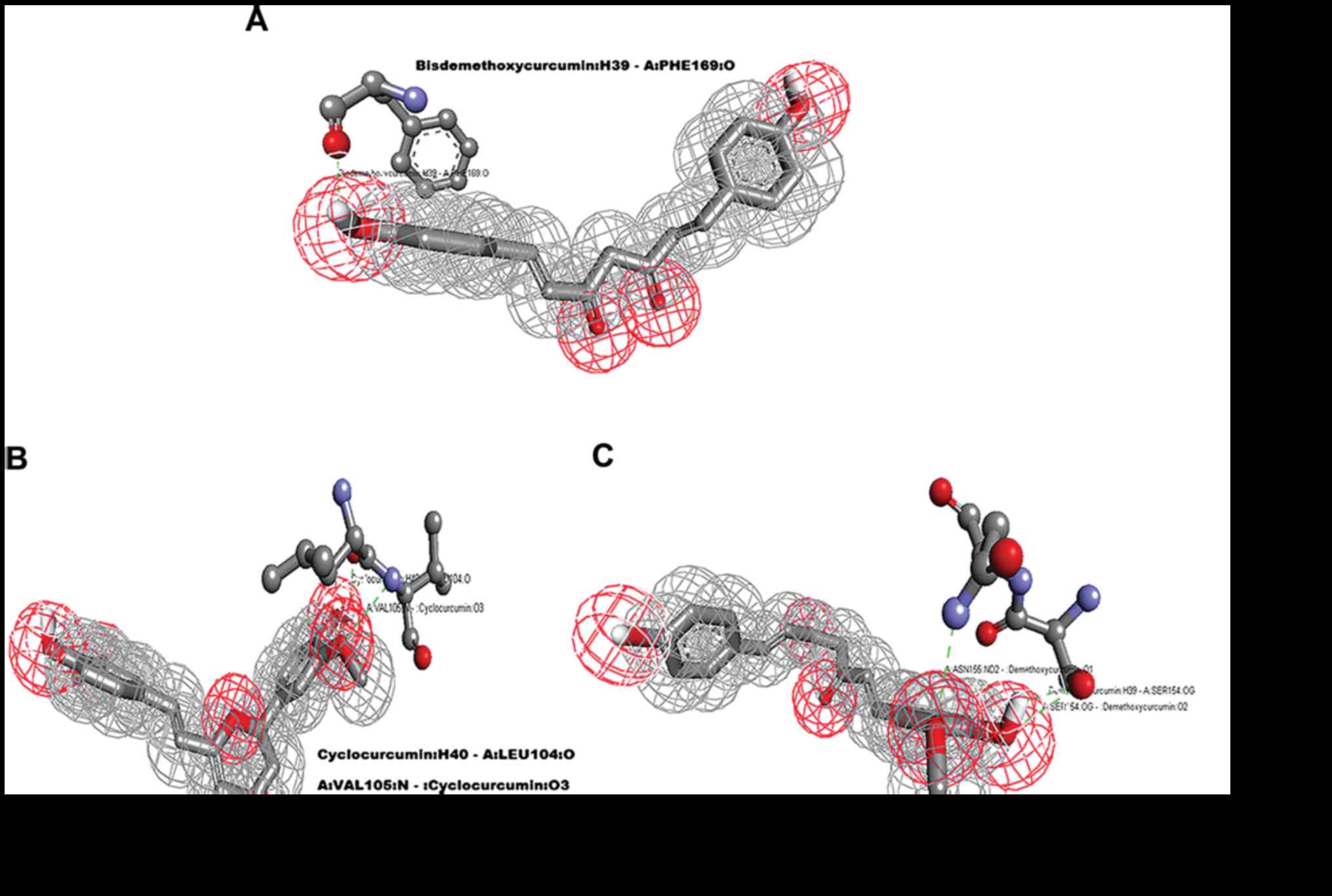
highest calculated among the studied complexes. The
bisdemethoxycurcumin-p38α complex formed one hydrogen bond
(Fig. 4A). The interacting
residue PHE169 of p38α is involved in active p38α inhibition. The
oxygen atom of PHE169 formed a physical non-covalent interaction
with bisdemethoxycurcumin, and the distance of the hydrogen bond
formed was 2.36 A°. The binding pocket (Fig. 3B) of the bisdemethoxycurcumin-p38α
complex involved LEU75, LEU74, THR106, ASP168, GLY170, GLU71,
ILE84, PHE169, LEU167, MET109, GLY110, ASP112, ALA111 and ALA157.
The third complex was that of cyclocurcumin with p38α protein
kinase. The binding energy of the complex was calculated to be
−6.12 kcal/mol. In the cyclocurcumin-p38α complex the results
showed two hydrogen bonds (Fig.
3C) between the protein and ligand atoms. The interaction
residues included LEU104 and VAL105 involved in active p38α
inhibition. The binding pose of hit CID 69879809 clearly suggested
effective inhibition. The atom 'O2' of the key residue
LEU104 interacted with cyclocurcumin through hydrogen bond
interactions (Fig. 4B). The key
residue VAL105 of the active binding site cavity also interacted
with cyclocurcumin oxygen atoms. Overall, the number of hydrogen
bonds confirmed the high affinity of cyclocurcumin towards
inhibition of p38α. In the case of the demethoxycurcumin-p38α
complex, the results showed 3 hydrogen bonds between p38α and
demethoxycurcumin (Fig. 3D). The
interaction residues included SER154 and ASN155 (Fig. 4C). The interaction of an 'O' atom
of SER154 and two 'O' atoms in the demethoxycurcumin aromatic ring
showed a strong interaction pattern. The 'O' atom in both sides of
the demethoxycurcumin aromatic ring was more potent in p38α
inhibition.
 | Table IBinding energy score of curcumin and
its derivatives with p38α. |
Table I
Binding energy score of curcumin and
its derivatives with p38α.
| Name | CID | ΔG kcal/mol |
|---|
| Curcumin | 969516 | −6.67 |
|
Bisdemethoxycurcumin | 5315472 | −6.38 |
|
Curcuminglucuronide | 71315012 | −3.15 |
| Cyclocurcumin | 69879809 | −6.12 |
|
Demethoxycurcumin | 5469424 | −5.88 |
|
Dihydrocurcumin | 10429233 | −4.48 |
|
Hexahydocurcumin | 5318039 | −4.81 |
|
Tetrahydrocurcumin | 124072 | −3.33 |
| Table IIAutodock analysis of the top four
curcumin-based compounds. |
Table II
Autodock analysis of the top four
curcumin-based compounds.
| Complexes | Ligand binding
pocket | Interaction |
|---|
| Curcumin-p38α | GLY110, ASN115,
ASP112, ASP168, VAL38, LEU104, GLU71, LYS53, VAL105, LEU75, LEU86,
VAL30, THR106, ALA111 | Nil |
|
Bisdemethoxycurcumin-p38α | LEU75, LEU74,
THR106, ASP168, GLY170, GLU71, ILE84, PHE169, LEU167, MET109,
GLY110, ASP112, ALA111, ALA157 | One
Bisdemethoxycurcumin: H39 - A:PHE169:O 2.36 A° |
|
Cyclocurcumin-p38α | ILE84, LEU75,
LEU86, LYS53, VAL105, GLY85, THR106, LEU104, VAL52, GLU71, ASP168,
ARG67, TYR35, PHE169 | Two
Cyclocurcumin:H40 - A:LEU104:O 2.13 A°
A:VAL105:N - Cyclocurcumin:O3 3.02 A° |
|
Demethoxycurcumin-p38α | THR106, LYS53,
LEU108, ILE84, LEU104, MET109, LEU167, VAL38, ASP112, HIS107,
ASP168, TYR35, ASN155, SER154, GLU71, LEU75 | Three
Demethoxycurcumin:H39 - A:SER154:OG 1.92 A°
ASN155:ND2 - :Demethoxycurcumin:O1 3.04 A°
A:SER154:OG - :Demethoxycurcumin:O2 2.67 A° |
Molecular dynamic simulation
analysis
After identification of the top 4 curcumin-based
compounds from the molecular docking, molecular dynamic simulation
(MDS) was employed to refine the top 4 complexes. MDS is a powerful
technique to monitor protein-ligand complexes over a period of
time. MDS run at a 100-nsec production was carried out under GROMOS
force field. The analysis was evaluated by group properties, RMSD,
hydrogen bond interactions and advance properties including PCA and
MM/PBSA free energy calculations using trajectory files. All the 4
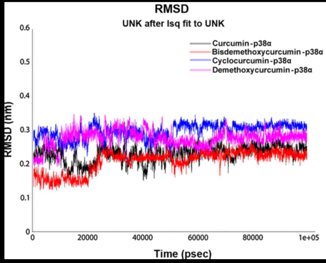
complexes were stable throughout the run. For each complex, RMSD
was evaluated for the convergence of the protein structure towards
an equilibrium state after lead binding. Fig. 5 shows the RMSD plot based on
backbone atoms. Curcumin and bisdemethoxycurcumin in complex with
p38α exhibited a jump at 30 nsec and equilibrated well and both of
the complexes converged with an RMSD range almost >0.2 nm. The
RMSD value of the bisdemethoxycurcumin-p38α complex was least among
the complexes. With an RMSD of 0.3 nm, cyclocurcumin showed the
highest deviation. From the RMSD plot of the demethoxycurcumin-p38α
complex, the structure equilibrated at 30 nsec and then started to
converge near 0.25 nm. The change in RMSD in all 4 complexes up to
30 nsec was due to ligand binding with the active site and after 30
nsec all of the structure obtained stability with less structural
deviation at the end of the 100-nsec simulation. After ligand
binding, the structural deviation of the bisdemethoxycurcumin-p38α
complex was the most stable among the 4 under consideration.
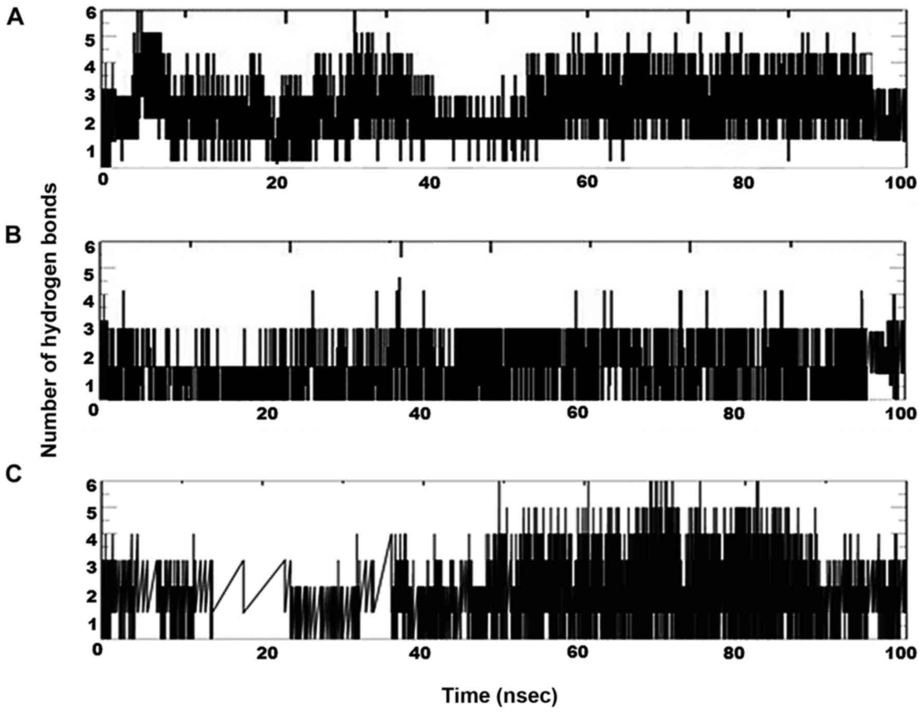
Inter-hydrogen bond interactions between protein and ligands were
evaluated for the p38α-curcumin-based complexes (Fig. 6). In the case of the curcumin
complex (Fig. 6A), NH plot
results showed a range of 4–6 hydrogen bond interactions observed
throughout the 100-nsec simulation and a maximum of 8 hydrogen
bonds. NH analysis confirmed strong inhibition of p38α by curcumin
in dynamic system similar to the docking results inferred with 6
hydrogen bonds. In the case of the bisdemethoxycurcumin complex
(Fig. 6B), the results showed a
range of 3–4 hydrogen bond interaction found throughout the
100-nsec simulation and a maximum of 6 hydrogen bonds similar to
the docking results. In the case of the cyclocurcumin complex
(Fig. 6C), the results showed a
range of 1–5 hydrogen bond interactions found throughout the
100-nsec simulation and a maximum of 5 hydrogen bonds similar to
the docking results. The inter-hydrogen bond interaction pattern
suggested the plausible mode of the strong binding of the lead
candidates with p38α which favored the inhibition mechanism.
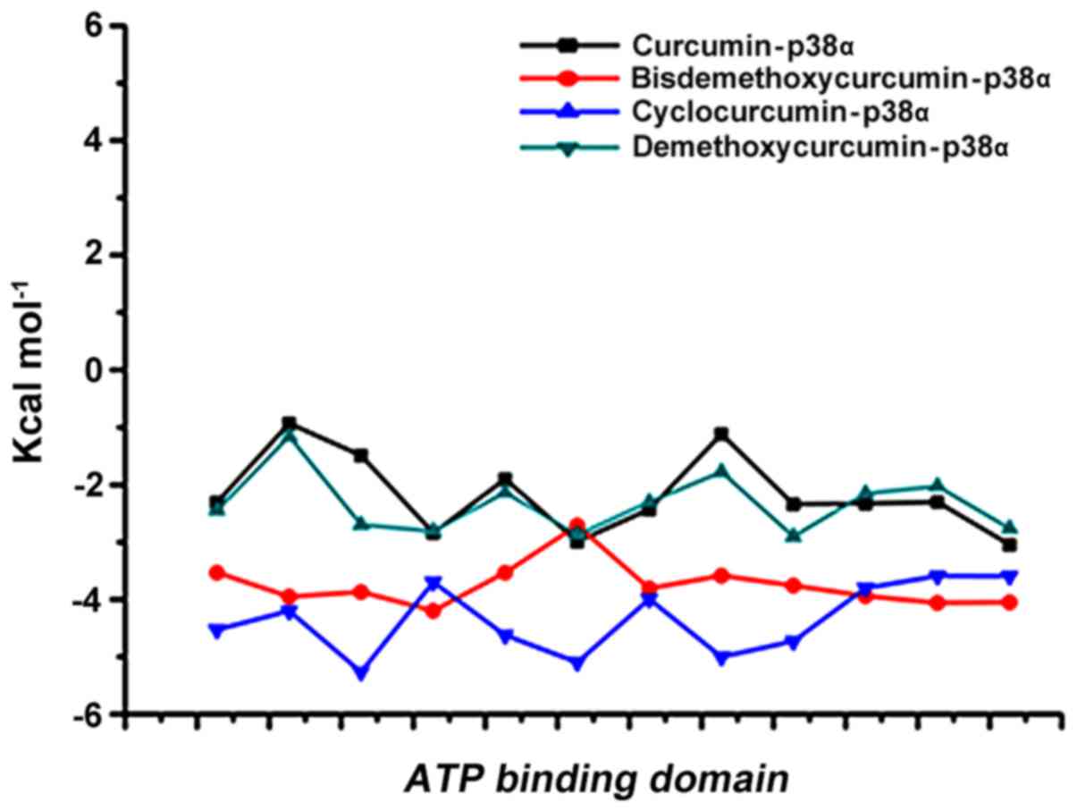
MM/PBSA calculation
The best 4 ligands in complex with p38α from MDS
were used to calculate the binding free energy using MM-PBSA
method. Snapshots were extracted at every 10-psec of stable
intervals from 100-nsec MD trajectory and served as input for
calculation. The binding free energy and its corresponding
components obtained from the MM/PBSA calculation of the p38α
curcumin-based complexes are listed. The results indicated that 4
curcumin-based compounds possessed a negative binding free energy
of −141.727, −132.758, −151.455 and −133.56 kcal/mol (Table III). Moreover, van der Waals,
electrostatic interactions and non-polar solvation energy
negatively contribute to the total interaction energy while only
polar solvation energy positively contributes to total free binding
energy. The relative binding free energies of the 4
p38α-curcumin-based complexes supported the strong binding in the
dynamic system. The interaction with the active site of p-p38α is
shown in Fig. 7.
| Table IIIMM-PBSA score of the top four
curcumin-based compounds. |
Table III
MM-PBSA score of the top four
curcumin-based compounds.
| Complex | ΔGbind
(kcal/mol) |
|---|
| Curcumin-p38α | −141.727 |
|
Bisdemethoxycurcumin-p38α | −132.758 |
|
Cyclocurcumin-p38α | −151.455 |
|
Demethoxycurcumin-p38α | −133.56 |
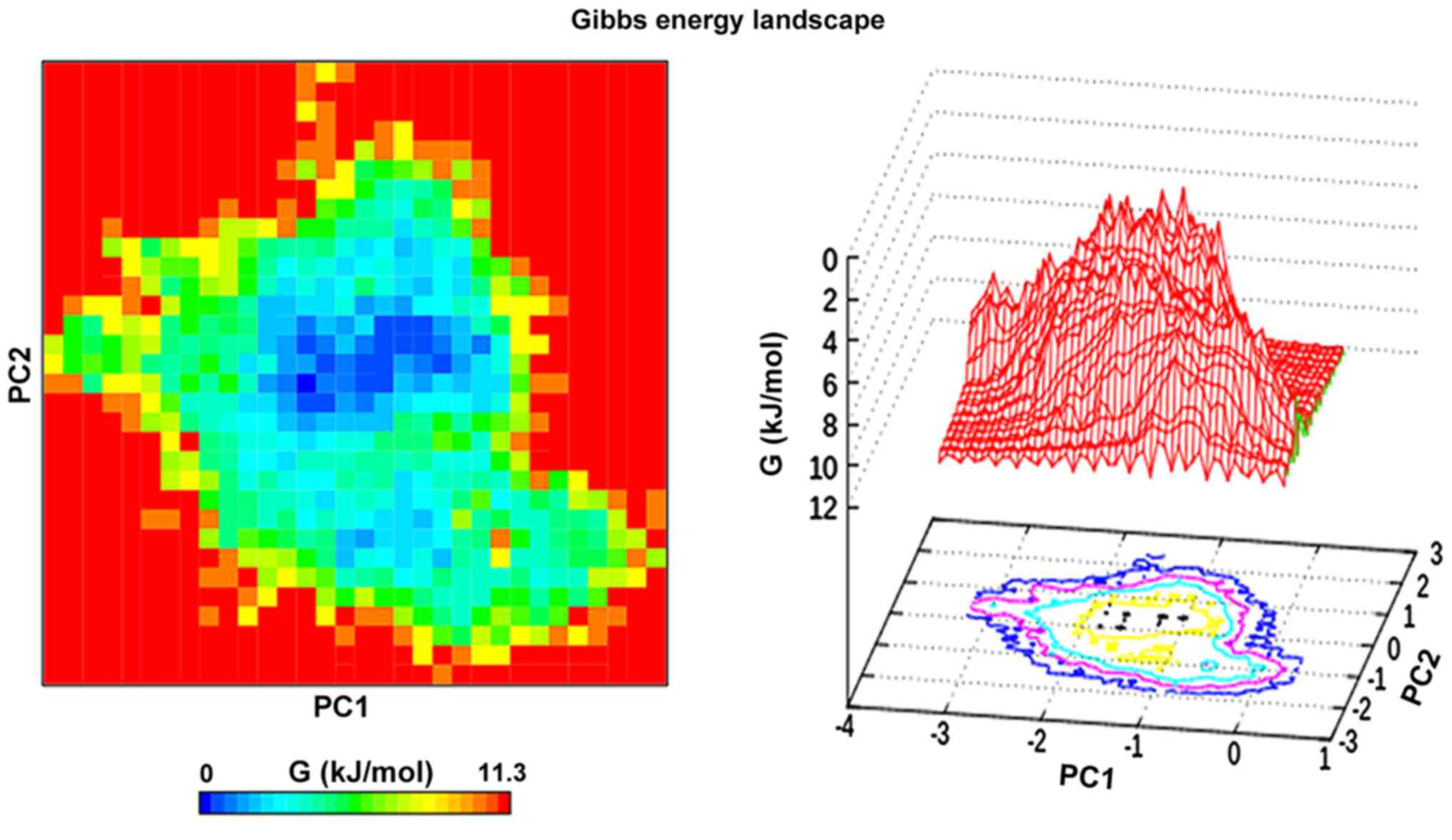
Principal component analysis
Principal component analysis or essential dynamics
was performed on the cyclocurcumin complex with p38α. The analysis
was used to monitor the overall strenuous motions of the complex.
The results showed that the first 6 eigenvectors formed 90% of the
total motion of the complex. The first 2 components of the
eigenvectors were projected at 300 K and analysis of this revealed
the clusters of stable states for the complex (41). The FEL was plotted over this
projection, which provided a clear description of the stability of
the complex (Fig. 8).
Cyclocurcumin inhibits the release of
TNF-α from human macrophages
TNF-α is a key factor in a variety of inflammatory
diseases. Keeping in view the role of p38α in regulating TNF-α
expression, we evaluated its expression in the culture supernatants
of human macrophages (which constitute the major TNF-α-producing
cells in highly relevant inflammatory disorders) treated with
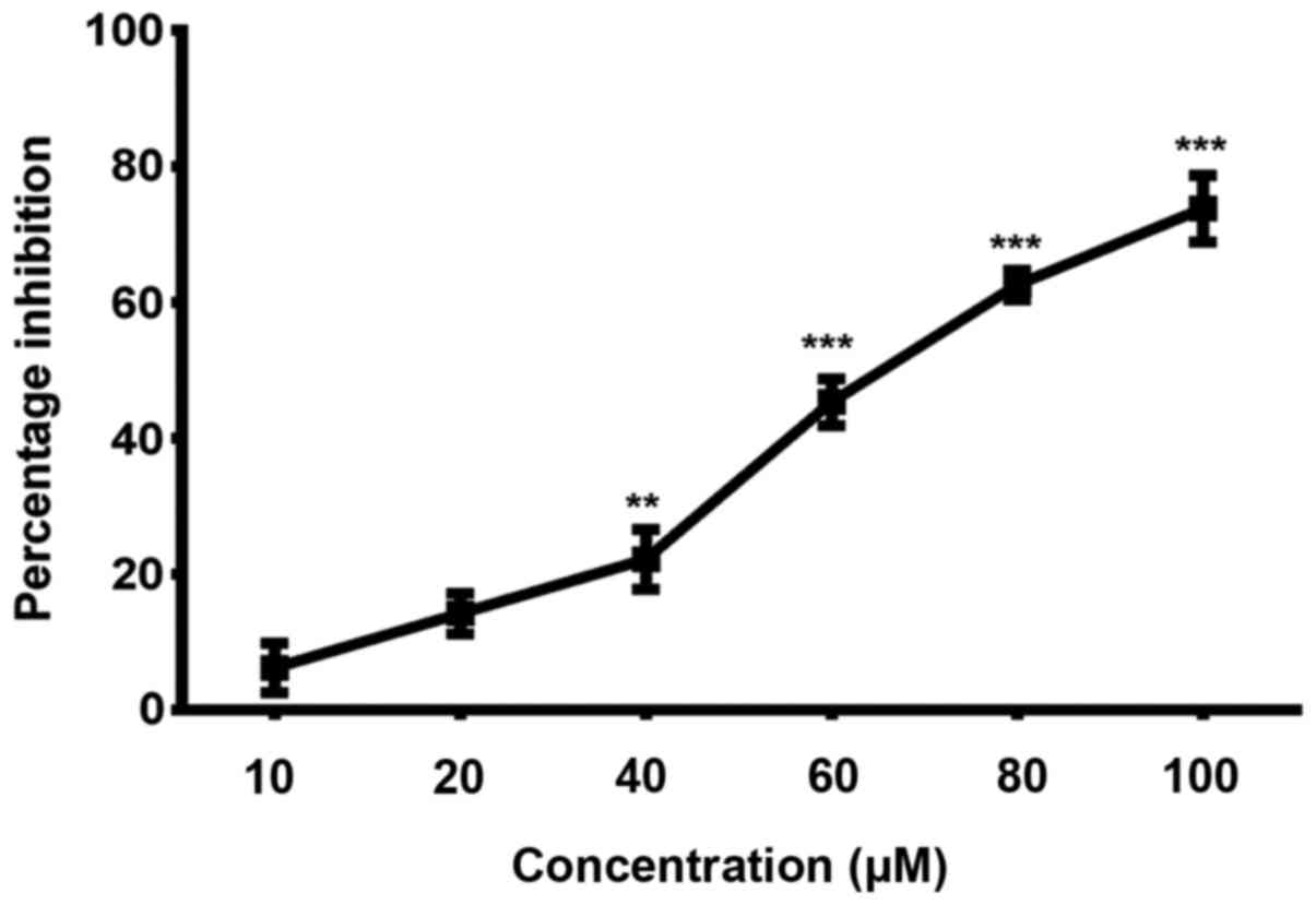
non-cytotoxic doses of cyclocurcumin. In order to determine the
suitable dosage of cyclocurcumin that result in less cytotoxicity,
macrophages (obtained with treatment of THP-1 monocytes with CSF),
were treated with different concentrations of cyclocurcumin (10–100
µM) for 24 h, and the viability was measured by MTT assay.
The results demonstrated that treatment with cyclocurcumin at
concentrations such as 10, 20 and 40 µM exhibited the least
cytotoxic effects (Fig. 9).
Therefore, these concentrations of cyclocurcumin, without cytotoxic
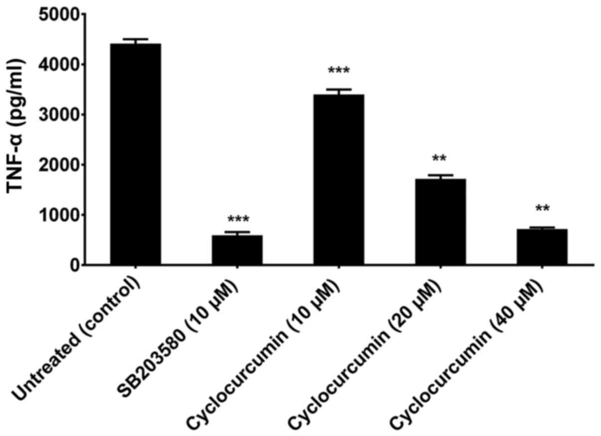
effects on macrophages were used. To test the effect of
cyclocurcumin, macrophages were treated with the indicated
concentrations of cyclocurcumin or 10 µm SB203580, a
selective inhibitor of p38 MAPK. This was followed by stimulation
with 10 ng/ml LPS for 18 h to induce the production of TNF-α. It
was observed that treatment of the LPS-stimulated macrophages with
cyclocurcumin led to significant inhibition in the release of TNF-α
in a dose-dependent manner, similar to the inhibition caused by
SB203580 (Fig. 10). Thus, these
data confirm the role of cyclocurcumin in overcoming p38α-induced
production of TNF-α and hence can be used as a therapeutic agent to
target RA.
In conclusion, natural products have been described
as a gold mine for arthritis treatment. These products can suppress
the expression of TNF-α, IL-1β, cyclooxygenase-2, lipooxygenase,
matrix metalloproteinases or adhesion molecules, or suppress the
activation of nuclear factor-κB (NF-κB). All are factors linked to
the treatment of RA (42).
Curcumin has long been reported to have an antirheumatoid effect.
Initial studies have linked it with the downregulation of NF-κB
(43). A plethora of research on
curcumin has given us an opportunity to investigate it and its
derivatives as potential kinase inhibitors for p38α. To the best of
our knowledge, our study is the first to use an advanced
computational approach to identify the possible DMRDs. MM-PBSA is a
potent tool in the field of drug design (44) and this approach was employed in
this study for short listing of compounds. From the molecular
docking and molecular dynamic simulation studies, the potential
binding mode of cyclocurcumin with p38α with stability was revealed
as a top compound for the treatment of RA. Finally, inhibition of
the release of TNF-α from LPS-stimulated macrophages by
cyclocurcumin treatment confirms its role as a potent p38α
inhibitor.
Abbreviations:
|
RA
|
rheumatoid arthritis
|
|
MAP
|
mitogen-activated protein
|
|
MDS
|
molecular dynamic simulation
|
|
RMSD
|
root mean square deviation
|
|
MM-PBSA
|
molecular mechanics-Poisson Boltzmann
surface area
|
|
FEL
|
free energy landscape
|
Acknowledgments
The authors would like to acknowledge the funding
support for this study by the Shandong Province Important Research
and Development Plan (no. 2015GSF118002), the Shandong Province
Natural Science Foundation of China Youth Fund Projects (no.
ZR2010HQ016) and the Natural Science Foundation of China Youth Fund
Projects (no. 81202824/H1008).
References
|
1
|
Grassi W, De Angelis R, Lamanna G and
Cervini C: The clinical features of rheumatoid arthritis. Eur J
Radiol. 27(Suppl 1): S18–S24. 1998. View Article : Google Scholar : PubMed/NCBI
|
|
2
|
Jacobson DL, Gange SJ, Rose NR and Graham
NM: Epidemiology and estimated population burden of selected
autoimmune diseases in the United States. Clin Immunol
Immunopathol. 84:223–243. 1997. View Article : Google Scholar : PubMed/NCBI
|
|
3
|
Kahlenberg JM and Fox DA: Advances in the
medical treatment of rheumatoid arthritis. Hand Clin. 27:11–20.
2011. View Article : Google Scholar :
|
|
4
|
Demoruelle MK, Deane KD and Holers VM:
When and where does inflammation begin in rheumatoid arthritis?
Curr Opin Rheumatol. 26:64–71. 2014. View Article : Google Scholar :
|
|
5
|
Nielen MM, van Schaardenburg D, Reesink
HW, Twisk JW, van de Stadt RJ, van der Horst-Bruinsma IE, de Gast
T, Habibuw MR, Vandenbroucke JP and Dijkmans BA: Increased levels
of C-reactive protein in serum from blood donors before the onset
of rheumatoid arthritis. Arthritis Rheum. 50:2423–2427. 2004.
View Article : Google Scholar : PubMed/NCBI
|
|
6
|
Masson-Bessière C, Sebbag M, Durieux JJ,
Nogueira L, Vincent C, Girbal-Neuhauser E, Durroux R, Cantagrel A
and Serre G: In the rheumatoid pannus, anti-filaggrin
autoantibodies are produced by local plasma cells and constitute a
higher proportion of IgG than in synovial fluid and serum. Clin Exp
Immunol. 119:544–552. 2000. View Article : Google Scholar : PubMed/NCBI
|
|
7
|
Snir O, Widhe M, Hermansson M, von Spee C,
Lindberg J, Hensen S, Lundberg K, Engström A, Venables PJ, Toes RE,
et al: Antibodies to several citrullinated antigens are enriched in
the joints of rheumatoid arthritis patients. Arthritis Rheum.
62:44–52. 2010. View Article : Google Scholar
|
|
8
|
Murray CJ, Vos T, Lozano R, Naghavi M,
Flaxman AD, Michaud C, Ezzati M, Shibuya K, Salomon JA, Abdalla S,
et al: Disability-adjusted life years (DALYs) for 291 diseases and
injuries in 21 regions, 1990–2010: A systematic analysis for the
Global Burden of Disease Study 2010. Lancet. 380:2197–2223. 2012.
View Article : Google Scholar : PubMed/NCBI
|
|
9
|
Gibofsky A: Overview of epidemiology,
pathophysiology, and diagnosis of rheumatoid arthritis. Am J Manag
Care. 18(Suppl 13): S295–S302. 2012.
|
|
10
|
Cross M, Smith E, Hoy D, Carmona L, Wolfe
F, Vos T, Williams B, Gabriel S, Lassere M, Johns N, et al: The
global burden of rheumatoid arthritis: Estimates from the global
burden of disease 2010 study. Ann Rheum Dis. 73:1316–1322. 2014.
View Article : Google Scholar : PubMed/NCBI
|
|
11
|
Clough J: The Cleveland Clinic Guide to
Arthritis. Kaplan Publishing; New York, NY: 2009
|
|
12
|
van Oosterhout M, Bajema I, Levarht EW,
Toes RE, Huizinga TW and van Laar JM: Differences in synovial
tissue infiltrates between anti-cyclic citrullinated
peptide-positive rheumatoid arthritis and anti-cyclic citrullinated
peptide-negative rheumatoid arthritis. Arthritis Rheum. 58:53–60.
2008. View Article : Google Scholar : PubMed/NCBI
|
|
13
|
van der Helm-van Mil AH and Huizinga TW:
Advances in the genetics of rheumatoid arthritis point to
subclassification into distinct disease subsets. Arthritis Res
Ther. 10:2052008. View
Article : Google Scholar : PubMed/NCBI
|
|
14
|
Scott DL, Wolfe F and Huizinga TW:
Rheumatoid arthritis. Lancet. 376:1094–1108. 2010. View Article : Google Scholar : PubMed/NCBI
|
|
15
|
Eastgate JA, Symons JA, Wood NC,
Grinlinton FM, di Giovine FS and Duff GW: Correlation of plasma
interleukin 1 levels with disease activity in rheumatoid arthritis.
Lancet. 2:706–709. 1988. View Article : Google Scholar : PubMed/NCBI
|
|
16
|
Feldmann M, Brennan FM and Maini RN:
Brennan, FM, and Maini, RN Rheumatoid Arthritis. Cell.
85:1277–1289. 1996. View Article : Google Scholar
|
|
17
|
Choy EH, Isenberg DA, Garrood T, Farrow S,
Ioannou Y, Bird H, Cheung N, Williams B, Hazleman B, Price R, et
al: Therapeutic benefit of blocking interleukin-6 activity with an
anti-interleukin-6 receptor monoclonal antibody in rheumatoid
arthritis: A randomized, double-blind, placebo-controlled,
dose-escalation trial. Arthritis Rheum. 46:3143–3150. 2002.
View Article : Google Scholar : PubMed/NCBI
|
|
18
|
van Roon JA, van Roy JL, Gmelig-Meyling
FH, Lafeber FP and Bijlsma JW: Prevention and reversal of cartilage
degradation in rheumatoid arthritis by interleukin-10 and
interleukin-4. Arthritis Rheum. 39:829–835. 1996. View Article : Google Scholar : PubMed/NCBI
|
|
19
|
Smolen JS, Landewé R, Breedveld FC, Buch
M, Burmester G, Dougados M, Emery P, Gaujoux-Viala C, Gossec L, Nam
J, et al: EULAR recommendations for the management of rheumatoid
arthritis with synthetic and biological disease-modifying
anti-rheumatic drugs: 2013 update. Ann Rheum Dis. 73:492–509. 2014.
View Article : Google Scholar
|
|
20
|
Smolen JS, van der Heijde D, Machold KP,
Aletaha D and Landewé R: Proposal for a new nomenclature of
disease-modifying antirheumatic drugs. Ann Rheum Dis. 73:3–5. 2014.
View Article : Google Scholar
|
|
21
|
Smolen JS, Aletaha D, Koeller M, Weisman
MH and Emery P: New therapies for treatment of rheumatoid
arthritis. Lancet. 370:1861–1874. 2007. View Article : Google Scholar : PubMed/NCBI
|
|
22
|
Dcodhar S, Sethi R and Srimal R:
Preliminary study on anti-rheumatic activity of curcumin
(diferuloyl methane). Indian J Med Res. 71:632–634. 1980.
|
|
23
|
Kuttan G, Hari Kumar K, Guruvayoorappan C
and Kuttan R: Antitumor, anti-invasion, and antimetastatic effects
of curcumin. The Molecular Targets and Therapeutic Uses of Curcumin
in Health and Disease. Aggarwal BB, Surh YJ and Shishodia S:
Springel; New York, NY: pp. 173–184. 2007, View Article : Google Scholar
|
|
24
|
Lev-Ari S, Strier L, Kazanov D, Elkayam O,
Lichtenberg D, Caspi D and Arber N: Curcumin synergistically
potentiates the growth-inhibitory and pro-apoptotic effects of
celecoxib in osteoarthritis synovial adherent cells. Rheumatology
(Oxford). 45:171–177. 2006. View Article : Google Scholar
|
|
25
|
Joe B, Rao UJ and Lokesh BR: Presence of
an acidic glycoprotein in the serum of arthritic rats: Modulation
by capsaicin and curcumin. Mol Cell Biochem. 169:125–134. 1997.
View Article : Google Scholar : PubMed/NCBI
|
|
26
|
Newton R and Holden N: Inhibitors of 38
mitogen-activated protein kinase: Potential as anti-inflammatory
agents in asthma? BioDrugs. 17:113–129. 2003. View Article : Google Scholar
|
|
27
|
Guex N and Peitsch MC: SWISS-MODEL and the
Swiss-PdbViewer: an environment for comparative protein modeling.
Electrophoresis. 18:2714–2723. 1997. View Article : Google Scholar
|
|
28
|
Morris GM, Huey R, Lindstrom W, Sanner MF,
Belew RK, Goodsell DS and Olson AJ: AutoDock4 and AutoDockTools4:
Automated docking with selective receptor flexibility. J Comput
Chem. 30:2785–2791. 2009. View Article : Google Scholar : PubMed/NCBI
|
|
29
|
Discovery Studio, version 3.5: Accelrys
Inc; San Diego, CA, USA: 2012
|
|
30
|
Hess B, Bekker H, Berendsen HJ and Fraaije
JG: LINCS: A linear constraint solver for molecular simulations. J
Comput Chem. 18:1463–1472. 1997. View Article : Google Scholar
|
|
31
|
Hess B, Kutzner C, van der Spoel D and
Lindahl E: GROMACS 4: Algorithms for highly efficient,
load-balanced, and scalable molecular simulation. J Chem Theory
Comput. 4:435–447. 2008. View Article : Google Scholar : PubMed/NCBI
|
|
32
|
Schüttelkopf AW and van Aalten DM: PRODRG:
A tool for high-throughput crystallography of protein-ligand
complexes. Acta Crystallogr D Biol Crystallogr. 60:1355–1363. 2004.
View Article : Google Scholar : PubMed/NCBI
|
|
33
|
Essmann U, Perera L, Berkowitz ML, Darden
T, Lee H and Pedersen LG: A smooth particle mesh Ewald method. J
Chem Phys. 103:8577–8593. 1995. View Article : Google Scholar
|
|
34
|
DeLano WL: The PyMOL Molecular Graphics
System. World Wide Web. http://www.pymol.org.
2002
|
|
35
|
Humphrey W, Dalke A and Schulten K: VMD:
Visual molecular dynamics. J Mol Graph. 14:33–38. 27–28. 1996.
View Article : Google Scholar : PubMed/NCBI
|
|
36
|
Laskowski RA and Swindells MB: LigPlot+:
Multiple ligand-protein interaction diagrams for drug discovery. J
Chem Inf Model. 51:2778–2786. 2011. View Article : Google Scholar : PubMed/NCBI
|
|
37
|
Turner P: XMGRACE, version 5.1.19. Center
for Coastal and Land-Margin Research. Oregon Graduate Institute of
Science and Technology; Beaverton, OR: 2005
|
|
38
|
Kumari R, Kumar R and Lynn A: Open Source
Drug Discovery Consortium: g_mmpbsa - a GROMACS tool for
high-throughput MM-PBSA calculations. J Chem Inf Model.
54:1951–1962. 2014. View Article : Google Scholar : PubMed/NCBI
|
|
39
|
Baker NA, Sept D, Joseph S, Holst MJ and
McCammon JA: Electrostatics of nanosystems: Application to
microtubules and the ribosome. Proc Natl Acad Sci USA.
98:10037–10041. 2001. View Article : Google Scholar : PubMed/NCBI
|
|
40
|
Amadei A, Linssen AB and Berendsen HJ:
Essential dynamics of proteins. Proteins. 17:412–425. 1993.
View Article : Google Scholar : PubMed/NCBI
|
|
41
|
Chikan NA and Vipperla B: KAISO
inhibition: An atomic insight. J Biomol Struct Dyn. 33:1794–1804.
2015. View Article : Google Scholar
|
|
42
|
Khanna D, Sethi G, Ahn KS, Pandey MK,
Kunnumakkara AB, Sung B, Aggarwal A and Aggarwal BB: Natural
products as a gold mine for arthritis treatment. Curr Opin
Pharmacol. 7:344–351. 2007. View Article : Google Scholar : PubMed/NCBI
|
|
43
|
Singh S, Aggarwal BB and Aggarwal B:
Activation of transcription factor NF-kappa B is suppressed by
curcumin (diferuloylmethane) [corrected]. J Biol Chem.
270:24995–25000. 1995. View Article : Google Scholar : PubMed/NCBI
|
|
44
|
Zhao FL, Yang GH, Xiang S, Gao DD and Zeng
C: In silico analysis of the effect of mutation on epidermal growth
factor receptor in non-small-cell lung carcinoma: from mutational
analysis to drug designing. J Biomol Struct Dyn. 35:427–434. 2017.
View Article : Google Scholar
|