Introduction
Graves' orbitopathy (GO) is an autoimmune
manifestation of Graves' disease that affects the orbit (1). While the pathogenesis of GO is not
yet fully understood, it is widely accepted that orbital
fibroblasts express thyroid-stimulating hormone receptor, which
interacts with the insulin-like growth factor-1 receptor (IGF-1-R),
facilitating the recruitment of autoreactive T and B cells into the
orbit (2,3). These infiltrating immune cells
stimulate orbital fibroblasts to secrete inflammatory modulators,
and to proliferate and differentiate into adipocytes (1,4).
The interaction between activated immune cells and orbital
fibroblasts exacerbates orbital inflammation and promotes the
deposition of extracellular matrix components, such as hyaluronan
(5). Consequently, chronic
inflammation and expansion of orbital connective tissue volume can
ultimately lead to erythema, swelling, proptosis and
sight-threatening compressive optic neuropathy (6).
Conventionally, high-dose systemic corticosteroid
and radiation therapy are considered as the first-line treatments
for GO (7); however, due to its
clinical heterogeneity and variable disease course, notable efforts
have been made to target the underlying pathophysiology at the
molecular level (8). Several
targeted biological and immunosuppressive agents, including those
targeting IL-6, CD20, fragment crystallizable receptors and
IGF-1-R, have been identified and are under pre-clinical and
clinical trials for use in GO (9–11).
Adenosine monophosphate-activated protein kinase
(AMPK) is a key phosphorylation enzyme involved in regulating fatty
acid and cholesterol synthesis, which can be activated under
various conditions of metabolic stress (12). AMPK has garnered substantial
attention as a potential therapeutic target for metabolic
disorder-related diseases, including diabetes, obesity, fatty liver
disease and cancer (13,14). Under nutrient-rich conditions,
AMPK is inactivated by an increased AMP/ATP ratio, driven by
glycolysis and mitochondrial oxidative phosphorylation (15). Compound C, also known as
dorsomorphin, is a cell permeable AMPK inhibitor, which also
functions as a broad-spectrum kinase inhibitor with
anti-proliferative effects (16).
Increasing evidence has suggested that compound C inhibits multiple
kinases beyond AMPK, which have been implicated in various
neurological diseases such as glioma (17,18). Therefore, the present study aimed
to investigate the effects of compound C on GO pathogenesis,
focusing on inflammation, fibrosis, hyaluronan production and
adipogenesis.
Materials and methods
Reagents and cell culture
The suppliers of reagents used in the present study
are listed in Table I. Orbital
tissue explants were obtained from patients with GO (n=12) during
orbital decompression surgery and healthy controls (n=12) during
upper or lower blepharoplasty (Table
II). Patients with GO were clinically inactive and had not
received systemic steroid treatment or radiation therapy 3 months
prior to the surgery. Normal healthy controls were free of thyroid
or autoimmune diseases. The present study was approved by the
Institutional Review Board of Severance Hospital (approval number
4-2024-0492; Seoul, South Korea) and adhered to the tenets of The
Declaration of Helsinki. All of the participants provided written
informed consent. Orbital fibroblasts were obtained from each
orbital tissue explant were cultured as previously described
(19). The harvested orbital
tissue explants were primarily cultured in DMEM/F12 1:1 containing
1% of penicillin (100 U/ml)/streptomycin (100 µg/ml) and 20% FBS to
promote fibroblast proliferation. Once cell proliferation was
confirmed, the monolayers were serially passaged using
trypsin/EDTA. The cells were stored in liquid nitrogen, and orbital
fibroblasts between the third and sixth passages were used for the
experiments.
 | Table I.Suppliers of study reagents. |
Table I.
Suppliers of study reagents.
| Reagent (cat.
no.) | Supplier |
|---|
| Compound C; Oil Red
O powder; MTT assay | Millipore
Sigma |
| DMEM; DMEM/F12;
penicillin-streptomycin; trypsin/EDTA | Welgene, Inc. |
| FBS;
Halt™ protease inhibitor cocktail | Thermo Fisher
Scientific, Inc. |
| Recombinant human
IL-1β; recombinant human TGF-β; hyaluronan | R&D Systems,
Inc. |
| ELISA kit
(DHYAL0) |
|
| Antibodies against
PPAR-γ (sc-7273), C/EBPβ (sc-7962), adiponectin | Santa Cruz
Biotechnology, Inc. |
| (sc-136131), leptin
(ob) (sc-48408), aP2 (sc-271529), SREBP-1 |
|
| (sc-365513), and
β-actin (sc-47778) |
|
| Antibodies against
IL-6 (12153), IL-8 (94407), MCP-1 (2027), ICAM-1 | Cell Signaling
Technology, Inc. |
| (4915), C/EBPα
(2295), p-AMPK (2535), t-AMPK (2532), p-YAP s127 |
|
| (13008), t-YAP
(4912), TAZ (70148), p-AKT (9271), t-AKT (9272), p-JNK |
|
| (9251), t-JNK
(9252), p-ERK (9101), t-ERK (9102), p-p38 (9211), t-p38 |
|
| (9212), p-STAT3
(94994), t-STAT3 (30835), p-SMAD1 (13820), p-SMAD2 |
|
| (3108), p-SMAD3
(9520), t-SMAD1 (9743), t-SMAD2 (5339), t-SMAD3 |
|
| (9523), t-SMAD4
(38454) |
|
| RIPA buffer | Biosesang |
| RNAiMAX; antibody
against p-SMAD4 (PA5-64712) | Invitrogen; Thermo
Fisher Scientific, Inc. |
| Silencer™ Select
Negative Control No. 1 siRNA (cat. no. 4390844) | Ambion; Thermo
Fisher Scientific, Inc. |
| Antibody against
fibronectin (610077) | BD Biosciences |
| Antibody against
collagen Ia (ab138492) | Abcam |
| AICAR; antibody
against α-SMA (A5228), DMSO | Sigma-Aldrich |
| Table II.Patient demographics. |
Table II.
Patient demographics.
| A, Patents with
Graves' orbitopathy |
|---|
|
|---|
| Age, years | Sex | CAS | Duration,
years | Proptosis R/L,
mm | Type of
surgery |
|---|
| 53 | F | 1 | 1.5 | 19/16 | Orbital wall
decompression |
| 42 | F | 0 | 2 | 22/22 | Orbital wall
decompression |
| 26 | M | 0 | 3 | 21/22.5 | Orbital wall
decompression |
| 29 | F | 2 | 2 | 21/21 | Orbital wall
decompression |
| 27 | M | 1 | 2.5 | 22/22 | Orbital wall
decompression |
| 31 | M | 0 | 3 | 23/22.5 | Orbital wall
decompression |
| 35 | F | 2 | 4 | 23/23 | Orbital wall
decompression |
| 43 | F | 1 | 2 | 20/20 | Orbital wall
decompression |
| 37 | M | 1 | 1 | 21/21.5 | Orbital wall
decompression |
| 41 | M | 0 | 1.5 | 22/22.5 | Orbital wall
decompression |
| 42 | F | 1 | 2 | 21/20 | Orbital wall
decompression |
| 48 | M | 1 | 3.5 | 21/21 | Orbital wall
decompression |
|
| B, Healthy
control patients |
|
| Age,
years | Sex | CAS | Duration,
years | Proptosis R/L,
mm | Type of
surgery |
|
| 43 | F |
|
|
| Upper lid
blepharoplasty |
| 71 | M |
|
|
| Upper lid
blepharoplasty |
| 65 | F |
|
|
| Lower lid
blepharoplasty |
| 53 | F |
|
|
| Lower lid
blepharoplasty |
| 42 | M |
|
|
| Upper lid
blepharoplasty |
| 48 | F |
|
|
| Upper lid
blepharoplasty |
| 52 | M |
|
|
| Upper lid
blepharoplasty |
| 59 | F |
|
|
| Lower lid
blepharoplasty |
| 61 | M |
|
|
| Lower lid
blepharoplasty |
| 54 | M |
|
|
| Lower lid
blepharoplasty |
| 59 | F |
|
|
| Upper lid
blepharoplasty |
| 39 | F |
|
|
| Upper lid
blepharoplasty |
Cell viability
To determine the non-toxic concentrations of
compound C on orbital fibroblasts, orbital fibroblasts extracted
from tissues obtained from patients with GO were seeded into
24-well culture plates (1×10⁴ cells/well) and were
treated with various concentrations of compound C (0–15 µM) for 72
h, 37°C under 5% humified CO2. After treatment, the
cells were washed twice with PBS and incubated in 5 mg/ml MTT
solution at 37°C for 3 h. Formazan was then dissolved using
ice-cold isopropanol and absorbance was measured at 560 nm with
background correction at 630 nm, using a microplate reader (EL 340
Bio Kinetics Reader; BioTek; Agilent Technologies, Inc.). Cell
viability was calculated as a percentage relative to untreated
control cells.
Western blotting
Confluent orbital fibroblasts were pretreated with
compound C (5 or 10 µM for 6 h) or 5-aminoimidazole-4-carboxamide
ribonucleotide (AICAR; AMPK agonist; Sigma-Aldrich; Merck KGaA); 5
or 10 µM for 6 h) followed by stimulation with either IL-1β (10
ng/ml for 24 h) or TGF-β (5 ng/ml for 24, 48 or 72 h) at 37°C to
assess the expression levels of inflammatory cytokines and
fibrosis-related proteins. The control group was treated with 0.1%
DSMO (Sigma-Aldrich; Merck KGaA) at 37°C for 6 h as each
experimental group and received no stimulation. In addition,
orbital fibroblasts were cultured in an adipogenic medium for 10
days with or without compound C treatment to evaluate the
expression levels of adipogenic transcription factors. Western blot
analysis was performed on orbital fibroblasts subjected to the
different study conditions. Proteins were extracted with RIPA
buffer (Biosesang) with Halt™ Protease Inhibitor
Cocktail (Thermo Fisher Scientific, Inc.). Protein concentration
was quantified by BCA assay (Pierce™ BCA Protein Assay
Kit; Thermo Fisher Scientific, Inc.). Equal amounts of protein from
orbital fibroblasts under different study conditions (50 µg) were
separated by electrophoresis on 8–15% SDS polyacrylamide gels.
Blocking was then performed in 5% skim milk for 24 h at 4°C. The
resolved proteins were then transferred onto nitrocellulose
membranes and were incubated with primary antibodies overnight at
4°C. The immunoreactive bands were detected using horseradish
peroxidase-conjugated secondary antibodies. After exposure to
Supersignal™ West Pico PLUS Chemiluminescent Substrate
(Thermo Fisher Scientific, Inc.), the membranes were visualized
using an X-ray film or image reader (LAS-4000 Mini; FUJIFILM Wako
Pure Chemical Corporation). The density of each band was measured
and semi-quantified using ImageJ software version 1.54 (National
Institutes of Health). The experimental values were normalized to
the β-actin levels of the same sample.
Adipogenesis
Orbital fibroblasts were cultured for 10 days in
adipogenic medium, which comprised high-glucose DMEM supplemented
with 10% FBS and 1% penicillin (100 U/ml)/streptomycin (100 µg/ml)
(Welgene, Inc.), 17 µM pantothenic acid (Sigma-Aldrich; Merck
KGaA), 33 µM biotin (Sigma-Aldrich; Merck KGaA), 10 µg/ml
transferrin (Sigma-Aldrich; Merck KGaA), 1 µM insulin (Roche
Diagnostics GmbH), 0.2 nM T3 (Sigma-Aldrich; Merck KGaA) and 0.2 µM
carbaprostaglandin (Calbiochem; Merck KGaA), as previously
described (20). Adipogenic
medium was replaced every 2-3 days. For the first 4 days of cell
culture, the differentiation medium contained 10 µM dexamethasone
and 0.1 mM 3-isobutyl-1-methylxanthine (IBMX); after day 5,
dexamethasone and IBMX were excluded. From the first day of
adipogenic differentiation, stimulation of adipogenesis was
enhanced by the addition of the peroxisome proliferator-activated
receptor γ (PPARγ) agonist rosiglitazone (Cayman, Ann Arbor, MI,
USA), IL-1β (10 ng/ml) or both. For treatment with compound C (5 or
10 µM), it was added to the adipogenic medium. To evaluate the
effects of yes-associated protein (YAP) and transcriptional
co-activator with PDZ-binding motif (TAZ) suppression on
adipogenesis, cells were transfected with YAP/TAZ small interfering
(si)RNA (10 nM) prior to the induction of adipogenesis. Adipogenic
differentiation was induced for 10 days at 37°C.
Oil Red O staining
Orbital fibroblasts under different study conditions
(with or without either IL-1β or compound C treatment, as
aforementioned) were stained with Oil Red O (Sigma-Aldrich; Merck
KGaA). Briefly, a stock solution of Oil Red O Solution was diluted
with 4 ml distilled water to prepare the working solution.
Subsequently, the working solution was filtered using a 0.2-µm
filter. Adipogenically differentiated cells were washed with PBS
and fixed with 10% formalin at 4°C for 1 h. After fixation, the
cells were washed with PBS, incubated with 60% isopropanol for 5
min and dried completely. The cells were then stained with Oil Red
O solution for 1-2 h at room temperature and washed with PBS.
Stained cells were observed under a light and confocal microscope
(IX73; Olympus Corporation). In addition, the Oil Red O stain was
solubilized with 100% isopropanol to quantify lipid accumulation
and optical density was measured using a spectrophotometer at 490
nm.
Silencing of YAP and TAZ
Orbital fibroblasts from patients with GO (50%
confluency) were transfected with siRNA (10 nM) using
Lipofectamine® RNAiMAX (Invitrogen; Thermo Fisher
Scientific, Inc.). Silencer™ Select Negative Control No. 1 siRNA
(si-control; 10 nM) was used as a control. Following transfection
at 37°C for 48 h in a humified incubator at 5% CO2, the
orbital fibroblasts were cultured in adipogenic medium to induce
adipogenesis, as aforementioned. The following siRNAs were used in
the present study: si-YAP: 5′-GACAUCUUCUGGUCAGAGADTDT-3′; si-TAZ:
5′-ACGUUGACUUAGGAACUUUDTDT-3′.
ELISA
Orbital fibroblasts from patients with GO were
pretreated with compound C (10 µM) for 6 h, and were subsequently
stimulated with either IL-1β (10 ng/ml) or TGF-β (5 ng/ml) for 96 h
at 37°C to determine hyaluronan production. Supernatants were
collected from orbital fibroblasts under various conditions, and
hyaluronan production levels were measured using a commercially
available ELISA kit according to the manufacturer's instructions.
Absorbance was measured at 450 nm, and the binding percentage of
each sample was calculated. A standard binding curve was generated
for quantification.
Statistical analysis
All experiments were performed in duplicate for each
of the three strains, which are derived from three different
individuals. After Kolmogorov-Smirnov test, Kruskal-Wallis test was
performed to compare quantitative data between the groups with or
without treatment with compound C, IL-1β or TGF-β, and to compare
quantitative data between groups with or without siRNA
transfection. For pairwise comparisons, Dunn's post hoc test was
applied after Kruskal-Wallis test to adjust for multiple
comparisons. Statistical analysis was performed using SPSS version
20.0 for Windows (IBM Corp.), and P<0.05 was considered to
indicate a statistically significant difference.
Results
Effect of compound C on the viability
of orbital fibroblasts
To determine the non-toxic concentration of compound
C in orbital fibroblasts, an MTT assay was conducted. Orbital
fibroblasts from patients with GO were exposed to various
concentrations of compound C (0–15 µM) for 72 h (Fig. S1). For cells treated with 10 µM
compound C, viability was >95%, therefore ≤10 µM compound C was
applied for further analysis.
Compound C inhibits AMPK activity and
YAP phosphorylation in orbital fibroblasts
After confirming the non-cytotoxic concentration of
compound C, the effects of compound C (AMPK antagonist) and AICAR
(AMPK agonist), on AMPK and YAP activation were assessed. Orbital
fibroblasts from patients with GO and healthy controls were treated
with 5 and 10 µM compound C or AICAR for 6 h, and phosphorylated
(p-)AMPK and p-YAP protein levels were determined using western
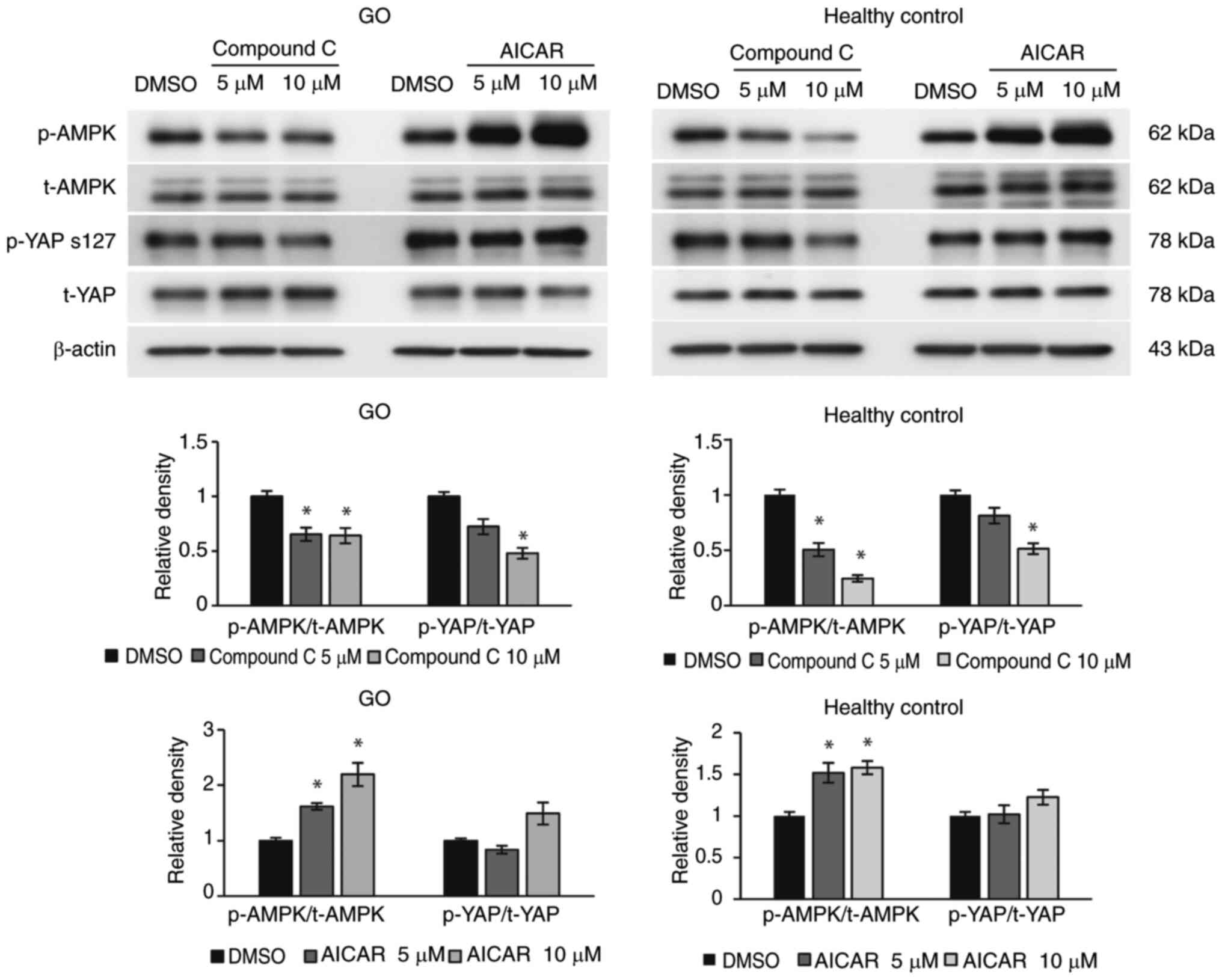
blotting. Western blot analysis revealed that compound C reduced
AMPK and YAP activation, whereas AICAR enhanced AMPK activation but
did not affect YAP activation, in both GO and normal orbital
fibroblasts, indicating the ability of compound C to inhibit AMPK
activity and YAP phosphorylation (Fig. 1).
Compound C reduces inflammatory
cytokine production
IL-1β is a key inflammatory mediator in GO;
therefore, orbital fibroblasts from patients with GO and healthy
controls were challenged with compound C (10 µM) for 6 h, followed
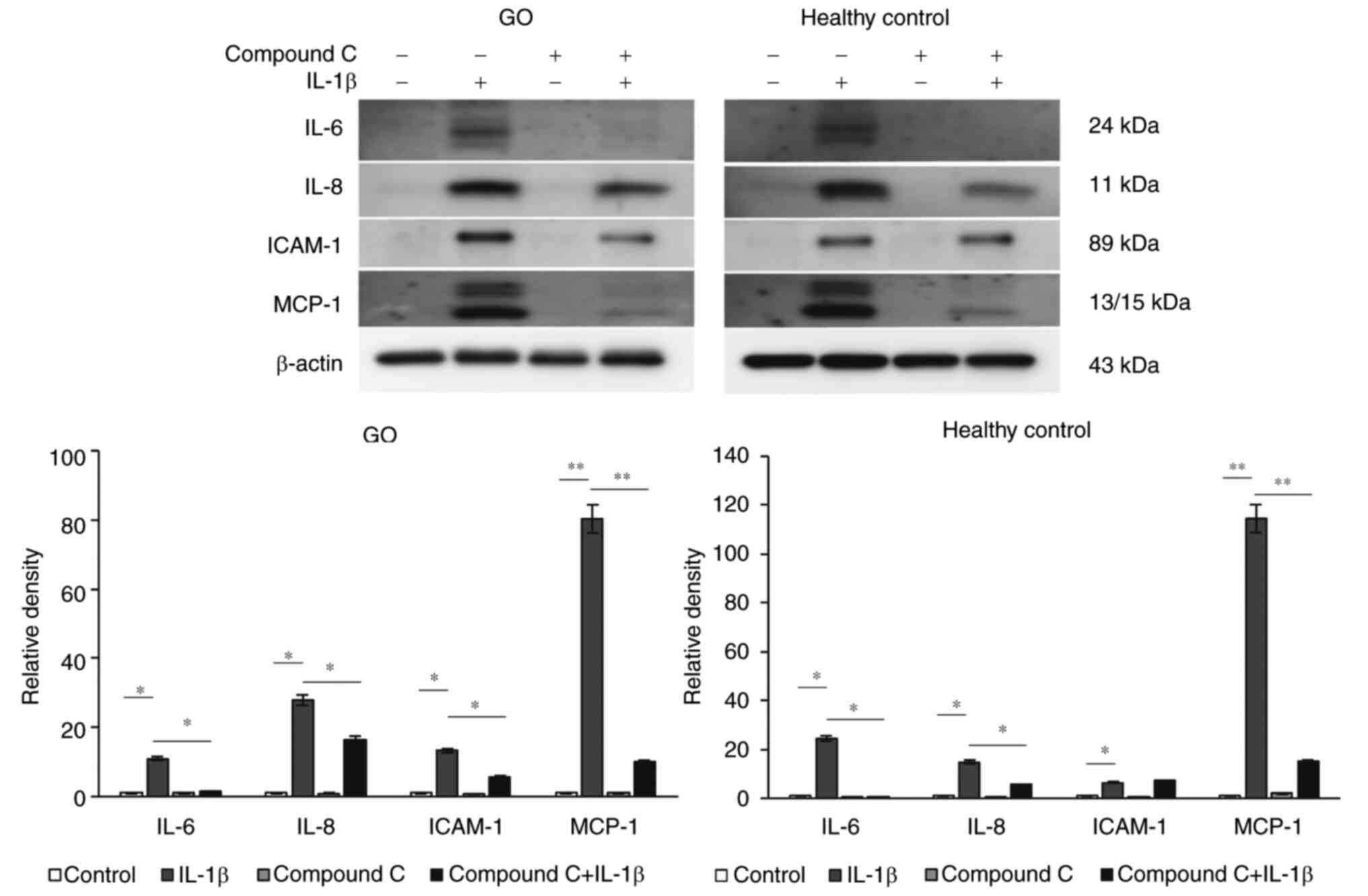
by IL-1β stimulation (10 ng/ml) for 24 h. Treatment with compound C
significantly reduced IL-1β-induced expression of IL-6, IL-8, and
monocyte chemoattractant protein (MCP)-1 in both GO and healthy
control orbital fibroblasts, and intracellular adhesion molecule
(ICAM)-1 in GO orbital fibroblasts, suggesting that compound C
suppressed inflammation in orbital fibroblasts (Fig. 2).
Effects of compound C on TGF-β-induced
fibrosis-related protein expression
TGF-β serves a crucial role in fibrosis and tissue
remodeling response in GO (21);
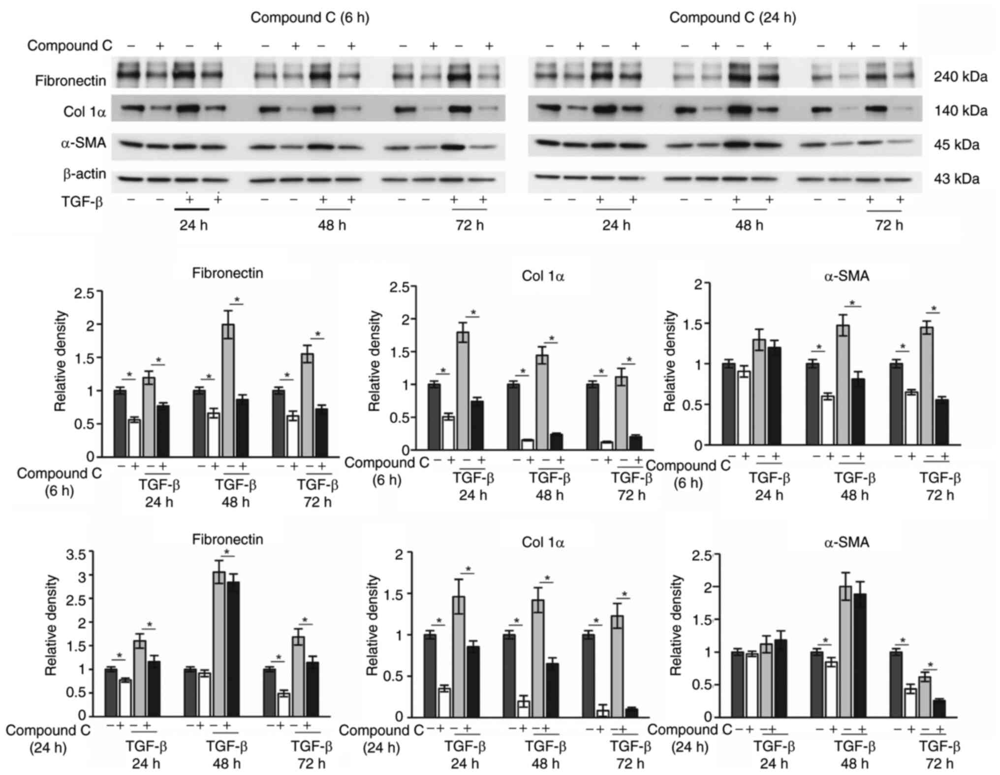
therefore, the GO orbital fibroblasts were pretreated with compound
C (10 µM) for 6 or 24 h prior to TGF-β (5 ng/ml) stimulation for
24, 48 or 72 h. Compound C treatment reduced TGF-β-induced
expression of fibronectin and collagen Iα in GO orbital fibroblasts
across all time points, and α-smooth muscle actin at 48 and 72 h,
indicating that compound C abrogated TGF-β-induced fibrotic
response in GO orbital fibroblasts (Fig. 3).
Effects of compound C on adipogenic
differentiation
To determine the effects of compound C on
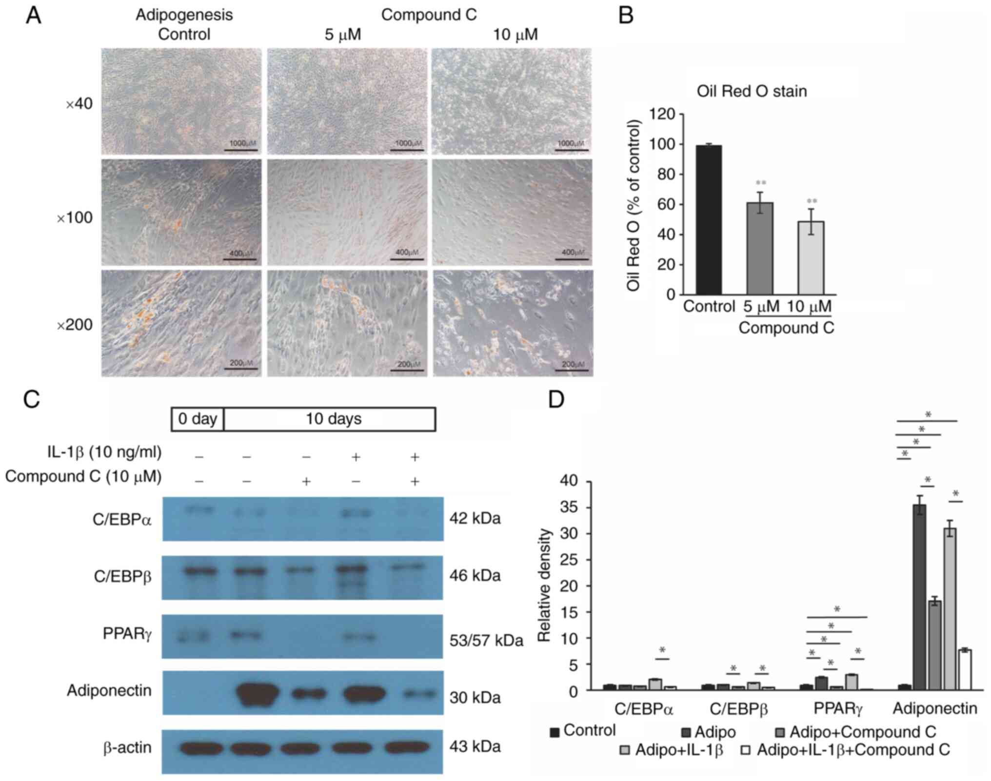
adipogenesis in GO, orbital fibroblasts from patients with GO were
treated with 5 or 10 µM compound C and were cultured for 10 days to
induce adipogenesis. Notably, compound C significantly inhibited
lipid accumulation (Fig. 4A and
B). At day 10 of adipogenesis, western blot analysis revealed
that 10 µM compound C markedly suppressed the expression of
adipogenic transcription factors, including CCAAT/enhancer-binding
protein (C/EBP)α/β, PPARγ and adiponectin in GO orbital
fibroblasts, suggesting that compound C suppressed adipogenic
differentiation in GO orbital fibroblasts (Fig. 4C and D).
Silencing YAP and TAZ inhibits
adipogenic differentiation
To investigate the effects of YAP and TAZ silencing
on adipogenesis, orbital fibroblasts from patients with GO were
transfected with si-control, si-YAP or si-TAZ, and cultured in
adipogenic medium for 10 days. Silencing of YAP, TAZ or both
suppressed adipogenic differentiation (Fig. 5A and C). Furthermore, YAP
silencing reduced the expression levels of C/EBPα/β, sterol
regulatory element-binding protein (SREBP)-1, aP2, adiponectin and
leptin. Similarly, TAZ silencing decreased the expression of PPARγ,
C/EBPα/β, SREBP-1, aP2, adiponectin and leptin (Fig. 5B and D). Taken together, these
findings indicated that silencing YAP and TAZ suppressed adipogenic
protein expression.
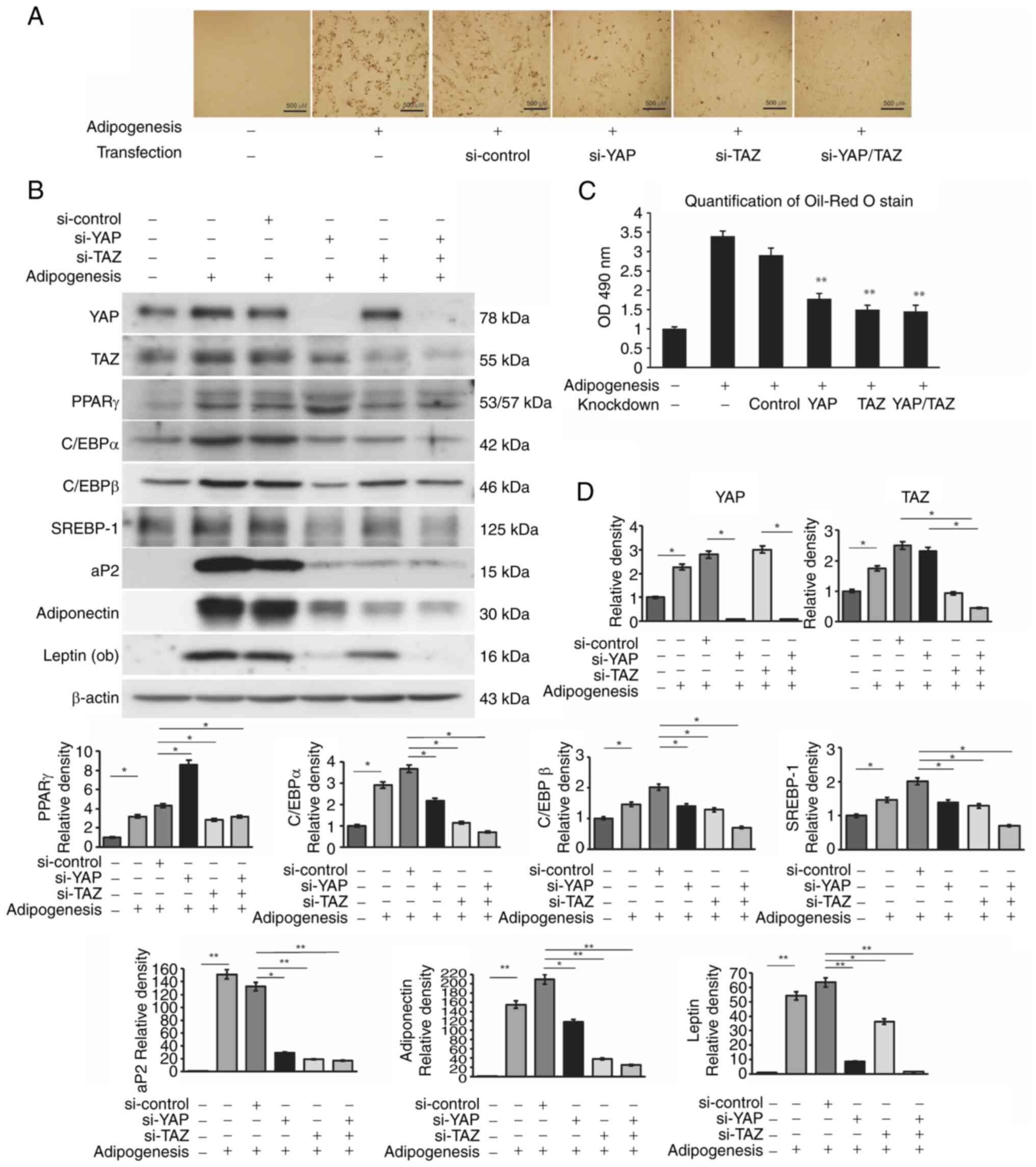 | Figure 5.Effects of YAP/TAZ silencing on
adipogenesis. YAP and TAZ siRNA transfection in orbital fibroblasts
from patients with GO (n=3) was performed to assess the effects of
YAP/TAZ knockdown on adipogenesis. (A) Representative Oil Red O
staining results. Cells were stained with Oil-Red O and examined
using a microscope (×40 magnification). (B) Western blot analysis
of adipogenic factors and hormones. Protein expression levels of
adipogenic transcription factors: PPARγ, C/EBPα, C/EBPβ, SREBP-1
and aP2, and hormones: Adiponectin and leptin, were analyzed by
western blotting on days 0 and 10 of differentiation with or
without si-control, si-YAP or si-TAZ transfection. (C) Oil-Red O
staining was quantified by measuring the optical density of cell
lysates at 490 nm. Silencing YAP or TAZ significantly suppressed
the adipogenesis compared with in the si-control group. (D) YAP
silencing reduced the expression levels of C/EBPα/β, SREBP-1, aP2,
adiponectin and leptin. Similarly, TAZ silencing decreased the
expression of PPARγ, C/EBPα/β, SREBP-1, aP2, adiponectin and
leptin. Data are presented as mean ± standard deviation.
*P<0.05, **P<0.01 vs. control or as indicated. GO, Graves'
orbitopathy; C/EBP, CCAAT/enhancer-binding protein; PPAR,
peroxisome proliferator-activated receptor; si, small interfering;
SREBP, sterol regulatory element-binding protein; TAZ,
transcriptional co-activator with PDZ-binding motif; YAP,
yes-associated protein. |
Compound C inhibits GO pathogenesis by
suppressing Akt and SMAD phosphorylation
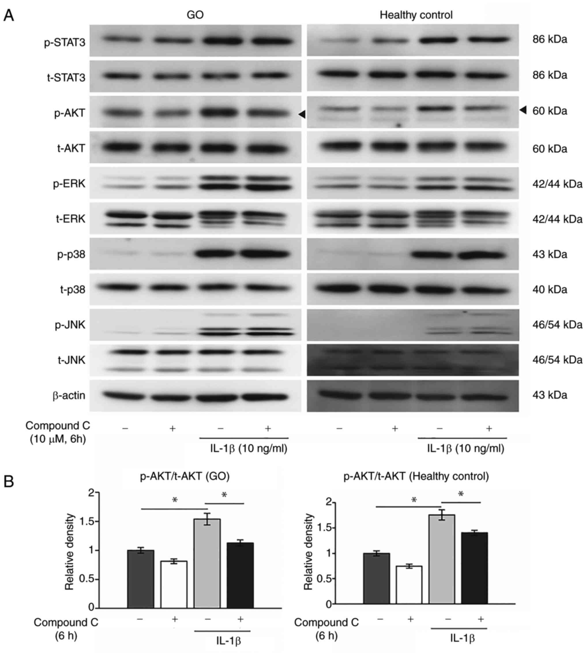
To evaluate the effect of high-dose compound C on
intracellular signaling pathways, the expression levels of p-STAT3,
p-Akt, p-ERK, p-p38 and p-JNK were assessed with or without
compound C treatment (10 µM) for 6 h in both GO and healthy control
orbital fibroblasts. Notably, treatment with IL-1β (10 ng/ml) for
30 min induced phosphorylation of Akt, which was significantly
suppressed by compound C treatment in both GO and healthy control
orbital fibroblasts, whereas other intracellular signaling pathways
were not affected (Fig. 6).
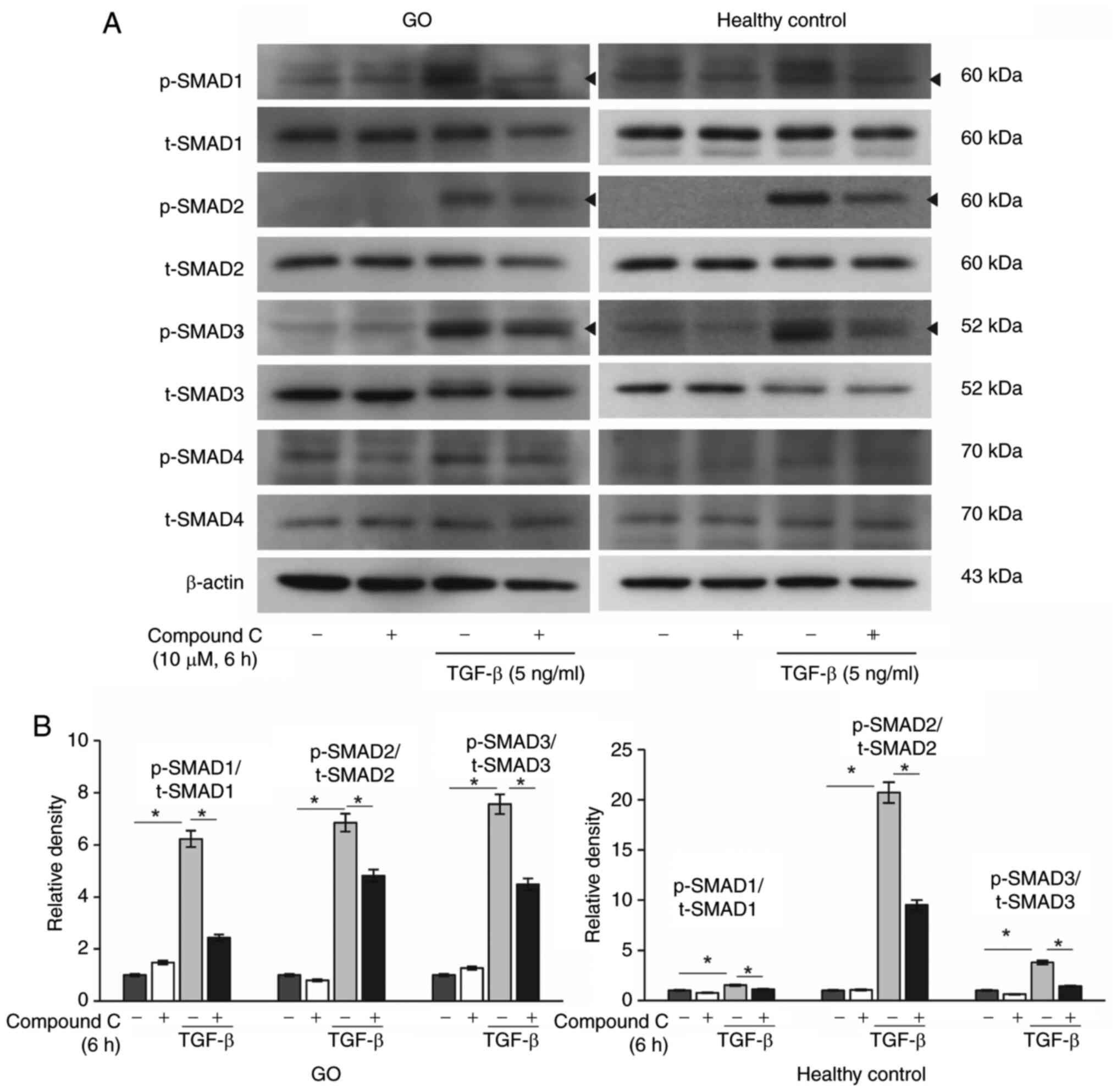
In a separate experiment, orbital fibroblasts from
both patients with GO and healthy controls were pretreated with
compound C (10 µM) for 6 h and were then stimulated with TGF-β (5
ng/ml) for 1 h. Compound C treatment significantly reduced SMAD1, 2
and 3 phosphorylation in both GO and normal orbital fibroblasts
(Fig. 7). Taken together, these
findings indicated that compound C may inhibit GO pathogenesis by
suppressing Akt and SMAD 1/2/3 phosphorylation.
Effects of compound C on hyaluronan
production
In addition to inflammation, adipogenesis and
fibrosis, excessive hyaluronan accumulation contributes to tissue
remodeling in GO (21). To
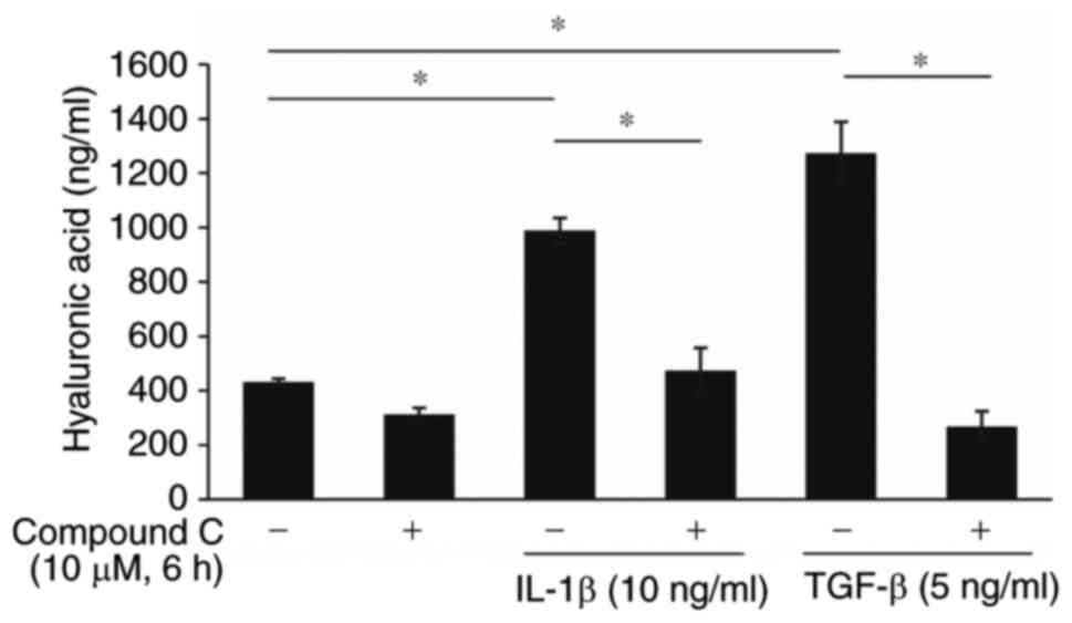
evaluate the effects of compound C on hyaluronan secretion, ELISA
was performed. GO orbital fibroblasts treated with IL-1β (10 ng/ml)
or TGF-β (5 ng/ml) for 96 h exhibited markedly increased hyaluronan
production, which was significantly reversed by treatment with
compound C (10 µM) for 6 h, indicating that compound C suppressed
hyaluronan production (Fig.
8).
Discussion
The present study indicated that compound C, a
widely used AMPK inhibitor, may effectively reverse inflammation,
adipogenesis and fibrosis in GO orbital fibroblasts. At a
concentration of 10 µM, compound C suppressed the phosphorylation
of YAP, a mechano-sensing signal protein, as well as AMPK. It also
inhibited IL-1β-induced pro-inflammatory cytokine production,
TGF-β-induced profibrotic protein expression and adipogenic
differentiation. Additionally, compound C attenuated IL-1β- and
TGF-β-stimulated hyaluronan secretion in GO orbital fibroblasts.
Notably, the silencing of YAP/TAZ also suppressed the expression of
adipogenic markers. Although the precise mechanism remains unclear,
it could be hypothesized that the suppression of various key GO
pathologies by compound C is related to YAP inactivation.
AMPK is a highly conserved energy sensor that serves
as a central regulator of cellular processes, including cell
growth, proliferation, survival and metabolic regulation (22). Decreased AMPK levels have been
reported in various metabolic diseases, such as diabetes and
obesity, and also in the aging process (23). Previous studies have partially
elucidated the role of AMPK in GO pathogenesis. In our earlier
research, it was demonstrated that metformin and phenformin can
exert anti-adipogenic effects and inhibit hyaluronan production via
the AMPK/ERK pathway in an in vitro model of GO (24). Similarly, Xu et al
(25) reported that metformin can
regulate IL-1β-mediated inflammation and TGF-β-mediated fibrosis in
GO orbital fibroblasts by activating the AMPK/mTOR pathway.
Contrary to these findings, the present study revealed that
compound C, an AMPK inhibitor, suppressed the pathological
mechanisms of GO, including inflammation, fibrosis and
adipogenesis. These findings suggested that compound C may exert
its therapeutic effects on GO via pathways independent of AMPK.
Compound C has protective effects against
inflammatory responses in various in vitro disease models.
In murine macrophages, compound C has been shown to inhibit
lipopolysaccharide-induced expression of inflammatory mediators,
including IL-6 and MCP-1 (26).
Furthermore, treatment with compound C has been shown to
downregulate the production of pro-inflammatory mediators in rat
microglial cells, such as IL-1β, IL-6, TNF-α, nitric oxide and
reactive oxygen species, while preserving the release of IL-10 and
TGF-β (27). In a mouse model of
formalin-induced inflammatory nociception, compound C has been
reported to reduce the levels of TNF-α, IL-1β, IFN-β and
cyclooxygenase-2 in the serum, spinal cord and inflamed paw tissues
(28). Consistent with these
findings, the present study demonstrated that compound C suppressed
IL-1β-induced expression of IL-6, IL-8, ICAM-1 and MCP-1.
Considering that these mediators are the major pro-inflammatory
cytokines implicated in the pathogenesis of GO, these findings
suggested that compound C could exert a protective effect against
the inflammatory processes associated with GO.
The direct effect of compound C on fibrotic response
has been less frequently studied. Peyton et al (29) reported that compound C can inhibit
vascular smooth muscle cell proliferation and migration in an
AMPK-independent manner. In human adipose-derived stem cells,
compound C has been shown to suppress the phosphorylation of
SMAD1/5/8 and SMAD2 (30).
Additionally, in a murine mesenchymal precursor cell line, compound
C could inhibit both SMAD and non-SMAD pathways, including p38 and
Akt (31). The results of the
current study align with these findings, and demonstrated that
compound C significantly blocked phosphorylation of signaling
factors downstream of TGF-β, including SMAD1, 2 and 3. The SMAD
pathway, which is primarily activated by TGF-β, serves a pivotal
role in myofibroblast differentiation and collagen deposition,
contributing to fibrotic tissue remodeling and adipogenesis in GO
(32,33). These findings suggested that
compound C may effectively mitigate fibrosis through its modulation
of TGF-β signaling.
Adipogenesis and hyaluronan deposition are major
contributors to volume expansion during GO pathogenesis. The
effects of compound C on adipogenesis have previously been
partially elucidated. In a previous study, in mouse preadipocyte
3T3-L1 cells, compound C was shown to inhibit differentiation,
C/EBPα and PPARγ mRNA levels, and to attenuate late adipogenic
markers, such as aP2 (34). The
Hippo signaling pathway is a key determinant of organ size, which
has a central role in mediating cellular mechanotransduction
(35). In a mouse model of
obesity, the inhibition of YAP/TAZ activity has been reported to
significantly alleviate adipose tissue fibrosis, and Hippo pathway
inactivation was revealed to be coupled with TGF-β-stimulation to
promote fibrosis (36). Notably,
our previous study demonstrated that YAP is strongly expressed in
GO tissue, and YAP silencing and pharmacological YAP inhibition
could suppress TGF-β-induced myofibroblast differentiation and
collagen formation, thus indicating that the YAP/TAZ pathway serves
as a critical mediator in the transition from inflammation to
fibrosis in GO pathogenesis (37). Furthermore, in the present study,
it was revealed that compound C abrogated YAP phosphorylation and
silencing YAP/TAZ suppressed adipogenic differentiation in GO
orbital fibroblasts. Collectively, these findings strongly
suggested that compound C may inhibit inflammation and
adipogenesis, thereby preventing volume expansion in GO by blocking
the Hippo pathway.
Several studies have reported that compound C exerts
AMPK-independent pharmacological effects on multiple cell types.
For example, 3 and 5 µM compound C has been shown to inhibit
macrophage chemotaxis in murine macrophages and to enhance insulin
sensitivity in murine adipocytes, independent of AMPK suppression
(26). Furthermore, 10 µM
compound C can inhibit the proliferation and glioma formation of
human glioma cells via multiple mechanisms, including activation of
the calpain/cathepsin pathway and inhibition of the Akt and mTOR
pathways, both of which are AMPK-independent (17,38). Additionally, compound C has been
reported to reduce serum-induced activation of Akt and glycogen
synthase kinase-3β, as well as platelet-derived growth factor
(PDGF)-induced phosphorylation of Akt, PI3K, PLCγ1, ERK1/2 and
PDGFR in human diploid fibroblasts (39). Compound C and AICAR have also been
shown to inhibit T-cell cytokine production by suppressing the mTOR
signaling pathway in mouse lymph node cells in an AMPK-independent
manner (40).
In the present study, compound C suppressed Akt
phosphorylation in both GO and healthy control orbital fibroblasts.
In glioma cells, 5 and 10 µM compound C has been shown to inhibit
Akt phosphorylation, while MAPK/ERK activity remained unaffected by
compound C treatment (17).
Additionally, in cancer cells, including human glioma cells, 10 µM
compound C has been reported to induce protective autophagy through
the Akt/mTOR pathway, which operates independently of AMPK
(18). Xu et al (25) demonstrated that the AMPK activator
metformin exerts anti-inflammatory and anti-fibrotic responses in
GO orbital fibroblasts, whereas 100 nM compound C does not reverse
the preventive effect of metformin on Akt phosphorylation.
Consistent with these previous reports, the present findings
revealed that compound C reversed the IL-1β-induced phosphorylation
of Akt in both GO and healthy control cells, suggesting that
compound C may directly inhibit the Akt/PI3K pathway in orbital
fibroblasts. The Akt/PI3K pathway is associated with adipogenesis
and hyaluronan production in GO, which is one of the key
pathological intracellular signaling pathway in GO (41).
Activated Akt can enhance SMAD3 transcriptional
activity, thereby leading to increased collagen I expression
(42). Akt and YAP signaling are
closely interconnected, with Akt regulating YAP activity through
phosphorylation-mediated cytoplasmic retention or nuclear
translocation. Furthermore, YAP can bind to SMAD1 and support
SMAD1-dependent phosphorylation; in response to TGF-β signaling,
TAZ and YAP have been shown to associate with heterometric
SMAD2/3/4 complexes and dictate their intracellular localization
(43,44). Thus, YAP interacts with SMAD
proteins to enhance TGF-β-driven fibrotic responses, forming a
positive feedback loop that amplifies fibrosis in GO. The present
findings suggested that compound C modulated these pathways,
thereby inhibiting TGF-β-induced myofibroblast differentiation and
collagen formation. Although compound C has shown promising results
in various disease models, it has not yet been approved for
clinical use and is currently utilized exclusively in in
vitro models. Moreover, the absence of a well-established in
vivo model for GO hinders the evaluation of the therapeutic
effects of compound C in a physiological setting. Furthermore, the
present study focused on the therapeutic effects of compound C on
the pathological phenotypes of GO, rather than examining the
specific responses of different immune biomarkers or subtypes of
autoreactive B and T cells; future studies exploring the roles of
various immune components could provide additional insights.
Further investigations using in vivo models and clinical
settings are necessary to validate therapeutic potential of
compound C.
In summary, the present study demonstrated that
compound C may inhibit IL-1β- and TGF-β-induced inflammation,
adipogenesis, fibrosis and hyaluronan secretion in primary cultured
orbital fibroblasts through modulation of the Akt, Hippo/YAP and
SMAD signaling pathways. While most recent drug development efforts
have focused on AMPK activation, unilateral AMPK inactivation
offers limited advantages for drug development. The present results
suggested that the therapeutic effects of compound C were not
solely mediated through AMPK inhibition, but also involved
modulation of other key signaling pathways, such as Akt and YAP.
These findings provide insights into the pathological mechanisms of
GO and suggest that the therapeutic effects of compound C serves as
a foundation for future research aimed at developing drugs that
directly target the YAP or Akt pathways. Furthermore, given the
critical role of these pathways in other fibrotic and inflammatory
diseases, compound C may have therapeutic potential beyond GO, such
as in inflammatory arthritis or inflammatory metabolic diseases,
making it a promising candidate for further exploration in various
pathological contexts.
Supplementary Material
Supporting Data
Acknowledgements
Not applicable.
Funding
This work was supported by a National Research Foundation of
Korea grant funded by the Korean government (MSIT, Ministry of
Science and ICT; grant no. RS-2023-00208570) and by a faculty
research grant from Yonsei University College of Medicine (grant
no. 4-2023-1454).
Availability of data and materials
The data generated in the present study may be
requested from the corresponding author.
Authors' contributions
HYP, HSL and JSY conceptualized the study. SHC
performed experiments, while HYP, JSY and JK confirmed the
authenticity of all the raw data. HYP, JSY and JK analyzed and
interpreted the data. JSY and JK supplied the laboratory resources,
reagents and materials, and JSY procured the funding. HYP, SHC and
HSL drafted the manuscript, and JSY and JK reviewed and edited it.
All authors have read and approved the final version of the
manuscript.
Ethics approval and consent to
participate
The study protocol was reviewed and approved by the
Institutional Review Board of Severance Hospital (approval no.
4-2024-0492) and conducted in accordance with the principles of The
Declaration of Helsinki. Written informed consent was obtained from
all participants.
Patient consent for publication
Not applicable.
Competing interests
The authors declare that they have no competing
interests.
Glossary
Abbreviations
Abbreviations:
|
AMPK
|
adenosine monophosphate-activated
protein kinase
|
|
C/EBP
|
CCAAT/enhancer-binding protein
|
|
GO
|
Graves' orbitopathy
|
|
IBMX
|
3-isobutyl-1-methylxanthine
|
|
ICAM-1
|
intracellular adhesion molecule-1
|
|
IGF-1-R
|
insulin-like growth factor-1
receptor
|
|
MCP-1
|
monocyte chemoattractant protein-1
|
|
PDGF
|
platelet-derived growth factor
|
|
PPAR
|
peroxisome proliferator-activated
receptor
|
|
SMA
|
smooth muscle actin
|
|
TAZ
|
transcriptional co-activator with
PDZ-binding motif
|
|
YAP
|
yes-associated protein
|
References
|
1
|
Bahn RS: Graves' ophthalmopathy. N Engl J
Med. 362:726–738. 2010. View Article : Google Scholar : PubMed/NCBI
|
|
2
|
Khoo TK and Bahn RS: Pathogenesis of
Graves' ophthalmopathy: The role of autoantibodies. Thyroid.
17:1013–1018. 2007. View Article : Google Scholar : PubMed/NCBI
|
|
3
|
Cui X, Wang F and Liu C: A review of TSHR-
and IGF-1R-related pathogenesis and treatment of Graves'
orbitopathy. Front Immunol. 14:10620452023. View Article : Google Scholar : PubMed/NCBI
|
|
4
|
Huang Y, FangSLi D, Zhou H, Li B and Fan
X: The involvement of T cell pathogenesis in thyroid-associated
ophthalmopathy. Eye (Lond). 33:176–182. 2019. View Article : Google Scholar : PubMed/NCBI
|
|
5
|
Zhang L, Bowen T, Grennan-Jones F, Paddon
C, Giles P, Webber J, Steadman R and Ludgate M: Thyrotropin
receptor activation increases hyaluronan production in
preadi-pocyte fibroblasts: Contributory role in hyaluronan
accumu-lation in thyroid dysfunction. J Biol Chem. 284:26447–26455.
2009. View Article : Google Scholar : PubMed/NCBI
|
|
6
|
Weiler DL: Thyroid eye disease: A review.
Clin Exp Optom. 100:20–25. 2017. View Article : Google Scholar : PubMed/NCBI
|
|
7
|
Genere N and Stan MN: Current and emerging
treatment strategies for Graves' orbitopathy. Drugs. 79:109–124.
2019. View Article : Google Scholar : PubMed/NCBI
|
|
8
|
Bartalena L, Kahaly GJ, Baldeschi L, Dayan
CM, Eckstein A, Marcocci C, Marinò M, Vaidya B and Wiersinga WM:
EUGOGO† The 2021 European Group on Graves' orbitopathy
(EUGOGO) clinical practice guidelines for the medical management of
Graves' orbitopathy. Eur J Endocrinol. 185:G43–G67. 2021.
View Article : Google Scholar : PubMed/NCBI
|
|
9
|
Kamboj A, Harrison AR and Mokhtarzadeh A:
Emerging therapies in the medical management of thyroid eye
disease. Front Ophthalmol (Lausanne). 3:12959022023. View Article : Google Scholar : PubMed/NCBI
|
|
10
|
Douglas RS, Kahaly GJ, Patel A, Sile S,
Thompson EHZ, Perdok R, Fleming JC, Fowler BT, Marcocci C, Marinò
M, et al: Teprotumumab for the treatment of active thyroid eye
disease. N Engl J Med. 382:341–352. 2020. View Article : Google Scholar : PubMed/NCBI
|
|
11
|
Park JW and Yoon JS: A review of novel
medical treatments for thyroid eye disease. Korean J Ophthalmol.
38:249–259. 2024. View Article : Google Scholar : PubMed/NCBI
|
|
12
|
Garcia D and Shaw RJ: AMPK: Mechanisms of
cellular energy sensing and restoration of metabolic balance. Mol
Cell. 66:789–800. 2017. View Article : Google Scholar : PubMed/NCBI
|
|
13
|
Umezawa S, Higurashi T and Nakajima A:
AMPK: Therapeutic target for diabetes and cancer prevention. Curr
Pharm Des. 23:3629–3644. 2017. View Article : Google Scholar : PubMed/NCBI
|
|
14
|
Huang R, Guo F, Li Y, Liang Y, Li G, Fu P
and Ma L: Activation of AMPK by triptolide alleviates nonalcoholic
fatty liver disease by improving hepatic lipid metabolism,
inflammation and fibrosis. Phytomedicine. 92:1537392021. View Article : Google Scholar : PubMed/NCBI
|
|
15
|
Kahn BB, Alquier T, Carling D and Hardie
DG: AMP-activated protein kinase: Ancient energy gauge provides
clues to modern understanding of metabolism. Cell Metab. 1:15–25.
2005. View Article : Google Scholar : PubMed/NCBI
|
|
16
|
Zhou G, Myers R, Li Y, Chen Y, Shen X,
Fenyk-Melody J, Wu M, Ventre J, Doebber T, Fujii N, et al: Role of
AMP-activated protein kinase in mechanism of metformin action. J
Clin Invest. 108:1167–1174. 2001. View
Article : Google Scholar : PubMed/NCBI
|
|
17
|
Liu X, Chhipa RR, Nakano I and Dasgupta B:
The AMPK inhibitor compound C is a potent AMPK-independent
antiglioma agent. Mol Cancer Ther. 13:596–605. 2014. View Article : Google Scholar : PubMed/NCBI
|
|
18
|
Vucicevic L, Misirkic M, Janjetovic K,
Vilimanovich U, Sudar E, Isenovic E, Prica M, Harhaji-Trajkovic L,
Kravic-Stevovic T, Bumbasirevic V and Trajkovic V: Compound C
induces protective autophagy in cancer cells through AMPK
inhibition-independent blockade of Akt/mTOR pathway. Autophagy.
7:40–50. 2011. View Article : Google Scholar : PubMed/NCBI
|
|
19
|
Yoon JS, Lee HJ, Choi SH, Chang EJ, Lee SY
and Lee EJ: Quercetin inhibits IL-1β-induced inflammation,
hyaluronan production and adipogenesis in orbital fibroblasts from
Graves' orbitopathy. PLoS One. 6:e262612011. View Article : Google Scholar : PubMed/NCBI
|
|
20
|
Kim SE, Lee JH, Chae MK, Lee EJ and Yoon
JS: The role of sphingosine-1-phosphate in adipogenesis of Graves'
orbitopathy. Invest Ophthalmol Vis Sci. 57:301–311. 2016.
View Article : Google Scholar : PubMed/NCBI
|
|
21
|
Longo CM and Higgins PJ: Molecular
biomarkers of Graves' ophthalmopathy. Exp Mol Pathol. 106:1–6.
2019. View Article : Google Scholar : PubMed/NCBI
|
|
22
|
Hardie DG: AMP-activated protein kinase: A
cellular energy sensor with a key role in metabolic disorders and
in cancer. Biochem Soc Trans. 39:1–13. 2011. View Article : Google Scholar : PubMed/NCBI
|
|
23
|
Wang D, Yang L and Liu Y: Targeting AMPK
signaling in the liver: Implications for obesity and type 2
diabetes mellitus. Curr Drug Targets. 23:1057–1071. 2022.
View Article : Google Scholar : PubMed/NCBI
|
|
24
|
Han YE, Hwang S, Kim JH, Byun JW, Yoon JS
and Lee EJ: Biguanides metformin and phenformin generate
therapeutic effects via AMP-activated protein
kinase/extracellular-regulated kinase pathways in an in vitro model
of Graves' orbitopathy. Thyroid. 28:528–536. 2018. View Article : Google Scholar : PubMed/NCBI
|
|
25
|
Xu Z, Ye H, Xiao W, Sun A, Yang S, Zhang
T, Sha X and Yang H: Metformin attenuates inflammation and fibrosis
in thyroid-associated ophthalmopathy. Int J Mol Sci. 23:155082022.
View Article : Google Scholar : PubMed/NCBI
|
|
26
|
Lee Y, Park BH and Bae EJ: Compound C
inhibits macrophage chemotaxis through an AMPK-independent
mechanism. Biochem Biophys Res Commun. 469:515–520. 2016.
View Article : Google Scholar : PubMed/NCBI
|
|
27
|
Labuzek K, Liber S, Gabryel B, Bułdak L
and Okopień B: Ambivalent effects of compound C (dorsomorphin) on
inflammatory response in LPS-stimulated rat primary microglial
cultures. Naunyn Schmiedebergs Arch Pharmacol. 381:41–57. 2010.
View Article : Google Scholar : PubMed/NCBI
|
|
28
|
Xinqiang Y, Yuanyuan J, Zhipeng Y, Jie K,
Xiao T, Yumeng H, Chenxi Z, Shiyu D, Mingpeng Y, Yanlin Z, et al:
Systemic administration of dorsomorphin relieves inflammatory
nociception in the mouse formalin test. Int Immunopharmacol.
113:1093372022. View Article : Google Scholar : PubMed/NCBI
|
|
29
|
Peyton KJ, Yu Y, Yates B, Shebib AR, Liu
XM, Wang H and Durante W: Compound C inhibits vascular smooth
muscle cell proliferation and migration in an AMP-activated protein
kinase-independent fashion. J Pharmacol Exp Ther. 338:476–484.
2011. View Article : Google Scholar : PubMed/NCBI
|
|
30
|
Madhu V, Dighe AS, Cui Q and Deal DN: Dual
inhibition of activin/Nodal/TGF-β and BMP signaling pathways by
SB431542 and dorsomorphin induces neuronal differentiation of human
adipose derived stem cells. Stem Cells Int. 2016:10353742016.
View Article : Google Scholar : PubMed/NCBI
|
|
31
|
Boergermann JH, Kopf J, Yu PB and Knaus P:
Dorsomorphin and LDN-193189 inhibit BMP-mediated Smad, p38 and Akt
signalling in C2C12 cells. Int J Biochem Cell Biol. 42:1802–1807.
2010. View Article : Google Scholar : PubMed/NCBI
|
|
32
|
Shin HA, Park M, Banga JP and Lew H:
TGFβ-treated placenta-derived mesenchymal stem cells selectively
promote anti-adipogenesis in thyroid-associated ophthalmopathy. Int
J Mol Sci. 23:56032022. View Article : Google Scholar : PubMed/NCBI
|
|
33
|
Jang SY, Park SJ, Chae MK, Lee JH, Lee EJ
and Yoon JS: Role of microRNA-146a in regulation of fibrosis in
orbital fibroblasts from patients with Graves' orbitopathy. Br J
Ophthalmol. 102:407–414. 2018. View Article : Google Scholar : PubMed/NCBI
|
|
34
|
Gao Y, Zhou Y, Xu A and Wu D: Effects of
an AMP-activated protein kinase inhibitor, compound C, on
adipogenic differentiation of 3T3-L1 cells. Biol Pharm Bull.
31:1716–1722. 2008. View Article : Google Scholar : PubMed/NCBI
|
|
35
|
Dupont S, Morsut L, Aragona M, Enzo E,
Giulitti S, Cordenonsi M, Zanconato F, Le Digabel J, Forcato M,
Bicciato S, et al: Role of YAP/TAZ in mechanotransduction. Nature.
474:179–183. 2011. View Article : Google Scholar : PubMed/NCBI
|
|
36
|
Shen H, Huang X, Zhao Y, Wu D, Xue K, Yao
J, Wang Y, Tang N and Qiu Y: The Hippo pathway links adipocyte
plasticity to adipose tissue fibrosis. Nat Commun. 13:60302022.
View Article : Google Scholar : PubMed/NCBI
|
|
37
|
Ko J, Kim YJ, Choi SH, Lee CS and Yoon JS:
Yes-associated protein mediates the transition from inflammation to
fibrosis in Graves' orbitopathy. Thyroid. 33:1465–1475. 2023.
View Article : Google Scholar : PubMed/NCBI
|
|
38
|
Ríos M, Foretz M, Viollet B, Prieto A,
Fraga M, Costoya JA and Señarís R: AMPK activation by oncogenesis
is required to maintain cancer cell proliferation in astrocytic
tumors. Cancer Res. 73:2628–2638. 2013. View Article : Google Scholar : PubMed/NCBI
|
|
39
|
Kwon HJ, Kim GE, Lee YT, Jeong MS, Kang I,
Yang D and Yeo EJ: Inhibition of platelet-derived growth factor
receptor tyrosine kinase and downstream signaling pathways by
compound C. Cell Signal. 25:883–897. 2013. View Article : Google Scholar : PubMed/NCBI
|
|
40
|
Rao E, Zhang Y, Li Q, Hao J, Egilmez NK,
Suttles J and Li B: AMPK-dependent and independent effects of AICAR
and compound C on T-cell responses. Oncotarget. 7:33783–33795.
2016. View Article : Google Scholar : PubMed/NCBI
|
|
41
|
Kumar S, Nadeem S, Stan MN, Coenen M and
Bahn RS: A stimulatory TSH receptor antibody enhances adipogenesis
via phosphoinositide 3-kinase activation in orbital preadipocytes
from patients with Graves' ophthalmopathy. J Mol Endocrinol.
46:155–163. 2011. View Article : Google Scholar : PubMed/NCBI
|
|
42
|
Runyan CE, Schnaper HW and Poncelet AC:
The phosphatidylinositol 3-kinase/Akt pathway enhances
Smad3-stimulated mesangial cell collagen I expression in response
to transforming growth factor-beta1. J Biol Chem. 279:2632–2639.
2004. View Article : Google Scholar : PubMed/NCBI
|
|
43
|
Varelas X, Sakuma R, Samavarchi-Tehrani P,
Peerani R, Rao BM, Dembowy J, Yaffe MB, Zandstra PW and Wrana JL:
TAZ controls Smad nucleocytoplasmic shuttling and regulates human
embryonic stem-cell self-renewal. Nat Cell Biol. 10:837–848. 2008.
View Article : Google Scholar : PubMed/NCBI
|
|
44
|
Alarcón C, Zaromytidou AI, Xi Q, Gao S, Yu
J, Fujisawa S, Barlas A, Miller AN, Manova-Todorova K, Macias MJ,
et al: Nuclear CDKs drive Smad transcriptional activation and
turnover in BMP and TGF-beta pathways. Cell. 139:757–769. 2009.
View Article : Google Scholar : PubMed/NCBI
|