Introduction
Epithelial cell adhesion molecule (EpCAM) is a
transmembrane protein that was first proposed to function as a
homophilic cell adhesion molecule that can interfere with
cadherin-mediated cell-to-cell contact (1). EpCAM is composed of a large
extracellular domain (EpEx), a single transmembrane domain and a
short 26-amino acid intracellular domain (EpICD) (1,2).
EpCAM is frequently expressed in normal epithelial cells, and is
reported to be widely upregulated in various cancers and
cancer-initiating cells (3–5). The
high expression of membranous EpCAM in a variety of cancers
analyzed by immunohistochemistry has rendered EpCAM as an ideal
immunotherapeutic target (2,3).
However, results from clinical trials of EpCAM-specific monoclonal
antibodies in various cancers have shown limited efficacy (6,7). The
cleavage of EpEx by the protease tumor necrosis factor α-converting
enzyme (TACE) and its shedding have been shown to result in the
release of its intracellular domain EpICD (8). Released EpICD associates with
four-and-a-half LIM domain protein 2 (FHL2), forms a nuclear
complex with components of the Wnt signal pathway (β-catenin,
Lef-1), and induces gene transcription, resulting in activation of
oncogenic signaling (8). This
proposed mechanism of regulated intramembranous proteolysis
(RIP)-mediated loss of EpCAM from the cancer cell membrane and
activation of oncogenic signaling via translocated nuclear EpICD
might account for the restricted therapeutic efficacy of the
EpCAM-targeted immunotherapy using monoclonal antibodies to target
EpEx. Indeed, nuclear expression of EpICD in some types of cancer
has been reported (9), and EpICD
has been proposed as a new therapeutic target for cancer cells
(9,10). However, little is known about the
roles of EpICD in carcinogenesis and cancer progression.
β-catenin plays an important role in both adhesion
and signal transduction of epithelial cells, depending on the
intracellular localization. β-catenin forms adherens junctions
through conjugation with α-catenin and E-cadherin at the plasma
membrane. However, β-catenin can also act as a main regulator of
transcription through DNA-binding proteins, such as TCF/LEF family
members in the nucleus (11).
Previous studies suggest that the activation of the Wnt/β-catenin
signaling pathway plays an important role in human cancer invasion
and metastasis (11–14). Nuclear expression of β-catenin, a
hallmark of an active β-catenin-dependent Wnt pathway, was found
predominantly at the invasive front of cancers (12–14).
Extrahepatic cholangiocarcinoma (ECC) arises from
cholangiocytes of extrahepatic bile ducts. ECC has a devastating
prognosis, and its poor prognosis is associated with extensive
local tumor invasion and metastasis (15,16).
Despite advances in screening for early detection and in therapy
over recent decades, the overall survival rates for patients with
ECC have not been improved significantly (17). Thus, to improve the prognosis of
ECC patients, identification of new molecular mechanisms of
invasion and metastasis of ECC is essential. Based on the
observation of nuclear translocation of EpICD in a complex with
β-catenin and Lef, which are both components of the Wnt signaling
pathway, we hypothesized that spatial localization of EpICD and its
mutual interaction with β-catenin may be important to ECC cancer
cell invasion and metastasis.
In the present study, we examined: i) the
localization and expression of EpEx, EpICD and β-catenin in
surgical specimens of ECCs from 79 patients with a focus on the
relationship between nuclear expression of EpICD and β-catenin; ii)
the effect of EpICD in cholangiocarcinoma (CC) cell growth and
proliferation; iii) the effect of EpICD in EpCAM target gene
expression; and iv) the role of EpICD in the migration and invasion
of CC cells. We also examined whether EpCAM silencing by small
interfering RNA (siRNA) could affect cell growth, migration and
invasiveness in CC cells.
Materials and methods
Materials
This study was approved by the Institutional Review
Board (IRB) of Chonbuk National University Hospital. Surgical
specimens of 79 formalin-fixed, paraffin-embedded ECCs were
obtained from Surgical Pathology Archives of Chonbuk National
University Hospital, Korea between 1998 and 2010. Of the 79
patients with ECC, 56 (70.9%) were men and 23 (29.1%) were women.
The tumors were histologically graded as 26 cases (23.9%) of
well-differentiated, 46 cases (58.3%) of moderately-differentiated
and seven cases (8.8%) of poorly-differentiated ECC.
We used the four human CC cell lines, designated as
JCK, Cho-CK, Choi-CK and SCK, which were established in our
laboratory (18). In addition, the
RBE cell line was purchased from the RIKEN BRC cell bank (Tsukuba,
Japan). All CC cell lines were maintained in Dulbecco’s modified
Eagle’s medium (DMEM) supplemented with penicillin/streptomycin and
10% fetal bovine serum (Gibco BRL, Gaithersburg, MD, USA). The
cells were cultured at 37°C in a 5% CO2 humidified
incubator.
Immunohistochemical staining
For immunohistochemical staining, we used a polymer
intense detection system using the Bond-Max Automatic stainer
(Leica, Bannockburn, IL, USA) in accordance with the manufacturer’s
instructions. Briefly, after deparaffinization, the tissue sections
were treated with a microwave antigen retrieval procedure in 0.01 M
sodium citrate buffer (pH 6.0) for 12 min. The samples were
incubated with anti-EpCAM (Calbiochem, La Jolla, CA, USA) for the
extracellular domain of EpCAM (EpEX), anti-EpICD for the
intracellular domain of EpCAM (C-terminus, cytoplasmic domain of
EpCAM; 1144-1) (Epitomics, Burlingame, CA, USA) and anti-β-catenin
(BD Biosciences, San Jose, CA, USA) antibodies for 20 min.
Peroxidase activity was detected with the enzyme substrate
3-amino-9-ethyl carbazole. Samples with EpEx, EpICD and β-catenin
staining of ≥10% of the tumor cells with strong intensity were
defined as positive.
Plasmid cDNA and small interfering RNA
transfection
For EpICD plasmid cDNA transfection, EpICD cDNA
(NCBI accession number NM_002354.2; EpCAM cytoplasmic domain) was
synthesized from CosmoGenetech Co., Ltd. (Seoul, Korea) and
inserted into the XhoI and BamHI sites of pEGFP-N1
vector (Clontech, Palo Alto, CA, USA). Transfection of EpICD
plasmid DNA was performed with lipofectamine 2000 transfection
reagent (Invitrogen, Carlsbad, CA, USA) following the
manufacturer’s protocol. At 48 h after transfection, the cells were
collected and used for further experiments.
Small interfering RNA (siRNA) was used for silencing
EpCAM expression as previously described (19). EpCAM siRNA and negative control
were purchased from Bioneer Corp. (Daejeon, Korea). Sequences for
EpCAM-specific siRNA and negative control siRNA were as follows:
sense: 5′-GUGAGAACCUACUGGAUCA(dTdT)-3′, antisense:
5′-UGAUCCAGUAGGUUCUCAC(dTdT)-3′; and negative control: sense:
5′-CCUACGCCACCAAUUUCGU(dTdT)-3′, antisense:
5′-ACGAAAUUGGUGGCGUAGG(dTdT)-3′. Transfection of siRNA was
performed with lipofectamine RNAiMAX transfection reagent
(Invitrogen) following the manufacturer’s protocol.
Western blotting
Western blotting of EpEx (Calbiochem) and EpICD
(Epitomics) in CC cell lines was performed as previously described
(20). To determine the effect of
forced expression of EpICD, we also examined the expression profile
of E-cadherin (BD Biosciences), active β-catenin (Merck Millipore,
Billerica, MA, USA), c-Myc (Abcam, Cambridge, UK), and cyclin D1
(Santa Cruz Biotechnology, Santa Cruz, CA, USA) in
EpICD-transfected CC cell lines. Blots were developed using
secondary antibody, and immune complexes were visualized using an
enhanced chemiluminescence detection system (Amersham Biosciences,
Buckinghamshire, UK), prior to exposure to a luminescent image
analyzer (LAS-3000, Fuji Film, Tokyo, Japan).
Cell growth and proliferation assay
The effects of the EpICD gene transfection and
silencing of EpCAM on cell proliferation were assessed by MTT
(3-(4,5-dimethylthiazol-2-yl)-2,5-diphenyltetrazolium bromide)
(Sigma, St. Louis, MO, USA) and BrdU cell proliferation assay, as
described previously (19).
In vitro assay for cellular migration and
invasion
Cell migration was assessed in a 24-Transwell
migration assay (Corning Life Sciences, Acton, MA, USA) and the
cell invasion assay was performed using a 24-Transwell BioCoat
Matrigel invasion chamber (BD Biosciences), as previously described
(20).
Statistical analysis
Statistical analysis was performed using SPSS
version 15.0 (SPSS, Chicago, IL, USA). Statistical significance was
determined by Chi-square test and appropriate Student’s t-test. All
experiments were repeated a minimum of three times. A P-value of
<0.05 was considered statistically significant.
Results
EpEx, EpICD and β-catenin expression in
extrahepatic cholangiocarcinoma
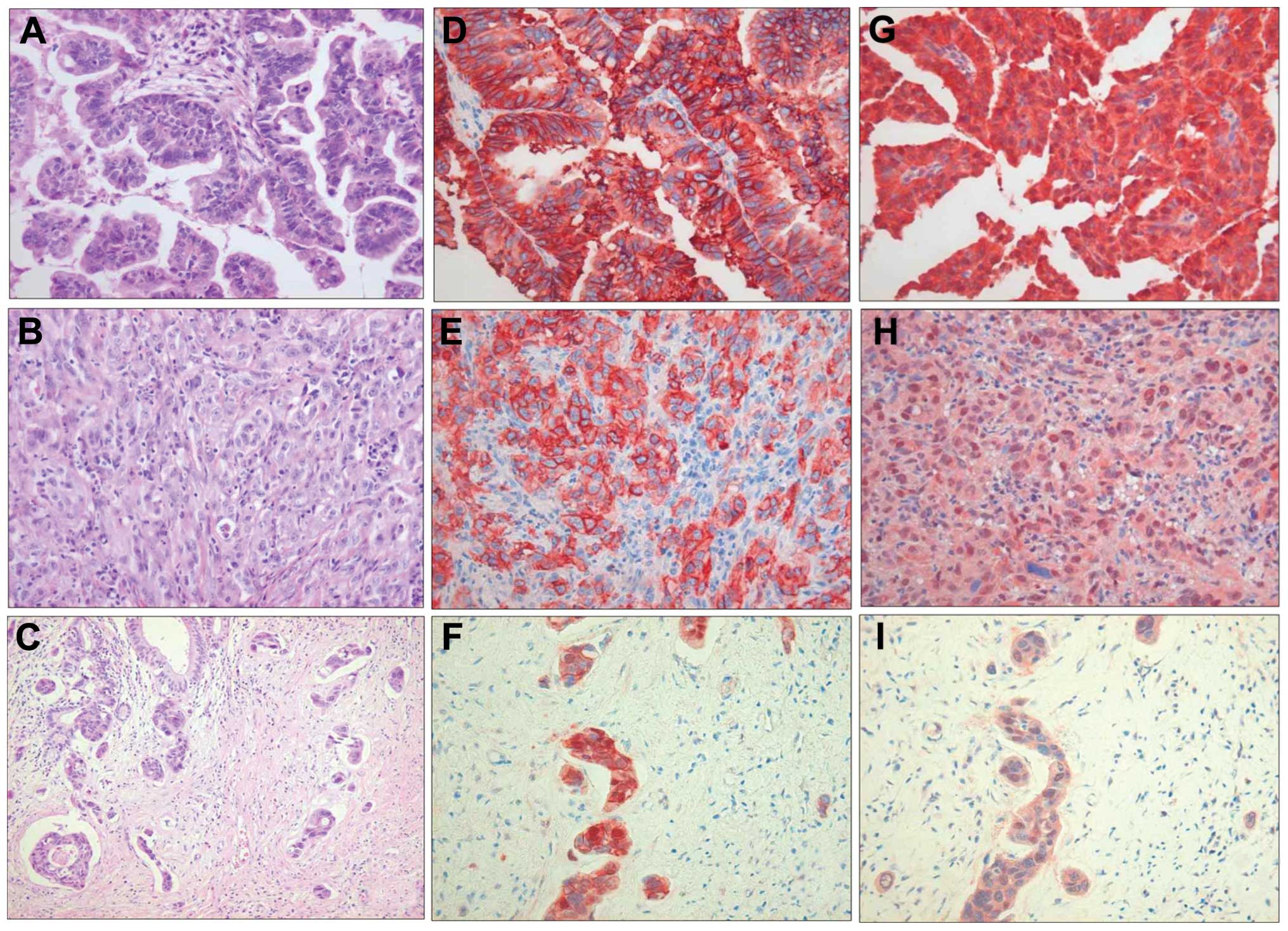
The positive expression of EpEx was observed in
98.7% (78 of 79) of ECC tissues. In ECC cells, EpEx expression was
predominantly localized to the membrane (Fig. 1A, B, D and E). EpEx was not
detected in any of the nuclei of tumor cells. Increased nuclear
and/or cytoplasmic expression with reduced or absent membrane
expression of EpICD was found in ECC cells (Fig. 1C and F). Nuclear expression of
EpICD was observed in 27.8% (22 of 79) of ECC tissues. No nuclear
expression of EpICD was present in non-neoplastic bile duct cells.
Nuclear expression of β-catenin was observed in 12.6% (10 of 79) of
ECC tissues. Frequent nuclear co-localization of EpICD and
β-catenin was observed in cancer cells forming the invasive front
(Fig. 1G, H and I). Nuclear
translocation of EpICD was significantly higher in poorly and
moderately differentiated tumor cells compared to
well-differentiated tumor cells (P=0.021) (Table I). There was a significant
correlation between the nuclear translocation of EpICD and nuclear
expression of β-catenin (P=0.015) (Table I). Aberrant nuclear expression, and
often co-localization, of EpICD and β-catenin in cancer cells
forming the invasive front suggests that they are engaged in local
interactions and play important roles in ECC invasion.
 | Table IAssociation between nuclear
expression of β-catenin, histologic tumor grade and nuclear
translocation of EpICD in extrahepatic cholangiocarcinoma. |
Table I
Association between nuclear
expression of β-catenin, histologic tumor grade and nuclear
translocation of EpICD in extrahepatic cholangiocarcinoma.
|
Characteristics | EpICD nuclear
translocation | EpICD no
translocation | P-value |
|---|
| Tumor grade | | | <0.05 |
| Well | 5 | 21 | |
| Moderately | 12 | 34 | |
| Poorly | 5 | 2 | |
| β-catenin
expression | | | <0.05 |
| Nuclear
translocation | 6 | 4 | |
| No
translocation | 16 | 53 | |
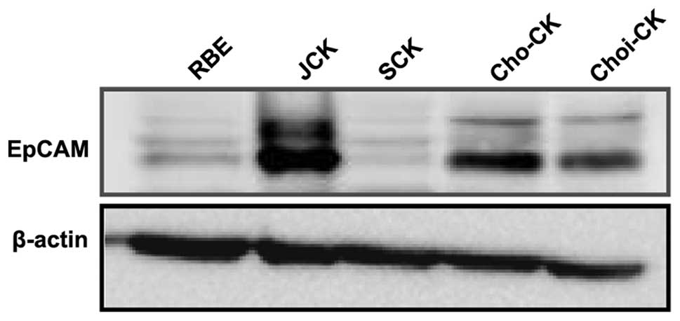
EpCAM expression level in
cholangiocarcinoma cell lines
The expression level of EpCAM protein was higher in
the JCK and Cho-CK cells than in the other cell lines. Choi-CK
cells showed low EpCAM protein level that was barely detected in
the SCK and RBE cells (Fig.
2).
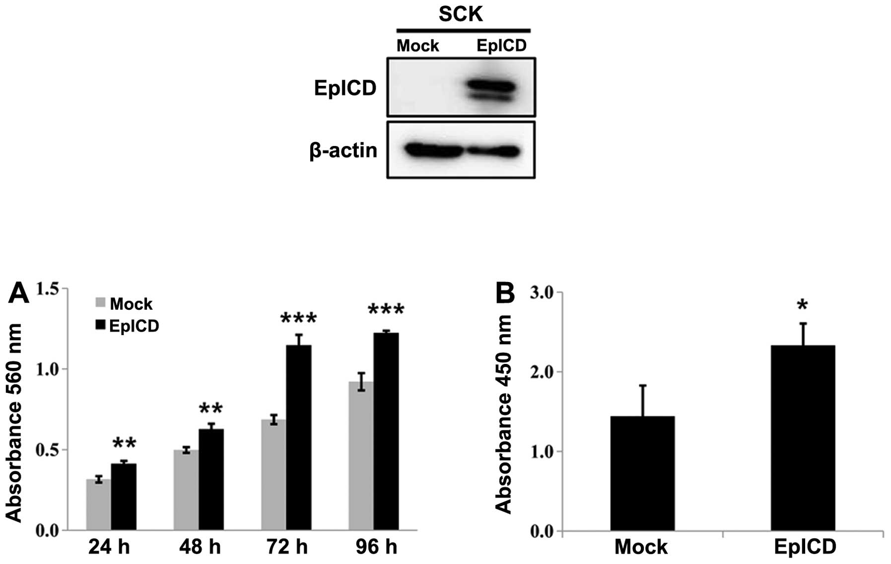
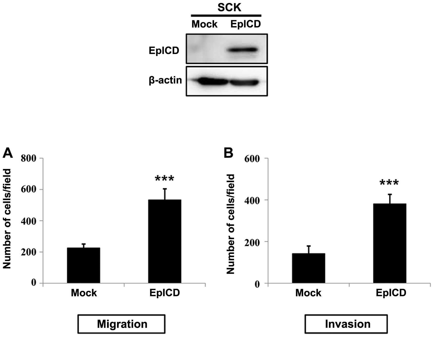
Effect of EpICD expression on cell
growth, proliferation, migration and invasion
SCK cells (minimal EpCAM protein expression) were
transiently transfected with EpICD cDNA. The EpICD-overexpressed
SCK cells revealed a significant time-dependent increase in cell
growth when compared to control cells (P<0.01; P<0.001)
(Fig. 3A). In EpICD-overexpressing
SCK cells, there was a significant increase in the BrdU
incorporation when compared to that of control cells (P<0.05)
(Fig. 3B). Overexpression of EpICD
in SCK cells leads to significantly increased cell migration and
invasion when compared to control cells (P<0.001) (Fig. 4A and B).
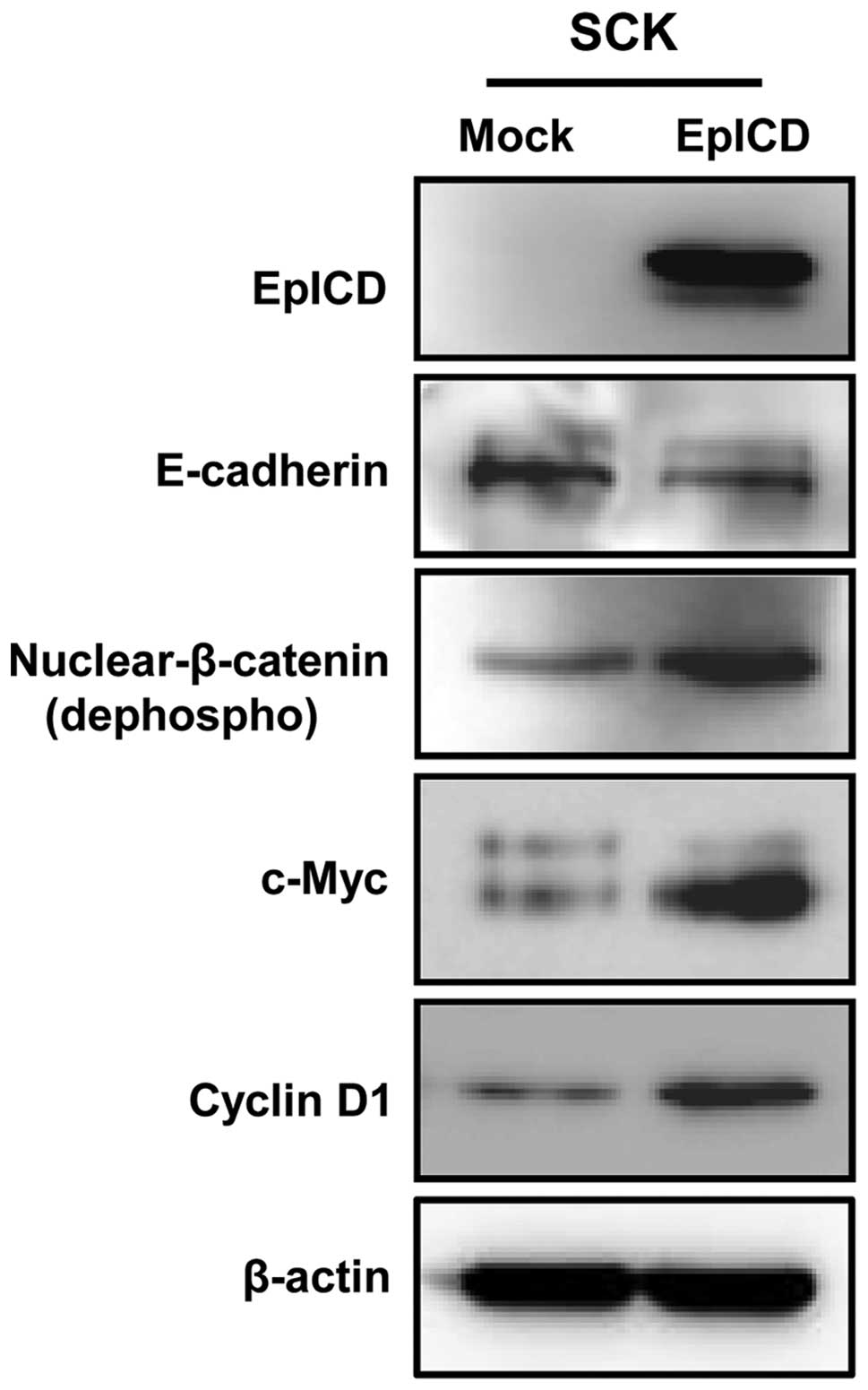
Overexpression of EpICD induces
expression of EpCAM target genes and nuclear β-catenin
The expression levels of EpICD and the active form
β-catenin (nuclear form) were significantly increased by 11- and
2.3-fold, respectively, in the EpICD-transfected SCK cells. Also,
the expression of representative EpCAM target genes, such as
c-myc and cyclin D1, were increased by 3.1- and
2.5-fold, respectively. However, overexpression of EpICD in the SCK
cells decreased the expression level of E-cadherin by 1.3-fold
(Fig. 5).
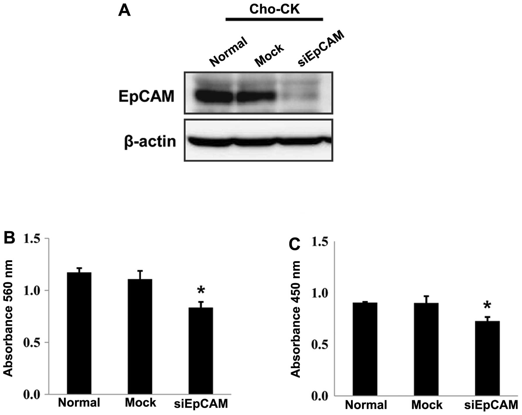
Silencing EpCAM decreases cell growth and
proliferation
Cho-CK and JCK cells (high EpCAM protein expression)
were transiently transfected with EpCAM-specific siRNA as well as
scrambled siRNA. Transfection with EpCAM siRNA resulted in a marked
decrease in the expression of EpCAM at 48 h post-transfection in
Cho-CK cells (Fig. 6A). Silencing
EpCAM gene expression in Cho-CK cells by EpCAM siRNA resulted in
significant inhibition of cell growth when compared to that of the
control (P<0.05) (Fig. 6B). In
EpCAM siRNA-transfected Cho-CK cells, there was a significant
decrease in the BrdU incorporation when compared to that of control
(P<0.05) (Fig. 6C). Silencing
EpCAM gene expression in JCK cells by EpCAM siRNA also decreased
the cell growth and BrdU incorporation compared to those of the
control, but the difference was not statistically significant.
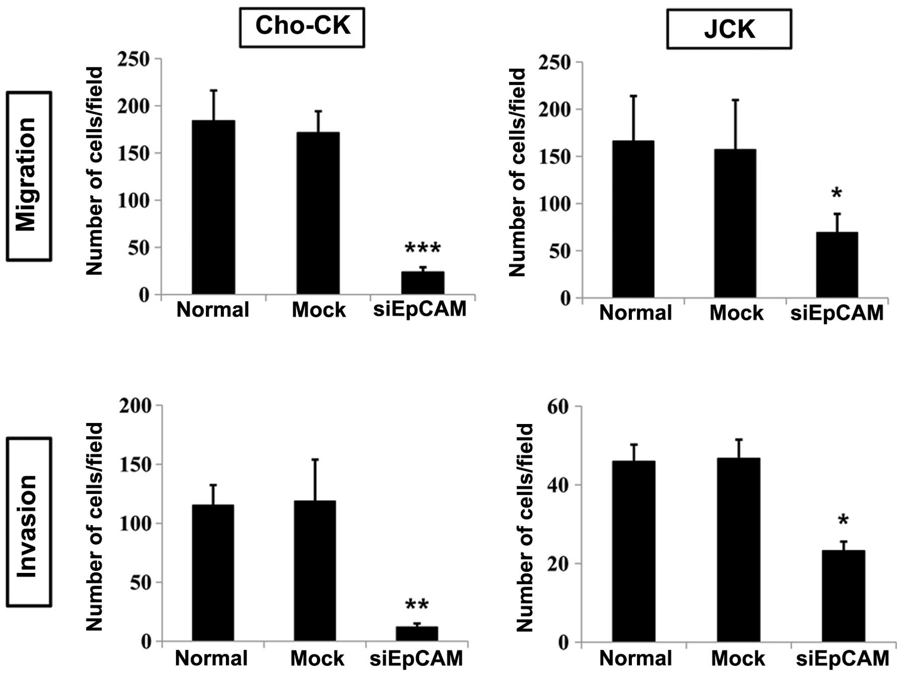
Silencing EpCAM suppresses cell migration
and invasion
Silencing EpCAM gene expression in Cho-CK and JCK
cells significantly inhibited migratory ability when compared to
control (P<0.001 and P<0.05, respectively). Silencing EpCAM
gene expression in Cho-CK and JCK cells also significantly
inhibited the invasiveness when compared to that of the control
(P<0.01 and P<0.05, respectively) (Fig. 7).
Discussion
EpCAM is highly upregulated in most human epithelial
cancers, and several biologic roles of EpCAM in carcinomas have
been reported (2–5). EpCAM is primarily a cell adhesion
molecule that inhibits cell scattering, thereby preventing tumor
cell invasion and metastasis (1,21).
Many studies indicate that the role of EpCAM is not limited to cell
adhesion; it has been shown to be widely involved in cell
proliferation, migration, invasion and cell signaling (4,5,8,10,22,23).
It has been proposed that formation of EpICD complex with the
scaffolding protein FHL2 plus Lef1 and β-catenin accounts for
activation of the oncogenic potential of EpCAM (4,8,10).
Nuclear localization of EpICD was first reported in colon cancer
cells (8); subsequently, its
nuclear expression has been frequently observed in various cancers
(9). Since the roles of EpCAM on
the biological behavior of cancer cells depend on the cancer
phenotype, we investigated the significance of nuclear EpICD and
its potential relationship with β-catenin in ECC.
This study was the first to demonstrate the
following findings in regards to EpICD expression in ECC: i)
nuclear expression of EpICD and β-catenin was detected in 27.8% and
12.6% of the 79 ECC patients, respectively, ii) a significant
correlation was observed between the nuclear expression of EpICD
and β-catenin in ECC cells with frequent co-localization of EpICD
and β-catenin in cancer cells forming the invasive front, iii)
nuclear expression of EpICD was significantly associated with high
tumor grade, iv) forced overexpression of EpICD in CC cells
increased the expression levels of the active form of β-catenin and
EpCAM target genes, such as c-myc and cyclin D1, v) the
overexpression of EpICD significantly increased cell growth and
proliferation in CC cells, and vi) the overexpression of EpICD
enhanced the cell motility and invasiveness of CC cells.
Furthermore, inhibition of the EpCAM expression in the EpCAM
high-expressing Cho-CK and JCK cells by siRNA significantly
decreased cell proliferation, migration and invasion. These
findings clearly indicate the important role of nuclear
co-expression of EpICD and β-catenin and their local interactions
in ECC progression and invasion.
The morbidity and mortality of ECC patients
predominantly depends on the degree of invasion and metastasis
(15–17). To investigate whether nuclear EpICD
is involved in ECC cell invasion in cooperation with β-catenin, we
performed immunohistochemistry using antibodies to EpICD and
β-catenin in ECC specimens. In this study, we found that budding
ECC cells of the invasive margin displayed increased translocation
of EpICD into the nucleus, which was frequently concurrent with
nuclear β-catenin expression. Notably, nuclear EpICD staining of
ECC specimens correlated significantly with nuclear expression of
β-catenin and de-differentiation of tumors. We also found that the
forced overexpression of EpICD in ECC cells increased expression
levels of the active form of β-catenin (nuclear β-catenin) and
enhanced the cell motility and invasiveness of CC cells.
Furthermore, consistent with the promoting role of EpICD in cell
invasion, knockdown of EpCAM by siRNA dramatically decreases cell
migration and invasiveness of ECC cells. These findings are in
agreement with results from a previous study demonstrating that the
oncogenic potential of EpCAM is activated by release of the EpICD
into the cytoplasm, which can signal into the nucleus by engagement
of elements of the Wnt pathway (4,8).
Evidence for the important role of EpICD in cancer
progression and invasion is increasing (25–28).
Concomitant nuclear expression of EpICD and β-catenin in anaplastic
and aggressive thyroid cancers has been reported and nuclear EpICD
expression has been shown to be correlated with poor overall
survival of thyroid cancer patients (24). Expression of EpCAM signaling
pathway members including EpICD and β-catenin in gastric carcinoma
also has been previously described (25). Fong et al have shown that
loss of membranous EpICD is frequently observed in pancreatic
cancer and can predict poor prognosis of pancreatic cancer patients
(26), and the expression level of
EpICD in colon cancer is associated with tumor grade (27). Similar results have been reported
by Gosens et al, who described a focal loss of membranous
EpEx at the invasive margin in colorectal cancer (28). Based on the above observations, our
findings of nuclear translocation of EpICD in parallel with
aberrant nuclear expression of β-catenin in cancer cells forming
the invasive front and increased expression of nuclear β-catenin by
EpICD suggest that activation of EpICD signaling can induce Wnt
signaling, thus accounting for the ECC de-differentiation and
increased invasive potential of tumor cells. However, how EpICD
promotes tumor cell invasion remains elusive. Matrix
metalloproteinases (MMPs) are a major group of proteolytic enzymes
that are implicated in migration and invasion of cancer cells
(29). Recently, Denzel et
al reported that MMP7 is a novel target gene of EpCAM
signaling, and transcription of the mmp7 gene is dependent
on nuclear translocation of EpICD and Wnt signaling through
β-catenin and Lef-1 consensus sites (30).
This study showed that overexpression of EpICD
resulted in a significant increase in the rate of cell
proliferation of CC cells. We also demonstrated that overexpression
of EpICD upregulated the expression of proto-oncogene c-myc
and the cell cycle regulating gene cyclin D1. These findings
are in agreement with those from a previous study showing that
EpICD regulates the activity of reprogramming genes, including
c-myc (27). Litvinov et
al first showed that EpCAM expression was correlated with the
proliferative activity of tumor cells as demonstrated by Ki-67
expression in cervical intraepithelial neoplasia (31). Accumulating evidence suggests that
EpCAM has a critical role in the generation of proliferative
signals into the nucleus via RIP to release EpICD (4,8–10,27,30,32).
Expression of EpICD in the absence of EpCAM is sufficient to induce
proliferation signals both in vitro and in vivo
(8). EpICD alone induces a
significant increase in cell proliferation, whereas EpCAM silencing
results in a significant decrease in cell number, which is reversed
upon EpICD co-transfection in hypopharynx carcinoma cells (32). Our findings, together with those
from previous studies, suggest that nuclear translocation of EpICD
is necessary and sufficient for cell cycle progression in ECC. This
notion is supported by the fact that the ability to rapidly
upregulate the proto-oncogene c-myc as well as cyclin A and E can
be mediated solely by EpICD (8,27,35).
Additionally, it has been shown that EpICD upregulates the
expression of cyclin D1 at the transcriptional level (34). In addition, we found that EpCAM
gene silencing by siRNA dramatically reduced CC cell proliferation
capacity. This is confirmation of earlier findings that EpCAM
blockage with siRNA inhibits cell proliferation and tumorigenicity
in various cancers (19,32–34).
In conclusion, our data indicate that EpICD is
translocated into the nucleus in a proportion of ECCs and
significantly correlates with nuclear expression of β-catenin,
especially in cancer cells forming the invasive front. The
overexpression of EpICD in the CC cells significantly enhanced cell
proliferation, migration and invasiveness with a concurrent
increase in the expression of nuclear β-catenin and EpCAM target
genes. Moreover, silencing of EpCAM gene expression significantly
inhibits tumor cell proliferation and decreases the migration and
invasiveness of CC cells. These findings strongly suggest the
importance of nuclear co-expression of EpICD and β-catenin and
their mutual interactions in ECC progression and invasion. EpCAM
has been targeted in clinical trials using monoclonal antibodies
directed against EpEx in various cancers (2,3,6,7). We
believe that the EpICD-mediated EpCAM nuclear signaling pathway is
the main contributor to the oncogenic signaling pathway. Nuclear
expression of EpICD and its role in oncogenic signaling, cancer
cell proliferation and invasiveness provide a strong basis for
further investigation of anti-EpICD-targeted therapy.
Acknowledgements
This study was supported by the National Research
Foundation of Korea (NRF) grant funded by the Korean Government
(MSIP) (no. 2008-0062279).
References
|
1
|
Litvinov SV, Balzar M, Winter MJ, et al:
Epithelial cell adhesion molecule (Ep-CAM) modulates cell-cell
interactions mediated by classic cadherins. J Cell Biol.
139:1337–1348. 1997. View Article : Google Scholar : PubMed/NCBI
|
|
2
|
Baeuerle PA and Gires O: EpCAM (CD326)
finding its role in cancer. Br J Cancer. 96:417–423. 2007.
View Article : Google Scholar : PubMed/NCBI
|
|
3
|
Went PT, Lugli A, Meier S, et al: Frequent
EpCam protein expression in human carcinomas. Hum Pathol.
35:122–128. 2004. View Article : Google Scholar : PubMed/NCBI
|
|
4
|
Munz M, Baeuerle PA and Gires O: The
emerging role of EpCAM in cancer and stem cell signaling. Cancer
Res. 69:5627–5629. 2009. View Article : Google Scholar : PubMed/NCBI
|
|
5
|
Patriarca C, Macchi RM, Marschner AK and
Mellstedt H: Epithelial cell adhesion molecule expression (CD326)
in cancer: a short review. Cancer Treat Rev. 38:68–75. 2012.
View Article : Google Scholar : PubMed/NCBI
|
|
6
|
Schmidt M, Scheulen ME, Dittrich C, et al:
An open-label, randomized phase II study of adecatumumab, a fully
human anti-EpCAM antibody, as monotherapy in patients with
metastatic breast cancer. Ann Oncol. 21:275–282. 2010. View Article : Google Scholar : PubMed/NCBI
|
|
7
|
Niedzwiecki D, Bertagnolli MM, Warren RS,
et al: Documenting the natural history of patients with resected
stage II adenocarcinoma of the colon after random assignment to
adjuvant treatment with edrecolomab or observation: results from
CALGB 9581. J Clin Oncol. 29:3146–3152. 2011. View Article : Google Scholar
|
|
8
|
Maetzel D, Denzel S, Mack B, et al:
Nuclear signalling by tumour-associated antigen EpCAM. Nat Cell
Biol. 11:162–171. 2009. View
Article : Google Scholar : PubMed/NCBI
|
|
9
|
Ralhan R, He HC, So AK, et al: Nuclear and
cytoplasmic accumulation of Ep-ICD is frequently detected in human
epithelial cancers. PLoS One. 5:e141302010. View Article : Google Scholar : PubMed/NCBI
|
|
10
|
Carpenter G and Red Brewer M: EpCAM:
another surface-to-nucleus missile. Cancer Cell. 15:165–166. 2009.
View Article : Google Scholar : PubMed/NCBI
|
|
11
|
Schmalhofer O, Brabletz S and Brabletz T:
E-cadherin, beta-catenin, and ZEB1 in malignant progression of
cancer. Cancer Metastasis Rev. 28:151–166. 2009. View Article : Google Scholar : PubMed/NCBI
|
|
12
|
Polette M, Mestdagt M, Bindels S, et al:
Beta-catenin and ZO-1: shuttle molecules involved in tumor
invasion-associated epithelial-mesenchymal transition processes.
Cells Tissues Organs. 185:61–65. 2007. View Article : Google Scholar
|
|
13
|
Vincan E and Barker N: The upstream
components of the Wnt signalling pathway in the dynamic EMT and MET
associated with colorectal cancer progression. Clin Exp Metastasis.
25:657–663. 2008. View Article : Google Scholar : PubMed/NCBI
|
|
14
|
Paul S and Dey A: Wnt signaling and cancer
development: therapeutic implication. Neoplasma. 55:165–176.
2008.PubMed/NCBI
|
|
15
|
Khan SA, Thomas HC, Davidson BR and
Taylor-Robinson SD: Cholangiocarcinoma. Lancet. 366:1303–1314.
2005. View Article : Google Scholar : PubMed/NCBI
|
|
16
|
Blechacz B and Gores GJ:
Cholangiocarcinoma: advances in pathogenesis, diagnosis, and
treatment. Hepatology. 48:308–321. 2008. View Article : Google Scholar : PubMed/NCBI
|
|
17
|
van der Gaag NA, Kloek JJ, de Bakker JK,
et al: Survival analysis and prognostic nomogram for patients
undergoing resection of extrahepatic cholangiocarcinoma. Ann Oncol.
23:2642–2649. 2012.PubMed/NCBI
|
|
18
|
Kim DG, Park SY, You KR, et al:
Establishment and characterization of chromosomal aberrations in
human cholangiocarcinoma cell lines by cross-species color banding.
Genes Chromosomes Cancer. 30:48–56. 2001. View Article : Google Scholar : PubMed/NCBI
|
|
19
|
Bae JS, Noh SJ, Jang KY, et al: Expression
and role of epithelial cell adhesion molecule in dysplastic nodule
and hepatocellular carcinoma. Int J Oncol. 41:2150–2158.
2012.PubMed/NCBI
|
|
20
|
Kwon CY, Kim KR, Choi HN, et al: The role
of serum response factor in hepatocellular carcinoma: Implications
for disease progression. Int J Oncol. 37:837–844. 2010.PubMed/NCBI
|
|
21
|
Litvinov SV, Velders MP, Bakker HA,
Fleuren GJ and Warnaar SO: Ep-CAM: a human epithelial antigen is a
homophilic cell-cell adhesion molecule. J Cell Biol. 125:437–446.
1994. View Article : Google Scholar : PubMed/NCBI
|
|
22
|
van der Gun BT, Melchers LJ, Ruiters MH,
de Leij LF, McLaughlin PM and Rots MG: EpCAM in carcinogenesis: the
good, the bad or the ugly. Carcinogenesis. 31:1913–1921.
2010.PubMed/NCBI
|
|
23
|
Trzpis M, McLaughlin PM, de Leij LM and
Harmsen MC: Epithelial cell adhesion molecule: more than a
carcinoma marker and adhesion molecule. Am J Pathol. 171:386–395.
2007. View Article : Google Scholar : PubMed/NCBI
|
|
24
|
Ralhan R, Cao J, Lim T, Macmillan C,
Freeman JL and Walfish PG: EpCAM nuclear localization identifies
aggressive thyroid cancer and is a marker for poor prognosis. BMC
Cancer. 10:3312010. View Article : Google Scholar : PubMed/NCBI
|
|
25
|
Warneke VS, Behrens HM, Haag J, et al:
Members of the EpCAM signalling pathway are expressed in gastric
cancer tissue and are correlated with patient prognosis. Br J
Cancer. 109:2217–2227. 2013. View Article : Google Scholar : PubMed/NCBI
|
|
26
|
Fong D, Moser P, Kasal A, et al: Loss of
membranous expression of the intracellular domain of EpCAM is a
frequent event and predicts poor survival in patients with
pancreatic cancer. Histopathology. 64:683–692. 2014. View Article : Google Scholar : PubMed/NCBI
|
|
27
|
Lin CW, Liao MY, Lin WW, Wang YP, Lu TY
and Wu HC: Epithelial cell adhesion molecule regulates tumor
initiation and tumorigenesis via activating reprogramming factors
and epithelial-mesenchymal transition gene expression in colon
cancer. J Biol Chem. 287:39449–39459. 2012. View Article : Google Scholar
|
|
28
|
Gosens MJ, van Kempen LC, van de Velde CJ,
van Krieken JH and Nagtegaal ID: Loss of membranous Ep-CAM in
budding colorectal carcinoma cells. Mod Pathol. 20:221–232. 2007.
View Article : Google Scholar : PubMed/NCBI
|
|
29
|
Kessenbrock K, Plaks V and Werb Z: Matrix
metalloproteinases: regulators of the tumor microenvironment. Cell.
141:52–67. 2010. View Article : Google Scholar : PubMed/NCBI
|
|
30
|
Denzel S, Mack B, Eggert C, et al: MMP7 is
a target of the tumour-associated antigen EpCAM. Int J Exp Pathol.
93:341–353. 2012. View Article : Google Scholar : PubMed/NCBI
|
|
31
|
Litvinov SV, van Driel W, van Rhijn CM, et
al: Expression of Ep-CAM in cervical squamous epithelia correlates
with an increased proliferation and the disappearance of markers
for terminal differentiation. Am J Pathol. 148:865–875. 1996.
|
|
32
|
Chaves-Pérez A, Mack B, Maetzel D, et al:
EpCAM regulates cell cycle progression via control of cyclin D1
expression. Oncogene. 32:641–650. 2013.
|
|
33
|
Osta WA, Chen Y, Mikhitarian K, et al:
EpCAM is overexpressed in breast cancer and is a potential target
for breast cancer gene therapy. Cancer Res. 64:5818–5824. 2004.
View Article : Google Scholar : PubMed/NCBI
|
|
34
|
Yamashita T, Ji J, Budhu A, et al:
EpCAM-positive hepatocellular carcinoma cells are tumor-initiating
cells with stem/progenitor cell features. Gastroenterology.
136:1012–1024. 2009. View Article : Google Scholar : PubMed/NCBI
|
|
35
|
Münz M, Kieu C, Mack B, Schmitt B, Zeidler
R and Gires O: The carcinoma-associated antigen EpCAM upregulates
c-myc and induces cell proliferation. Oncogene. 23:5748–5758.
2004.PubMed/NCBI
|