Programmed cell death 5 (PDCD5), also designated
TF-1 cell apoptosis-related gene-19 (TFAR19), is an
apoptosis-related gene cloned in 1999 from the human leukemic cell
line TF-1 (accession number AF014955 in GenBank) (1). Human PDCD5 gene is located on
chromosome 19q12-q13.1 (2), and
the integrated PDCD5 protein contains 125 amino acid (aa) residues
(3). The wide expression pattern
of PDCD5 protein in various cell lines indicated that it is a
regulator in pathological and physiological processed (4). Decreased expression of PDCD5
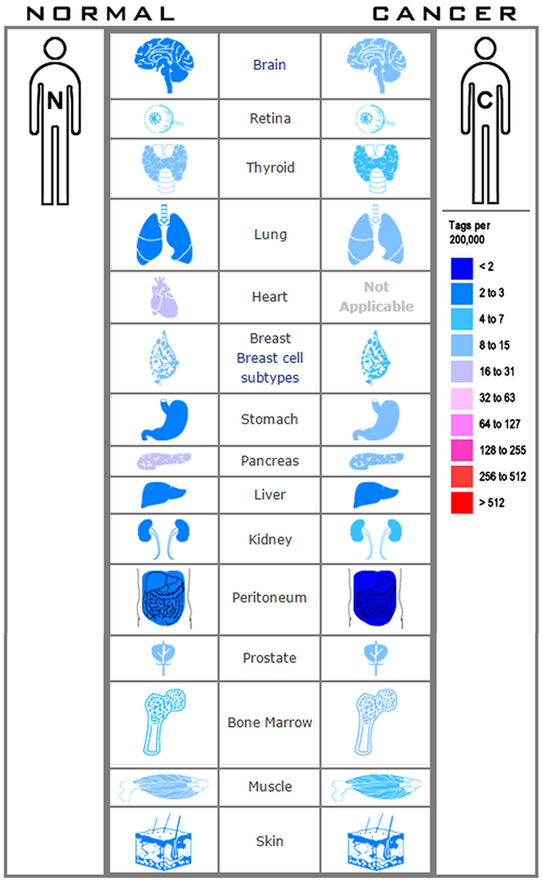
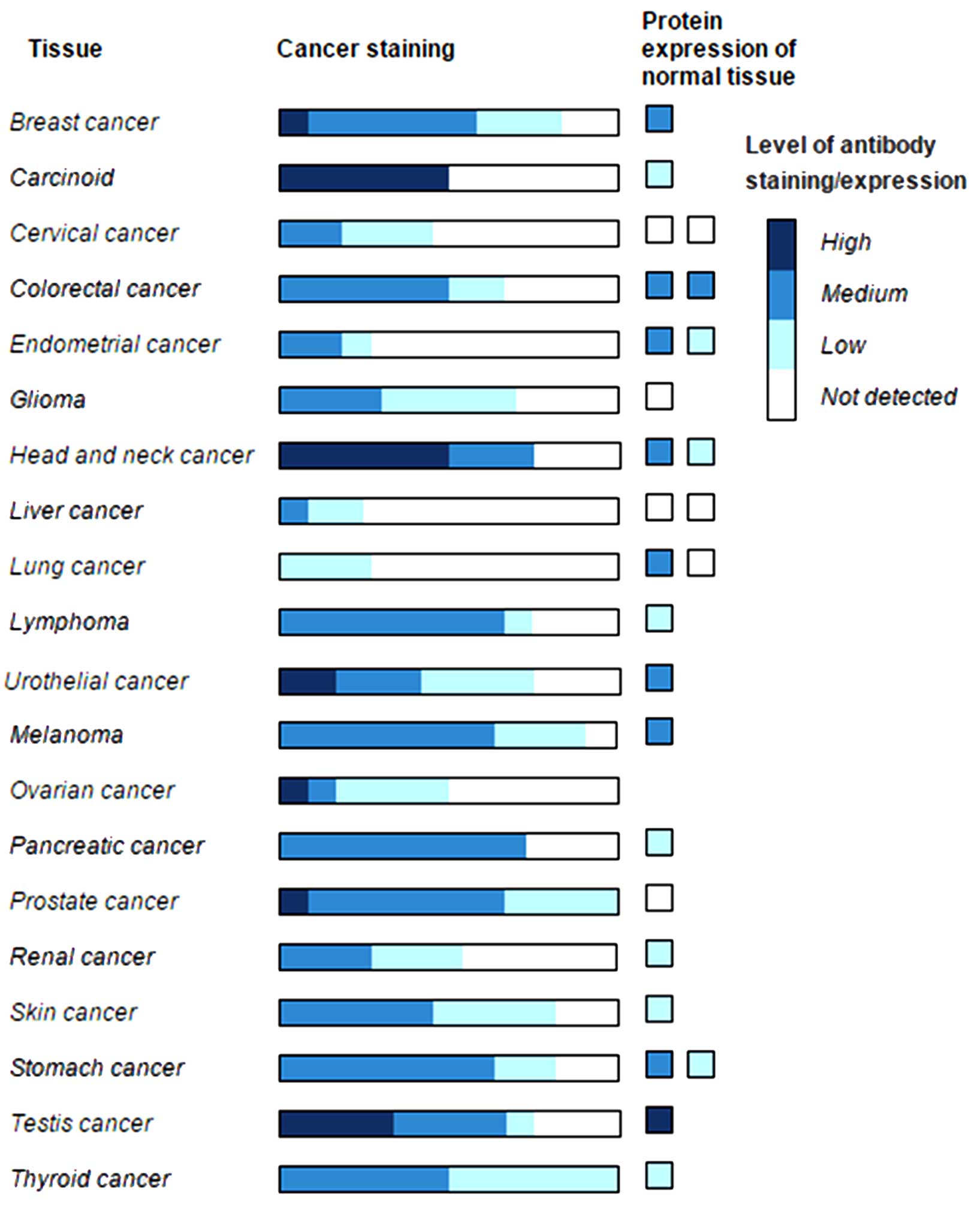
has been found in many human tumors, including breast (5), gastric cancer (6), astrocytic glioma (7), chronic myelogenous leukemia (8) and hepatocellular carcinoma (9) (Figs.
1 and 2). Previous studies
also showed that PDCD5 is involved in paraptosis (10), cell cycle regulation (11), ischemia/reperfusion (4), immunoregulation (12) and viral infection (13).
However, the molecular mechanism of PDCD5 regulation
during inflammation and cancer is very complex. According to the
published literature, PDCD5 is essential for inflammation and
cancer through regulating apoptosis. The present review is
structured to provide a comprehensive overview of the functions and
the mechanisms of PDCD5 in inflammation and cancer.
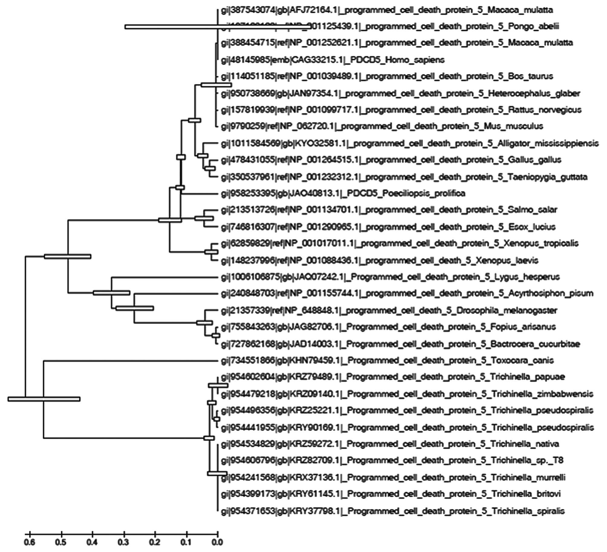
PDCD5 protein containing 125 amino acid (aa)
residues shares significant homology with the corresponding
proteins of species ranging from yeast to mice (1) (Fig.
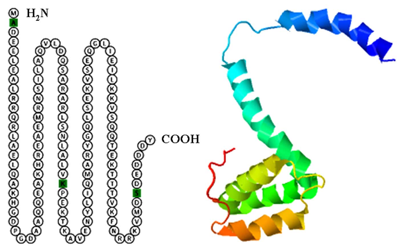
3). Human PDCD5 protein can be divided into three structural
regions: a rigid core region (residues 41-101 aa), C-terminal
residues (102-125 aa) and N-terminal residues (3-40 aa) (3) (Fig.
4). In the study of Liu et al (14), it was found that the sequence
region of PDCD5 (Asp3-Ala19) contains four
leucine residues, three alanine residues, and two polar amino acid
residues by using NMR methods. They also found that PDCD5 without
N-terminal residues significantly attenuates the
apoptosis-promoting effects on myeloblastic leukemia HL-60 cells
(14). The C-terminal region of
PDCD5 (Val109–Val116) includes four basic
residues, which mediate the ability of the protein to bind to
heparin, is important for PDCD5 to translocate through plasma
membranes (15). The nuclear
translocation of PDCD5 is an early event of the apoptotic process,
and may be a novel early marker for apoptosis (16).
Osteoarthritis (OA) is one of the most common
chronic health conditions and a leading cause of pain and
disability among adults (17). The
main cause of OA is that articular chondrocytes lose proliferative
capacity while maintaining the ability to produce proinflammatory
mediators and matrix degrading enzymes (18). Cheng et al (19) found the enhancement of PDCD5
expression in OA cartilage compared with that in normal healthy
cartilage. In the study of Yi et al (20) they also found that the level of
PDCD5 was negatively correlated with the rate of chondrocyte
apoptosis. These results indicated that PDCD5 is involved in the
pathogenesis of OA.
PDCD5 also plays an important role in rheumatoid
arthritis. Rheumatoid arthritis (RA) is a chronic systemic
inflammatory joint disease characterized by hyperplasia of synovial
tissue, inflammatory infiltrates and a progressive destruction of
cartilage and bone (21). PDCD5
expression was low in RA patient-derived synovial tissue and
cultured fibroblast-like synoviocytes (22). Plasma and synovial fluid PDCD5
abnormal expression and dysfunction may be correlated to tumor
necrosis factor-α (TNF-α) in RA patients (23). PDCD5 level was also negatively
correlated with proinflammatory cytokine interleukin (IL)-17 levels
both in serum and synovial fluid of RA patients (24). These results indicated that plasma
and synovial fluid PDCD5 could be useful for monitoring the
activity and progression of RA. Xiao et al (25) found that overexpression of PDCD5
increased the level of Foxp3 protein and percentage of
Foxp3+ regulatory T (Treg) cells, and suppressed Th17
and Th1 responses in PDCD5 transgenic mice. A recent study showed
that recombinant human PDCD5 (rhPDCD5) protein has prophylactic and
therapeutic properties in a mouse model of multiple sclerosis by
inhibiting Th1/Th17 differentiation and inducing apoptosis of
predominantly pathogenic T cells (13).
The results of the present studies confirmed that
PDCD5 serves as a guardian of immunological functions and that the
PDCD5-FOXP3-Treg axis may be a therapeutic target for
inflammation.
PDCD5 is apoptosis-related gene originally cloned in
1999 from TF-1 human leukemic cell line undergoing apoptosis
(1). From then on, PDCD5 was
demonstrated to promote apoptosis in cancer cells in many studies.
Ruan et al (8) found that
PDCD5 mRNA was lower in both acute myeloid leukemia (AML)
and chronic myeloid leukemia (CML) marrow cells than that in normal
donor marrow cells. They also observed a synergistic effect on
apoptotic cell death in human CML K562 cells after combination
therapy with adenovirus-mediated PDCD5 and idarubicin in
vitro and in vivo (26). Shi et al (27) found that rhPDCD5 protein sensitizes
K562 cells to apoptosis induced by chemotherapeutic drugs in
vitro and in vivo.
Human glioma is one of the most frequent primary
tumors of the central nervous system (7). A reduced expression of PDCD5
mRNA was found in glioma compared with normal brain tissue
(41). Li et al (42) confirmed that PDCD5 promotes
cisplatin-induced apoptosis in glioma cells (U87, U251 and T98G) by
activating mitochondrial apoptotic pathway. PDCD5 mRNA and protein
expression were downregulated in ovarian (43) and prostate cancer (44). rhPDCD5 protein increases
doxorubicin-induced apoptosis in ovarian cancer SKOV3 cells
(44). Notably, a recent study of
Gao et al (45) confirmed
that reduced PDCD5 protein is correlated with the degree of tumor
differentiation in endometrioid endometrial carcinoma.
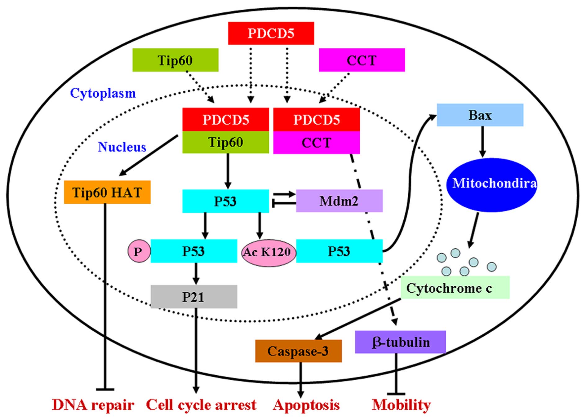
PDCD5 inhibits the proliferation of cancer cells by
the mitochondrial apoptotic pathway and the NF-κB pathway (54,55).
Collectively, these studies provide compelling
evidence of the antitumor mechanisms of PDCD5. Better understanding
of these mechanisms could create novel therapeutic opportunities in
treating cancer cells (Fig.
5).
To date, cancer is still a fatal disease, and
relapse rates in patients remain high regardless of the intensity
of treatment. In recent years, PDCD5 has received increased
attention in the molecular biology of cancer. Based on previous
results and concepts, PDCD5 could be used as a prognostic marker
for cancers. We believe that these data will open new perspectives
for a better understanding of PDCD5. However, we are only at the
beginning of understanding the precise roles of PDCD5 in cancer
cells. Current results did not have sufficient certainty to permit
their use in cancer patients. Thus, PDCD5 safety and efficacy in
cancer treatment may be significant issues in future studies.
The present review was supported by the Natural
Scientific Foundation of Liaoning Province (2014021036), the
General Project Scientific Research from the Department of
Education of Liaoning Province (L2012288) and the Natural
Scientific Foundation of China (81502107).
|
1
|
Liu H, Wang Y, Zhang Y, Song Q, Di C, Chen
G, Tang J and Ma D: TFAR19, a novel apoptosis-related gene cloned
from human leukemia cell line TF-1, could enhance apoptosis of some
tumor cells induced by growth factor withdrawal. Biochem Biophys
Res Commun. 254:203–210. 1999. View Article : Google Scholar : PubMed/NCBI
|
|
2
|
Spinola M, Meyer P, Kammerer S, Falvella
FS, Boettger MB, Hoyal CR, Pignatiello C, Fischer R, Roth RB,
Pastorino U, et al: Association of the PDCD5 locus with lung cancer
risk and prognosis in smokers. J Clin Oncol. 24:1672–1678. 2006.
View Article : Google Scholar : PubMed/NCBI
|
|
3
|
Liu D, Feng Y, Cheng Y and Wang J: Human
programmed cell death 5 protein has a helical-core and two
dissociated structural regions. Biochem Biophys Res Commun.
318:391–396. 2004. View Article : Google Scholar : PubMed/NCBI
|
|
4
|
Chen CH, Jiang Z, Yan JH, Yang L, Wang K,
Chen YY, Han JY, Zhang JH and Zhou CM: The involvement of
programmed cell death 5 (PDCD5) in the regulation of apoptosis in
cerebral ischemia/reperfusion injury. CNS Neurosci Ther.
19:566–576. 2013. View Article : Google Scholar : PubMed/NCBI
|
|
5
|
Hedenfalk I, Duggan D, Chen Y, Radmacher
M, Bittner M, Simon R, Meltzer P, Gusterson B, Esteller M,
Kallioniemi OP, et al: Gene-expression profiles in hereditary
breast cancer. N Engl J Med. 344:539–548. 2001. View Article : Google Scholar : PubMed/NCBI
|
|
6
|
Yang YH, Zhao M, Li WM and Lu YY, Chen YY,
Kang B and Lu YY: Expression of programmed cell death 5 gene
involves in regulation of apoptosis in gastric tumor cells.
Apoptosis. 11:993–1001. 2006. View Article : Google Scholar : PubMed/NCBI
|
|
7
|
Li H, Wang Q, Gao F, Zhu F, Wang X, Zhou
C, Liu C, Chen Y, Ma C, Sun W, et al: Reduced expression of PDCD5
is associated with high-grade astrocytic gliomas. Oncol Rep.
20:573–579. 2008.PubMed/NCBI
|
|
8
|
Ruan GR, Qin YZ, Chen SS, Li JL, Ma X,
Chang Y, Wang YZ, Fu JY and Liu YR: Abnormal expression of the
programmed cell death 5 gene in acute and chronic myeloid leukemia.
Leuk Res. 30:1159–1165. 2006. View Article : Google Scholar : PubMed/NCBI
|
|
9
|
Fu DZ, Cheng Y, He H, Liu HY and Liu YF:
PDCD5 expression predicts a favorable outcome in patients with
hepatocellular carcinoma. Int J Oncol. 43:821–830. 2013.PubMed/NCBI
|
|
10
|
Wang Y, Li X, Wang L, Ding P, Zhang Y, Han
W and Ma D: An alternative form of paraptosis-like cell death,
triggered by TAJ/TROY and enhanced by PDCD5 overexpression. J Cell
Sci. 117:1525–1532. 2004. View Article : Google Scholar : PubMed/NCBI
|
|
11
|
Xu L, Hu J, Zhao Y, Hu J, Xiao J, Wang Y,
Ma D and Chen Y: PDCD5 interacts with p53 and functions as a
positive regulator in the p53 pathway. Apoptosis. 17:1235–1245.
2012. View Article : Google Scholar : PubMed/NCBI
|
|
12
|
Xiao J, Liu C, Li G, Peng S, Hu J, Qu L,
Lv P, Zhang Y, Ma D and Chen Y: PDCD5 negatively regulates
autoimmunity by upregulating FOXP3+ regulatory T cells
and suppressing Th17 and Th1 responses. J Autoimmun. 47:34–44.
2013. View Article : Google Scholar : PubMed/NCBI
|
|
13
|
Li K, Zhou Z, Wang YO, Liu J, Zhao HB,
Yang J and Wang SQ: Pretreatment of mice with oligonucleotide prop5
protects them from influenza virus infections. Viruses. 6:573–581.
2014. View
Article : Google Scholar : PubMed/NCBI
|
|
14
|
Liu D, Yao H, Chen Y, Feng Y, Chen Y and
Wang J: The N-terminal 26-residue fragment of human programmed cell
death 5 protein can form a stable alpha-helix having unique
electrostatic potential character. Biochem J. 392:47–54. 2005.
View Article : Google Scholar : PubMed/NCBI
|
|
15
|
Colin S, Jeanny JC, Mascarelli F, Vienet
R, Al-Mahmood S, Courtois Y and Labarre J: In vivo involvement of
heparan sulfate proteoglycan in the bioavailability,
internalization, and catabolism of exogenous basic fibroblast
growth factor. Mol Pharmacol. 55:74–82. 1999.PubMed/NCBI
|
|
16
|
Chen Y, Sun R, Han W, Zhang Y, Song Q, Di
C and Ma D: Nuclear translocation of PDCD5 (TFAR19): An early
signal for apoptosis? FEBS Lett. 509:191–196. 2001. View Article : Google Scholar : PubMed/NCBI
|
|
17
|
Johnson VL and Hunter DJ: The epidemiology
of osteoarthritis. Best Pract Res Clin Rheumatol. 28:5–15. 2014.
View Article : Google Scholar : PubMed/NCBI
|
|
18
|
Loeser RF: Aging and osteoarthritis: The
role of chondrocyte senescence and aging changes in the cartilage
matrix. Osteoarthritis Cartilage. 17:971–979. 2009. View Article : Google Scholar : PubMed/NCBI
|
|
19
|
Cheng AX, Lou SQ, Zhou HW, Wang Y and Ma
DL: Expression of PDCD5, a novel apoptosis related protein, in
human osteoarthritic cartilage. Acta Pharmacol Sin. 25:685–690.
2004.PubMed/NCBI
|
|
20
|
Yi C, Ma C, Xie Z, Zhang G, Song W, Zhou X
and Cao Y: Downregulation of programmed cell death 5 by
insulin-like growth factor 1 in osteoarthritis chondrocytes. Int
Orthop. 37:937–943. 2013. View Article : Google Scholar : PubMed/NCBI
|
|
21
|
Firestein GS: Evolving concepts of
rheumatoid arthritis. Nature. 423:356–361. 2003. View Article : Google Scholar : PubMed/NCBI
|
|
22
|
Wang N, Lu HS, Guan ZP, Sun TZ, Chen YY,
Ruan GR, Chen ZK, Jiang J and Bai CJ: Involvement of PDCD5 in the
regulation of apoptosis in fibroblast-like synoviocytes of
rheumatoid arthritis. Apoptosis. 12:1433–1441. 2007. View Article : Google Scholar : PubMed/NCBI
|
|
23
|
Wang J, Guan Z and Ge Z: Plasma and
synovial fluid programmed cell death 5 (PDCD5) levels are inversely
associated with TNF-_α and disease activity in patients with
rheumatoid arthritis. Biomarkers. 18:155–159. 2013. View Article : Google Scholar : PubMed/NCBI
|
|
24
|
Wang JF, Guan ZP, Zhang SL, Pei Z, Chen YY
and Pan H: Programmed cell death 5 correlates with disease activity
and interleukin-17 in serum and synovial fluid of rheumatoid
arthritis patients. Chin Med J (Engl). 126:296–299. 2013.
|
|
25
|
Xiao J, Liu W, Chen Y and Deng W:
Recombinant human PDCD5 (rhPDCD5) protein is protective in a mouse
model of multiple sclerosis. J Neuroinflammation. 12:1172015.
View Article : Google Scholar : PubMed/NCBI
|
|
26
|
Ruan GR, Zhao HS, Chang Y, Li JL, Qin YZ,
Liu YR, Chen SS and Huang XJ: Adenovirus-mediated PDCD5 gene
transfer sensitizes K562 cells to apoptosis induced by idarubicin
in vitro and in vivo. Apoptosis. 13:641–648. 2008. View Article : Google Scholar : PubMed/NCBI
|
|
27
|
Shi L, Song Q, Zhang Y, Lou Y and Wang Y,
Tian L, Zheng Y, Ma D, Ke X and Wang Y: Potent antitumor activities
of recombinant human PDCD5 protein in combination with chemotherapy
drugs in K562 cells. Biochem Biophys Res Commun. 396:224–230. 2010.
View Article : Google Scholar : PubMed/NCBI
|
|
28
|
Thompson RC Jr, Cheng EY, Clohisy DR,
Perentesis J, Manivel C and Le CT: Results of treatment for
metastatic osteosarcoma with neoadjuvant chemotherapy and surgery.
Clin Orthop Relat Res. 397:240–247. 2002. View Article : Google Scholar : PubMed/NCBI
|
|
29
|
Han XR, Sun Y and Bai XZ: The anti-tumor
role and mechanism of integrated and truncated PDCD5 proteins in
osteosarcoma cells. Cell Signal. 24:1713–1721. 2012. View Article : Google Scholar : PubMed/NCBI
|
|
30
|
Zhao H, Peng C, Ruan G, Zhou J, Li Y and
Hai Y: Adenovirus-delivered PDCD5 counteracts adriamycin resistance
of osteosarcoma cells through enhancing apoptosis and inhibiting
Pgp. Int J Clin Exp Med. 7:5429–5436. 2014.
|
|
31
|
Rozeman LB, Cleton-Jansen AM and
Hogendoorn PC: Pathology of primary malignant bone and cartilage
tumours. Int Orthop. 30:437–444. 2006. View Article : Google Scholar : PubMed/NCBI
|
|
32
|
Chen C, Zhou H, Xu L, Liu X, Liu Z, Ma D,
Chen Y and Ma Q: Prognostic significance of downregulated
expression of programmed cell death 5 in chondrosarcoma. J Surg
Oncol. 102:838–843. 2010. View Article : Google Scholar : PubMed/NCBI
|
|
33
|
Chen C, Zhou H, Xu L, Xu D, Wang Y, Zhang
Y, Liu X, Liu Z, Ma D, Ma Q, et al: Recombinant human PDCD5
sensitizes chondrosarcomas to cisplatin chemotherapy in vitro and
in vivo. Apoptosis. 15:805–813. 2010. View Article : Google Scholar : PubMed/NCBI
|
|
34
|
Nanba K, Toyooka S, Soh J, Tsukuda K,
Yamamoto H, Sakai A, Ouchida M, Kobayashi N, Matsuo K, Koide N, et
al: The allelic distribution of a single nucleotide polymorphism in
the PDCD5 gene locus of Japanese non-small cell lung cancer
patients. Mol Med Rep. 1:667–671. 2008.PubMed/NCBI
|
|
35
|
Xu F, Wu K, Zhao M, Qin Y and Xia M:
Expression and clinical significance of the programmed cell death 5
gene and protein in laryngeal squamous cell carcinoma. J Int Med
Res. 41:1838–1847. 2013. View Article : Google Scholar : PubMed/NCBI
|
|
36
|
Wang L, Wang C, Su B, Song Q, Zhang Y, Luo
Y, Li Q, Tan W, Ma D and Wang L: Recombinant human PDCD5 protein
enhances chemosensitivity of breast cancer in vitro and in vivo.
Biochem Cell Biol. 91:526–531. 2013. View Article : Google Scholar : PubMed/NCBI
|
|
37
|
Fan GL, Yao Y, Yao L and Li Y: PDCD5
transfection increases cisplatin sensitivity and decreases invasion
in hepatic cancer cells. Oncol Lett. 9:411–417. 2015.
|
|
38
|
Xu HY, Chen ZW, Pan YM, Fan L, Guan J and
Lu YY: Transfection of PDCD5 effect on the biological behavior of
tumor cells and sensitized gastric cancer cells to
cisplatin-induced apoptosis. Dig Dis Sci. 57:1847–1856. 2012.
View Article : Google Scholar : PubMed/NCBI
|
|
39
|
Yin A, Jiang Y, Zhang X, Zhao J and Luo H:
Transfection of PDCD5 sensitizes colorectal cancer cells to
cisplatin-induced apoptosis in vitro and in vivo. Eur J Pharmacol.
649:120–126. 2010. View Article : Google Scholar : PubMed/NCBI
|
|
40
|
Wang D, Wang W, Song CL and Xia P: The
roles of serum PDCD5 in circulating CD133 positive cells of the
patients with gastric cancer. Tumour Biol. Mar 31–2016.Epub ahead
of print.
|
|
41
|
Olischar M, Stavroudis T, Karp JK,
Kaufmann WE and Theda C: Medical and ethical challenges in the case
of a prenatally undiagnosed massive congenital brain tumor. J
Perinatol. 35:773–775. 2015. View Article : Google Scholar : PubMed/NCBI
|
|
42
|
Li H, Zhang X, Song X, Zhu F, Wang Q, Guo
C, Liu C, Shi Y, Ma C, Wang X, et al: PDCD5 promotes
cisplatin-induced apoptosis of glioma cells via activating
mitochondrial apoptotic pathway. Cancer Biol Ther. 13:822–830.
2012. View Article : Google Scholar : PubMed/NCBI
|
|
43
|
Gao L, Ye X, Ma RQ, Cheng HY, Han HJ, Cui
H, Wei LH and Chang XH: Low programmed cell death 5 expression is a
prognostic factor in ovarian cancer. Chin Med J (Engl).
128:1084–1090. 2015. View Article : Google Scholar
|
|
44
|
Du YJ, Xiong L, Lou Y, Tan WL and Zheng
SB: Reduced expression of programmed cell death 5 protein in tissue
of human prostate cancer. Chin Med Sci J. 24:241–245. 2009.
View Article : Google Scholar
|
|
45
|
Gao M, Gao W, Wang Z, Liu Y, Li Y, Wei C,
Sun Y, Guo C, Zhang L, Wei Z, et al: The reduced PDCD5 protein is
correlated with the degree of tumor differentiation in endometrioid
endometrial carcinoma. Springerplus. 5:9882016. View Article : Google Scholar : PubMed/NCBI
|
|
46
|
Chen LN, Wang Y, Ma DL and Chen YY: Short
interfering RNA against the PDCD5 attenuates cell apoptosis and
caspase-3 activity induced by Bax overexpression. Apoptosis.
11:101–111. 2006. View Article : Google Scholar
|
|
47
|
Xu L, Chen Y, Song Q, Xu D, Wang Y and Ma
D: PDCD5 interacts with Tip60 and functions as a cooperator in
acetyltransferase activity and DNA damage-induced apoptosis.
Neoplasia. 11:345–354. 2009. View Article : Google Scholar : PubMed/NCBI
|
|
48
|
Zhuge C, Chang Y, Li Y, Chen Y and Lei J:
PDCD5-regulated cell fate decision after
ultraviolet-irradiation-induced DNA damage. Biophys J.
101:2582–2591. 2011. View Article : Google Scholar
|
|
49
|
Essers PB, Klasson TD, Pereboom TC, Mans
DA, Nicastro M, Boldt K, Giles RH and MacInnes AW: The von
Hippel-Lindau tumor suppressor regulates programmed cell death
5-mediated degradation of Mdm2. Oncogene. 34:771–779. 2015.
View Article : Google Scholar
|
|
50
|
Choi HK, Choi Y, Park ES, Park SY, Lee SH,
Seo J, Jeong MH, Jeong JW, Jeong JH, Lee PC, et al: Programmed cell
death 5 mediates HDAC3 decay to promote genotoxic stress response.
Nat Commun. 6:73902015. View Article : Google Scholar : PubMed/NCBI
|
|
51
|
Park SY, Choi HK, Choi Y, Kwak S, Choi KC
and Yoon HG: Deubiquitinase OTUD5 mediates the sequential
activation of PDCD5 and p53 in response to genotoxic stress. Cancer
Lett. 357:419–427. 2015. View Article : Google Scholar
|
|
52
|
Tracy CM, Gray AJ, Cuéllar J, Shaw TS,
Howlett AC, Taylor RM, Prince JT, Ahn NG, Valpuesta JM and
Willardson BM: Programmed cell death protein 5 interacts with the
cytosolic chaperonin containing tailless complex polypeptide 1
(CCT) to regulate β-tubulin folding. J Biol Chem. 289:4490–4502.
2014. View Article : Google Scholar : PubMed/NCBI
|
|
53
|
Fu DZ, Cheng Y, He H, Liu HY and Liu YF:
Recombinant human PDCD5 exhibits an antitumor role in
hepatocellular carcinoma cells via clathrin-dependent endocytosis.
Mol Med Rep. 12:8135–8140. 2015.PubMed/NCBI
|
|
54
|
Xu S, Sui G, Yuan L and Zou Z: Expression
of programmed cell death 5 protein inhibits progression of lung
carcinoma in vitro and in vivo via the mitochondrial apoptotic
pathway. Mol Med Rep. 10:2059–2064. 2014.PubMed/NCBI
|
|
55
|
Murshed F, Farhana L, Dawson MI and
Fontana JA: NF-κB p65 recruited SHP regulates PDCD5-mediated
apoptosis in cancer cells. Apoptosis. 19:506–517. 2014. View Article : Google Scholar
|